
PUK and moorens ulcer
Reading File
Finding Sources
Finding Sources
Searching PubMed
"peripheral ulcerative keratitis" AND "Mooren ulcer"
Searching the Web
peripheral ulcerative keratitis versus Mooren ulcer clinical differentiation management review
Reading File
Reading File
Reading Web Page
https://pmc.ncbi.nlm.nih.gov/articles/PMC9185208
Peripheral ulcerative keratitis (PUK) and Mooren ulcer
PUK is a painful, sight-threatening peripheral corneal ulceration with epithelial defect, stromal melt/thinning, and inflammation. It is a clinical syndrome that may signal an underlying systemic autoimmune vasculitis.
Mooren ulcer is an idiopathic autoimmune peripheral ulcerative keratitis, diagnosed only after excluding systemic autoimmune disease and infection.
| Feature | PUK associated with systemic disease | Mooren ulcer |
|---|---|---|
| Underlying cause | Often rheumatoid arthritis, granulomatosis with polyangiitis, relapsing polychondritis, SLE, PAN, etc. | Local corneal autoimmune process, no causal systemic autoimmune disease |
| Pain | Painful | Usually very painful, with photophobia and redness |
| Site and progression | Peripheral crescentic ulceration and corneal melt | Begins at limbus, often nasal/temporal interpalpebral cornea, spreads circumferentially then centrally |
| Sclera | Adjacent episcleritis/scleritis may occur | No scleritis. This is a key distinction |
| Ulcer edge | Peripheral stromal ulceration | Characteristic undermined, infiltrated advancing edge |
| Systemic significance | May be the first sign of a potentially life-threatening vasculitis | Diagnosis of exclusion |
| Laterality/course | Variable | Older patients: often unilateral, slower. Younger patients: often bilateral, aggressive |
| Treatment priority | Treat ocular disease and urgently control systemic disease | Local and sometimes systemic immunosuppression after excluding infection/systemic causes |
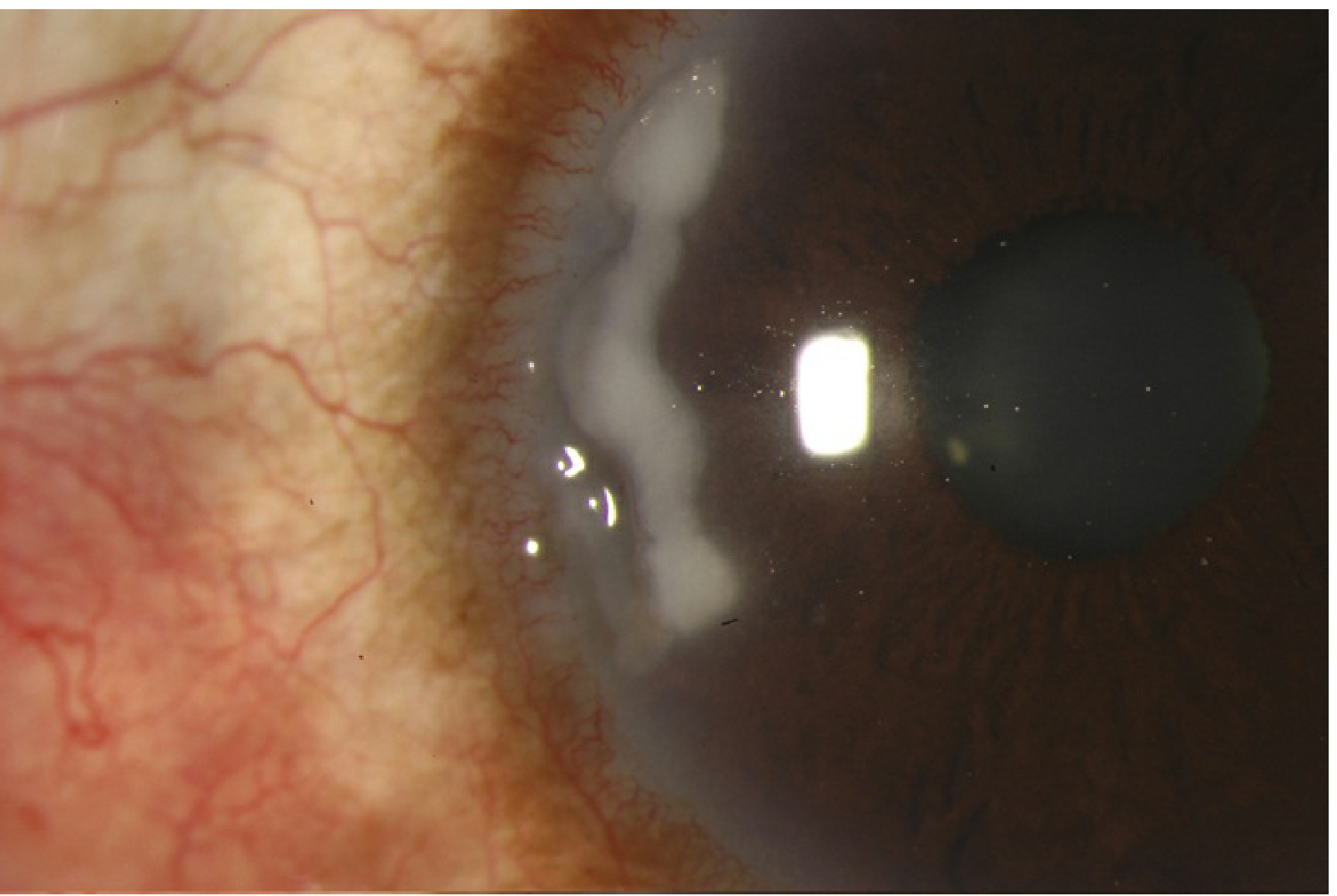
Peripheral ulcerative keratitis.
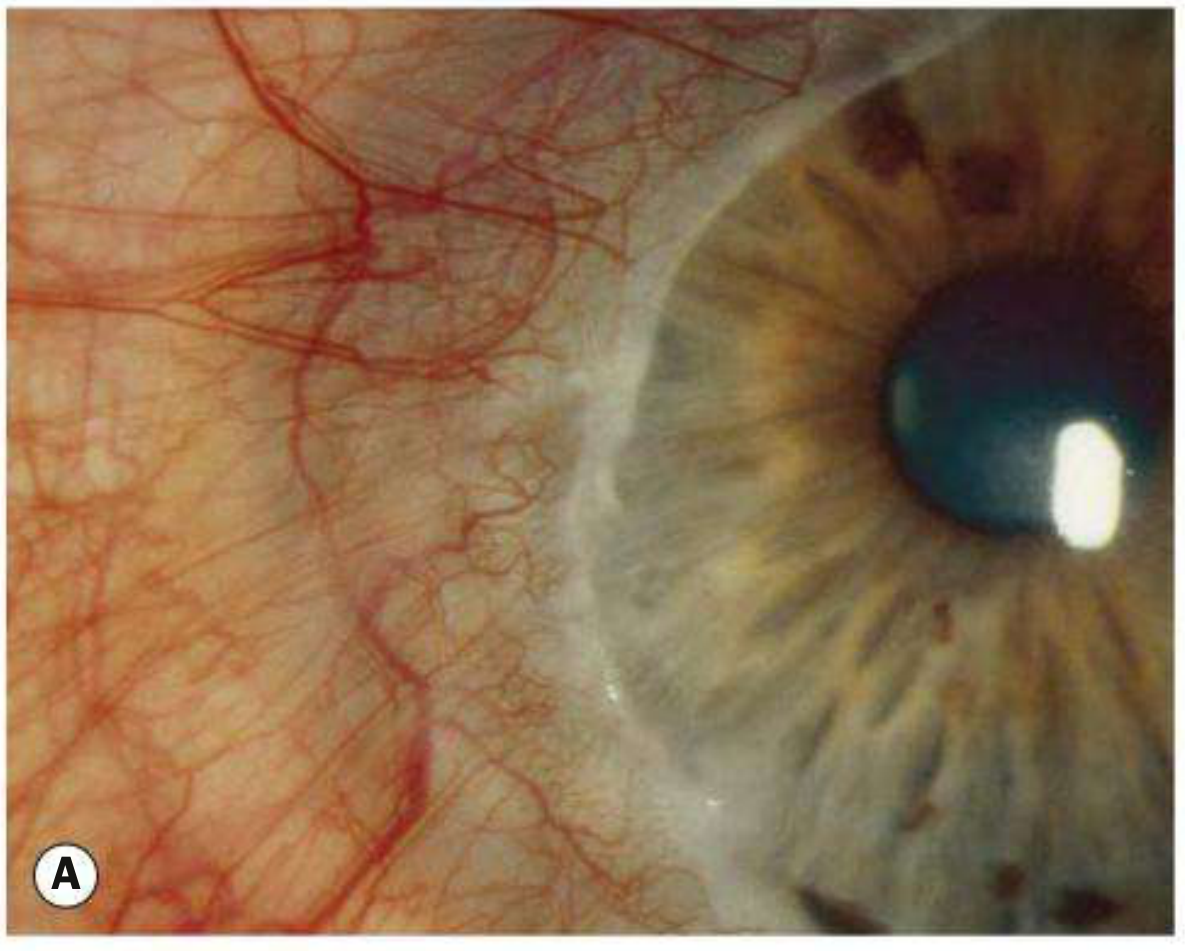
Mooren ulcer with peripheral ulceration and an advancing edge.
Diagnosis and work-up
- Urgent slit-lamp examination: ulcer extent, epithelial defect, stromal thinning, infiltrate, anterior chamber reaction, Seidel test, and adjacent scleral inflammation.
- Exclude infection first, especially bacterial, fungal, viral, and acanthamoeba keratitis. Culture/scrape when clinically indicated before immunosuppression.
- In apparent PUK, investigate for systemic inflammatory disease, commonly with rheumatology input: history and examination for arthritis, ENT/chest/renal symptoms, rash, neuropathy, oral ulcers; targeted tests may include RF, anti-CCP, ANCA, ANA, inflammatory markers, urinalysis, and renal function.
- Consider precipitating trauma, previous corneal surgery, ocular surface disease, and in suspected Mooren ulcer, relevant infectious associations such as hepatitis C where appropriate.
Management principles
- This is an ophthalmic emergency because rapid corneal melting and perforation can occur.
- Use intensive lubrication, protective measures, and therapy to limit collagen breakdown such as oral doxycycline or topical N-acetylcysteine when appropriate.
- Once infection is excluded, use topical corticosteroid and/or topical cyclosporine under ophthalmology supervision.
- Systemic PUK, especially with scleritis or vasculitis, often requires systemic corticosteroids plus steroid-sparing immunosuppression directed with rheumatology.
- In Mooren ulcer, topical therapy may suffice for milder unilateral disease, but aggressive/bilateral disease may require systemic immunosuppression.
- Progressive thinning or perforation may need tissue adhesive with bandage contact lens, amniotic membrane, conjunctival resection, lamellar patch graft, or keratoplasty.
Exam pearl:
Painful peripheral corneal ulcer + scleritis/systemic autoimmune disease = systemic disease-associated PUK.
Painful peripheral ulcer with undermined edge but no scleritis or systemic disease = Mooren ulcer.
Mooren ulcer is described as progressive circumferential peripheral stromal ulceration with later central extension; prominent pain and absence of scleritis help separate it from systemic PUK. Kanski’s Clinical Ophthalmology, 10th ed., p. 254. PUK may be associated with connective-tissue disease and can be its first manifestation, whereas Mooren ulcer is a diagnosis of exclusion. The Wills Eye Manual, 8th ed., p. 261. A recent PUK review likewise characterizes Mooren ulcer as an idiopathic form of PUK without scleral involvement.
Xerophthalmia
Reading File
Finding Sources
Searching PubMed
xerophthalmia AND vitamin A
Searching the Web
WHO xerophthalmia vitamin A deficiency eye signs treatment
Searching Images
xerophthalmia vitamin A deficiency Bitot spots corneal xerosis clinical photograph
Reading File
Finding Sources
Xerophthalmia
Xerophthalmia is the spectrum of ocular manifestations caused by vitamin A deficiency (VAD). It ranges from night blindness to corneal melting and irreversible blindness.
Pathogenesis
Vitamin A is needed for:
- Rhodopsin formation in retinal rods, so deficiency causes defective dark adaptation and night blindness.
- Maintenance of normal conjunctival and corneal epithelium. Deficiency causes loss of goblet cells, reduced mucin, keratinization, dryness, ulceration, and corneal melt.
WHO clinical stages
| WHO code | Manifestation | Key feature |
|---|---|---|
| XN | Night blindness | Difficulty seeing in dim light. Earliest symptom |
| X1A | Conjunctival xerosis | Dry, dull, wrinkled, non-wettable conjunctiva |
| X1B | Bitot spots | Triangular, foamy, pearly-white/yellow plaques on bulbar conjunctiva |
| X2 | Corneal xerosis | Dry, hazy, non-wettable cornea |
| X3A | Corneal ulceration/keratomalacia involving less than one-third of cornea | Severe disease |
| X3B | Corneal ulceration/keratomalacia involving one-third or more of cornea | Medical emergency, high risk of blindness |
| XS | Corneal scar | Residual scar after ulceration |
| XF | Xerophthalmic fundus | Retinal changes, uncommon |
Keratomalacia
Keratomalacia is rapid liquefactive necrosis or softening of the cornea due to severe vitamin A deficiency. The cornea may perforate or the globe may collapse within days. Treat as an emergency.
Risk factors
- Malnutrition and inadequate dietary vitamin A
- Measles and recurrent diarrhea
- Malabsorption: celiac disease, inflammatory bowel disease, pancreatic insufficiency, cholestatic liver disease
- Restricted diets and eating disorders
- Bariatric surgery
- Poverty and poor access to vitamin A-rich foods
Treatment
Requires urgent ophthalmology and pediatric/medical assessment, particularly with corneal involvement.
WHO therapeutic vitamin A regimen for clinical xerophthalmia:
- Children under 12 months: 100,000 IU orally on day 1 and day 2, then repeat after 2 weeks.
- Children over 12 months and adults: 200,000 IU orally on day 1 and day 2, then repeat after 2 weeks.
Also:
- Treat dehydration, diarrhea, measles, malnutrition, and malabsorption.
- Preservative-free lubricants.
- Topical antibiotic if epithelial defect/secondary infection is present.
- Protect the cornea and manage ulceration or perforation urgently.
Avoid giving high-dose vitamin A empirically to a pregnant person without specialist advice because excessive vitamin A can be teratogenic.
Prevention
- Exclusive breastfeeding in infancy where feasible
- Dietary diversification: liver, egg yolk, dairy, fish, and orange/green leafy vegetables
- Food fortification and public-health supplementation programs in high-risk communities
- Measles immunization and prompt treatment of diarrhea
Exam point:
Night blindness is the earliest functional sign.
Conjunctival xerosis is the earliest clinical ocular sign.
Bitot spots are foamy conjunctival plaques.
Keratomalacia is the most dangerous form and can cause rapid blindness.
The clinical spectrum and descriptions above are from Park’s Textbook of Preventive and Social Medicine, pp. 711-712. The emergency vitamin A replacement regimen is consistent with The Wills Eye Manual, 8th ed., and the WHO vitamin A guidance.
Explain keratomalacia management step by step
Reading File
Finding Sources
Finding Sources
Searching PubMed
xerophthalmia AND treatment
Searching the Web
keratomalacia emergency management vitamin A topical antibiotic guidelines
Reading File
Reading File
Reading Web Page
https://www.ncbi.nlm.nih.gov/sites/books/NBK431094
Keratomalacia: step-by-step management
Keratomalacia is corneal liquefactive necrosis from severe vitamin A deficiency. It can rapidly cause corneal perforation, loss of the eye, and is associated with severe malnutrition and systemic illness. Treat it as a medical and ophthalmic emergency.
1. Admit or urgently refer
- Arrange same-day ophthalmology assessment.
- Admit a child or medically unwell patient, especially with bilateral disease, corneal ulceration, inability to take oral treatment, dehydration, diarrhea, measles, or severe malnutrition.
- Examine both eyes for ulcer size, thinning, infiltrate, anterior chamber reaction, perforation, and a Seidel test for aqueous leakage.
2. Give vitamin A immediately
Do not wait for serum vitamin A results if keratomalacia is clinically suspected.
WHO regimen for clinical xerophthalmia:
| Patient | Vitamin A dose orally, preferred | Schedule |
|---|---|---|
| Infant under 12 months | 100,000 IU | Day 1, Day 2, and repeat at 2 weeks |
| Child over 12 months | 200,000 IU | Day 1, Day 2, and repeat at 2 weeks |
| Adult | 200,000 IU | Day 1, Day 2, and repeat at 2 weeks |
- If oral administration or absorption is unreliable, use a clinician-directed parenteral formulation.
- In pregnancy or women who may be pregnant, specialist advice is required because high-dose preformed vitamin A can be teratogenic. However, corneal involvement is vision-threatening, so treatment must not be delayed without urgent obstetric and ophthalmic input.
3. Protect and lubricate the ocular surface
- Use preservative-free artificial tears frequently, often every 15 to 60 minutes initially.
- Apply preservative-free lubricating ointment at night.
- Avoid eye patching when infection or perforation is a concern.
- Minimize manipulation of a severely softened cornea.
4. Exclude and treat superadded infection
Keratomalacia itself can be sterile, but the ulcerated cornea is highly vulnerable to bacterial infection.
- Obtain corneal scraping/culture if there is infiltrate, purulent discharge, significant epithelial defect, or diagnostic doubt.
- Start a topical broad-spectrum antibiotic when there is corneal epithelial loss or suspected secondary infection, as directed by ophthalmology.
- Do not start topical corticosteroids in an actively melting or potentially infected cornea unless an ophthalmologist specifically directs it.
5. Assess the severity of thinning and manage impending perforation
- Small epithelial defect or mild thinning: intensive medical treatment and close review, often daily.
- Descemetocele, severe thinning, or actual perforation: urgent corneal protection may require tissue adhesive with a bandage contact lens, amniotic membrane, conjunctival flap, lamellar patch graft, or therapeutic keratoplasty.
- Shield the eye rather than applying pressure if perforation is suspected.
6. Treat the systemic cause
Look for and correct the reason for vitamin A deficiency:
- Protein-energy malnutrition and poor dietary intake
- Recent measles or persistent diarrhea
- Fat malabsorption, such as celiac disease, inflammatory bowel disease, pancreatic insufficiency, cystic fibrosis, cholestatic liver disease, or prior bariatric/GI surgery
- Restrictive eating or chronic alcohol use
Management includes rehydration, treatment of infection, nutritional rehabilitation, and correction of associated deficiencies such as protein and zinc where indicated.
7. Monitor closely
- Recheck corneal integrity, ulcer size, and visual behavior/acuity frequently.
- Continue nutritional support and investigate malabsorption if the deficiency is not clearly dietary.
- After healing, assess residual corneal scar and visual rehabilitation needs. A visually significant scar may later need corneal surgery in selected eyes.
Practical emergency sequence
Recognize corneal melt -> give vitamin A immediately -> lubricate/protect -> culture and give antibiotic when ulcer/infection risk is present -> assess for perforation -> correct malnutrition and underlying illness -> close ophthalmic follow-up.
Keratomalacia is a marker of very severe vitamin A deficiency, may perforate, and warrants emergency treatment. Kanski’s Clinical Ophthalmology, 10th ed., p. 281. The vitamin A regimen and intensive preservative-free lubrication are described in The Wills Eye Manual, 8th ed., pp. 1045-1046. The NCBI clinical review also emphasizes immediate vitamin A and ophthalmology referral before irreversible corneal damage.
This is a shared conversation. Sign in to Orris to start your own chat.