
Make notes on nail changes in systemic diseases for me dermatology theory exam
koilonychia spoon nail iron deficiency
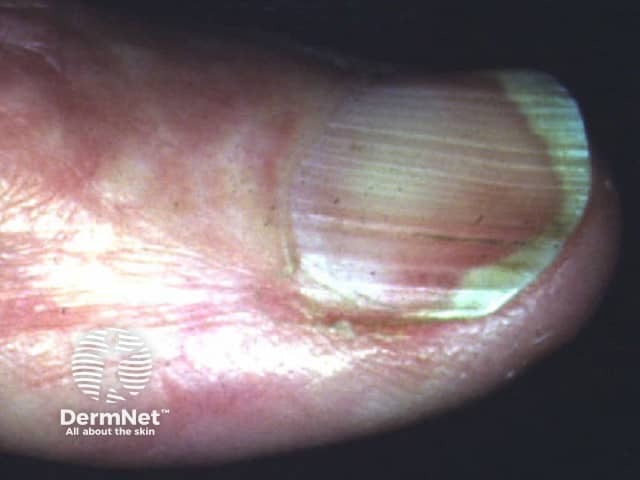
This is a high-resolution clinical photograph of a human fingernail demonstrating koilonychia, also called spoon nails. Imaging modality is clinical photography with macro/close-up technique to enhance surface and curvature details. The primary subject is the fingernail plate and surrounding nail folds, captured from a dorsal perspective for clear visualization of the nail curvature. The nail plate appears thin and concave, with a pronounced central depression producing a spoon-shaped contour. Lateral edges may be slightly tapered, and the distal third of the plate shows mild translucency. The surface is relatively smooth with minimal ridging, and the lunula is less conspicuous in this view. Surrounding cuticle and perionychial skin show mild erythema, possibly secondary to manipulation or irritation; no frank edema or pitting is evident. Clinically, koilonychia is a classic morphological clue associated with iron deficiency anemia and other nutritional or systemic disorders; in many cases nails become spoon-shaped due to chronic iron depletion. The diagnostic significance lies in recognizing a potentially reversible sign when iron stores are restored. This image is useful for dermatology, medical education, nursing training, and clinical scenario discussions focused on anemia screening, differential diagnosis of nail dystrophies, and patient education about nail changes as a diagnostic cue. Consider correlating with CBC and ferritin, and monitoring response to iron therapy.
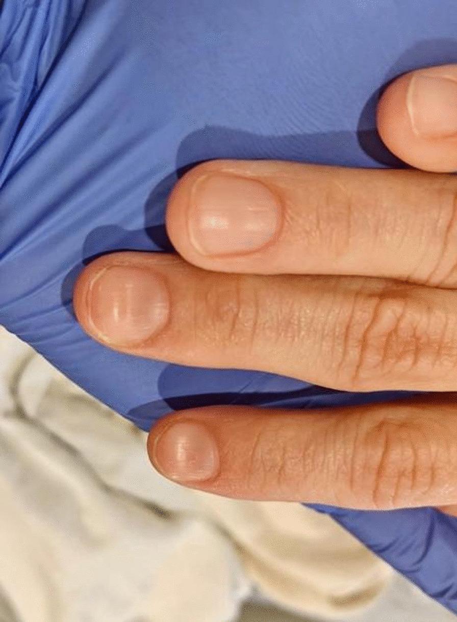
This clinical photograph displays the dorsal surface of three fingers from a patient's hand, demonstrating koilonychia. The nails exhibit a classic 'spoon-shaped' deformity characterized by the loss of normal convexity, resulting in a flattened or centrally concave nail plate with raised lateral and distal edges. The nail plates appear thin and slightly pale, consistent with hypochromic states. The surrounding periungual skin and cuticles appear intact without significant inflammation, paronychia, or splinter hemorrhages. This finding is a pathognomonic physical sign often associated with chronic iron deficiency anemia (Plummer-Vinson syndrome) or other systemic conditions affecting keratinization. The image serves as an educational reference for identifying dermatological manifestations of hematologic disorders during a physical examination.
splinter hemorrhages nail bed
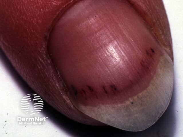
This is a high-resolution clinical macro photograph of a human fingernail demonstrating subungual splinter hemorrhages. Imaging modality: clinical photography; technique: macro digital photography of the distal fingertip in dorsal view. The primary subject is the nail unit of a finger with linear, slender, dark red to brown streaks running longitudinally beneath the nail plate along the distal two-thirds of the nail bed. The nail plate appears translucent with minimal surrounding edema; hemorrhages originate from superficial nail bed capillaries and project under the nail plate, producing discrete, parallel lines that follow the longitudinal axis. Notable features include uniform thickness of the streaks, consistent color, and absence of purulent material. Pathological findings are subungual capillary extravasations (splinter hemorrhages) that may reflect microvascular injury. Diagnostic significance is that splinter hemorrhages can be associated with trauma or, more clinically important, endocarditis and vasculitis; in isolation, they can occur with repetitive minor trauma or psoriatic nail disease. Differential considerations include traumatic nail bed injury, infective endocarditis (with embolic phenomena), vasculitis, psoriasis, systemic lupus erythematosus. Clinical correlation is essential: assess for fever, heart murmur, Janeway lesions, or cardiac risk; management focuses on treating underlying cause and monitoring for progression. This image is relevant for dermatology, clinical education, and medical sign identification.
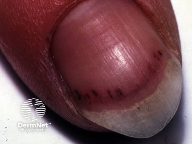
Imaging modality: Clinical close-up photography of the distal fingernail, captured with macro focus. The image shows a single fingernail with several slender, parallel splinter hemorrhages along the ventral edge of the nail bed. The hemorrhages appear as linear, red-brown to purplish streaks aligned with the longitudinal axis of the nail, extending proximally from the distal nail plate. Subungual microhemorrhages indicate capillary blood extravasation with otherwise intact surrounding tissue. The adjacent nail matrix and lunula appear non-edematous and without overt dystrophy or paronychia in this view. The pattern is characteristic of splinter hemorrhages and can result from minor trauma, vascular fragility, or microembolic phenomena. When multiple nails are involved or if systemic signs are present, differential considerations broaden to infectious endocarditis, vasculitis, autoimmune disease, or platelet/coagulation disorders. Clinically, splinter hemorrhages warrant correlation with history, cardiovascular examination, and, if indicated, cardiac imaging or laboratory evaluation. In educational settings, this image illustrates nail unit anatomy and the appearance of subungual hemorrhages, aiding dermatology, internal medicine, and medical microbiology teaching. The finding is not pathognomonic but serves as a meaningful clinical clue within a broader diagnostic framework. This image supports reproducible teaching and standardized exam prompts.
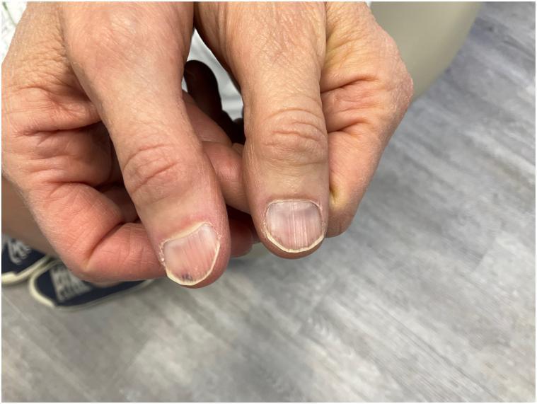
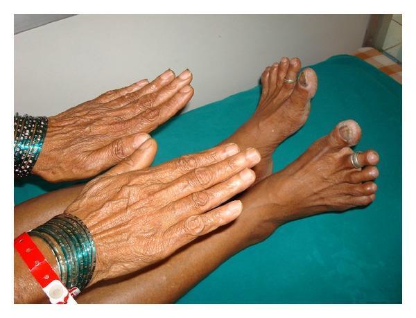
This clinical photograph displays the dorsal view of two thumbs, showcasing the nail plates. The primary finding is the presence of multiple longitudinal splinter hemorrhages. These appear as thin, reddish-brown to dark-maroon linear streaks located within the nail bed, oriented parallel to the long axis of the nail. The hemorrhages are most prominent on the distal portion of the nail plates. On one thumb, a small, more localized dark-purple subungual spot is visible near the distal edge. The surrounding periungual skin, including the proximal and lateral nail folds, appears normal without signs of inflammation, paronychia, or splintering. The nail surface is smooth, and there is no evidence of onycholysis (separation of the nail plate from the bed). Such findings are clinically significant as they can be associated with localized trauma, systemic diseases like infective endocarditis, or drug-induced reactions (e.g., nitrofurantoin). This image serves as a reference for dermatology and internal medicine residents to identify classic splinter hemorrhage morphology.
Nail Changes in Systemic Diseases – Dermatology Exam Notes
1. Basic Concept
| Site damaged | Clinical sign |
|---|---|
| Proximal matrix | Beau's lines, pitting, longitudinal ridging/fissuring, trachyonychia |
| Distal matrix | True leukonychia |
| Proximal + distal matrix | Onychomadesis, koilonychia, nail plate thinning, onychauxis |
| Nail bed | Onycholysis, subungual hyperkeratosis, apparent leukonychia, splinter hemorrhages |
- Dermatology 2-Volume Set, 5e, p. 1442
2. Nail Signs Classified by System
A. Cardiovascular / Respiratory
- Clubbing (Lovibond's sign - angle between proximal nail fold and nail plate >180°, bulbous distal digit): cyanotic congenital heart disease, pulmonary AVMs (hereditary hemorrhagic telangiectasia), infective endocarditis, congestive heart failure, bronchiectasis, lung cancer/mesothelioma, pulmonary fibrosis, cystic fibrosis, IBD, cirrhosis, thyroid disease (Graves), HIV.
- Yellow nail syndrome: thickened, over-curved (transverse + longitudinal) yellow-green nails with absent cuticle and arrested growth; triad with lymphedema and respiratory disease (chronic bronchitis, bronchiectasis, sinusitis, pleural effusion). Treatment: vitamin E 1200 IU/day, pulse itraconazole/fluconazole.
- Splinter hemorrhages: longitudinal red-brown streaks in nail bed; classic for infective endocarditis and vasculitis, but most often just trauma or psoriasis.
- Nail changes with lung disease (Murray & Nadel's Respiratory Medicine, p. 2572): cigarette staining, splinter hemorrhages, yellow nail syndrome; Beau's lines with dermatomyositis, sarcoidosis, systemic sclerosis.
B. Hepatic / Renal (apparent leukonychia group – blanch with pressure, unlike true leukonychia)
- Terry's nails: whole nail white except a 1-2 mm distal pink/brown band; seen in up to 80% of cirrhosis patients (also in normal aging).
- Muehrcke's lines: paired parallel transverse white bands parallel to the lunula; classic for hypoalbuminemia/nephrotic syndrome, also seen with chemotherapy.
- Half-and-half (Lindsay) nail: proximal half white/normal, distal half red-brown; seen in up to 25-40% of chronic renal failure/hemodialysis patients, thought due to melanin deposition; fingernails > toenails.
- Box: Nail changes in kidney disease (Brenner & Rector's The Kidney) — Lindsay nail, triangular lunulae (nail-patella syndrome), splinter hemorrhages, onychomycosis, koilonychia, onycholysis, Mees' lines, Muehrcke's lines, Beau's lines.
- Mees' lines (true leukonychia, does not blanch): transverse white bands from arsenic poisoning, also thallium, chemotherapy, severe illness.
C. Nutritional / Hematologic
- Koilonychia (spoon nails): thin, concave nail with everted edges; classic sign of chronic iron-deficiency anemia (Plummer-Vinson syndrome); also occupational chemical damage. Isolated koilonychia is unusual - often accompanies other dystrophy (e.g., trachyonychia in inflammatory disease).
D. Endocrine
- Thyrotoxicosis/Graves disease: onycholysis (Plummer's nails), brittle nails, clubbing/acropachy (thyroid acropachy - clubbing + soft tissue swelling + periosteal new bone), warm smooth skin.
- Hypothyroidism: brittle, slow-growing, striated nails; associated alopecia, lichen planopilaris.
- Diabetes mellitus: yellow discoloration, thickened slow-growing nails, onychomycosis (more frequent/severe), splinter hemorrhages.
E. Autoimmune Connective Tissue Disease (high-yield, capillaroscopy correlation)
- Dermatomyositis & Systemic sclerosis: ragged cuticles, periungual erythema/telangiectasia, hemorrhages, cuticle necrosis; capillaroscopy shows reduced capillary density with avascular areas alternating with dilated ("giant") capillary loops.
- SLE: capillaroscopy shows normal density but dilated, tortuous capillaries (distinguishes from scleroderma/DM pattern).
- Systemic sclerosis specific signs: ventral pterygium (pterygium inversus unguis - distal nail plate adherent to hyponychium, painful nail trimming) from impaired perfusion; parrot-beak nail - nail curves around a shortened, resorbed fingertip from acro-osteolysis.
F. Infective / Immunosuppression (HIV)
- Onychomycosis common (dermatophytes most common; also Candida, molds).
- Proximal subungual onychomycosis (T. rubrum) - opaque white proximal nail plate - considered a marker of immunosuppression (HIV, transplant recipients).
- True Candida onychomycosis - only occurs with immunosuppression (never in immunocompetent hosts).
- Longitudinal melanonychia (often multiple nails, with skin hyperpigmentation).
- HPV-induced periungual SCC (HPV-16) - suspect with longstanding periungual warts in HIV.
G. Genetic Syndromes
- Nail-patella syndrome (LMX1B mutation): triangular lunulae, hypoplastic/absent patellae, iliac horns.
- Also seen with renal dysfunction as above.
H. Drug-Induced (often mistaken for "systemic disease" signs in exams)
- Chemotherapy (cytotoxics): Beau's lines, onychomadesis, onycholysis, subungual hemorrhage/hematoma, Muehrcke's nails, melanonychia bands. Taxanes -> painful onycholysis with subungual hemorrhage.
- EGFR inhibitors: paronychia with pyogenic granuloma-like lesions.
- Retinoids: fragility, paronychia, periungual pyogenic granulomas.
I. Inflammatory Dermatoses with Systemic Overlap (for differentiation on exam)
- Psoriasis: irregular pitting, oil-drop sign, onycholysis, subungual hyperkeratosis, splinter hemorrhages - correlates with psoriatic arthritis risk.
- Alopecia areata: geometric/grid pitting, trachyonychia (20-nail dystrophy), punctate leukonychia.
- Lichen planus: thinning, longitudinal ridging, dorsal pterygium (fusion of proximal fold to matrix - scarring), can progress to anonychia.
3. Quick Revision Table for Exam
| Sign | Systemic Association |
|---|---|
| Clubbing | Cyanotic heart disease, bronchiectasis, lung cancer, IBD, cirrhosis, endocarditis |
| Yellow nail syndrome | Lymphedema + chronic respiratory disease |
| Terry's nails | Cirrhosis |
| Muehrcke's lines | Hypoalbuminemia/nephrotic syndrome, chemo |
| Half-and-half nail | Chronic renal failure |
| Mees' lines | Arsenic/heavy metal poisoning |
| Koilonychia | Iron deficiency anemia |
| Beau's lines | Any severe systemic illness (fever, MI, chemo) |
| Splinter hemorrhages | Endocarditis, vasculitis, trauma, psoriasis |
| Ventral pterygium, parrot-beak nail | Systemic sclerosis |
| Capillary loss + giant capillaries | Dermatomyositis/systemic sclerosis |
| Normal density, dilated tortuous capillaries | SLE |
| Triangular lunulae | Nail-patella syndrome |
| Proximal white subungual onychomycosis | HIV/immunosuppression |
Recent Evidence Check
Make notes on adverse effects of drugs on nails for md dermatology theory exam
Adverse Effects of Drugs on Nails – MD Dermatology Exam Notes
1. General Principles
-
Drug-induced nail disorders usually involve all 20 nails simultaneously (unlike localized trauma/infection which affects 1-2 nails) - a key differentiating clue in exams.
-
Onset takes weeks to months after starting the drug; resolution also takes months after withdrawal (nail growth is slow, ~0.1 mm/day fingernails).
-
Pathogenesis is most often toxic (direct effect on matrix/nail bed keratinocytes or melanocytes), sometimes vascular/ischemic, or immune-mediated.
-
Mechanistically map onto the same anatomic classification as systemic disease nail signs:
- Matrix toxicity → Beau's lines, onychomadesis, true leukonychia, brittleness/thinning
- Nail bed toxicity → onycholysis, subungual hemorrhage, hyperpigmentation
- Periungual tissue → paronychia, pyogenic granulomas
- Vascular → ischemia, splinter hemorrhages, pincer nails
-
Dermatology, 2-Volume Set 5e, p. 1452-1453; Fitzpatrick's Dermatology, Table 91-9
2. Nail Change → Causative Drug (Table format for quick recall)
| Nail Abnormality | Responsible Drug(s) |
|---|---|
| Beau's lines / onychomadesis | Chemotherapeutic agents (esp. taxanes), carbamazepine, lithium, retinoids |
| True leukonychia | Chemotherapeutic agents |
| Nail thinning/brittleness | Chemotherapeutic agents, retinoids |
| Onycholysis / photo-onycholysis | Taxanes, FGFR inhibitors, tetracyclines, psoralens, NSAIDs, fluoroquinolones |
| Apparent leukonychia (Muehrcke's lines) | Chemotherapeutic agents |
| Melanonychia (longitudinal/diffuse) | Chemotherapeutic agents (anthracyclines, taxanes, 5-FU), psoralens, zidovudine (AZT), acyclovir |
| Non-melanin discoloration | Minocycline (blue-gray), antimalarials, gold, silver |
| Paronychia + periungual pyogenic granulomas | Retinoids, antiretrovirals (indinavir, efavirenz, lamivudine), EGFR inhibitors (cetuximab, gefitinib, erlotinib, panitumumab), methotrexate, capecitabine, sirolimus/everolimus |
| Ischemic changes / digital ischemia | β-blockers, bleomycin |
| Splinter hemorrhages | Tyrosine kinase inhibitors, VEGF inhibitors |
| Pincer (trumpet) nails | β-blockers |
| Lunula discoloration | Tetracyclines (yellow), hydroxyurea (blue), AZT (blue), silver (blue-gray), lithium (brown-black) |
| Permanent nail loss | Bleomycin |
| Severe onycholysis | FGFR inhibitors |
| Brittle nails | BTK inhibitors (e.g., ibrutinib), retinoids |
- Dermatology, 2-Volume Set 5e, Table 71.9, p. 1453; Fitzpatrick's Dermatology, Table 91-9
3. Drug Class-Wise Details (exam-favorite groupings)
A. Cancer Chemotherapy (most frequent cause overall)
- Most common cause of drug-induced nail changes as a class.
- Effects: Beau's lines, onychomadesis, fragility, onycholysis, paronychia, subungual hemorrhage/hematoma, ischemia.
- Taxanes (paclitaxel, docetaxel): classic for painful onycholysis with subungual hemorrhage.
- Multiple bands of longitudinal/transverse melanonychia, diffuse pigmentation, and Muehrcke's nails also seen.
- Bleomycin: can cause permanent nail loss.
B. Targeted Cancer Therapies
- EGFR inhibitors (cetuximab, gefitinib, erlotinib, panitumumab): paronychia with (pseudo)pyogenic granulomas, appearing 1-3 months after starting, resolving after stopping drug.
- FGFR inhibitors: severe onycholysis.
- BTK inhibitors: brittle nails.
- Tyrosine kinase / VEGF inhibitors: splinter hemorrhages.
C. Retinoids (oral isotretinoin/acitretin)
- Nail fragility, paronychia, onychocryptosis (ingrown nail), periungual pyogenic granulomas, granulation tissue.
D. Antiretrovirals
- Zidovudine (AZT): melanonychia (longitudinal bands), blue lunula.
- Indinavir, efavirenz, lamivudine: paronychia and multiple periungual pyogenic granulomas.
E. β-Blockers
- Digital ischemia, lichenoid-psoriasiform nail changes, pincer nails.
F. Tetracyclines
- Onycholysis (esp. photo-onycholysis with sun exposure), yellow lunula discoloration.
G. Antimalarials (chloroquine, hydroxychloroquine)
- Nail bed pigmentation - transverse bands or diffuse blue-black/brown discoloration (also affects skin, mucosa).
H. Heavy Metals / Miscellaneous Pigment-Inducing Drugs
- Gold: blue-gray discoloration.
- Silver (argyria): blue-gray nail and skin discoloration.
- Minocycline: blue-gray nail bed pigmentation.
- Hydroxyurea: blue lunula/nail discoloration, diffuse pigmentation.
- Lithium: brown-black lunula discoloration, onychomadesis.
- Psoralens (PUVA): melanonychia, photo-onycholysis.
- Acyclovir: melanonychia.
I. Anticonvulsants
- Carbamazepine: onychomadesis.
- Lithium: onychomadesis, lunula discoloration.
4. Cytotoxic Drug-Induced Nail/Skin Pigmentation Patterns (useful cross-topic table)
| Pattern | Causative Drugs |
|---|---|
| Diffuse | Busulfan, cyclophosphamide, methotrexate, hydroxyurea, procarbazine |
| Dorsal extremities | Cisplatin, doxorubicin, mitoxantrone |
| Flexures/palms/soles | Ifosfamide, fluorouracil, tegafur, bleomycin, doxorubicin |
| Longitudinal nail bands (melanonychia) | Cisplatin, doxorubicin, idarubicin, fluorouracil, bleomycin, docetaxel, dacarbazine, hydroxyurea |
| Serpentine supravenous | Fotemustine, fluorouracil, vinorelbine, cisplatin, docetaxel |
| Flagellate | Fluorouracil, bleomycin |
- Fitzpatrick's Dermatology, Tables 45-5 and 45-6
5. Clinical/Exam Pearls
- "All 20 nails + months to resolve" = think drug-induced, not localized trauma or fungal infection.
- Taxane + painful onycholysis + subungual hemorrhage is a classic MCQ stem.
- EGFR inhibitor + paronychia + pyogenic granuloma is a very common oncology-dermatology overlap question.
- Zidovudine + longitudinal melanonychia is a classic HIV-pharmacology cross-link question.
- β-blocker + pincer nail/digital ischemia often tested against Raynaud's/scleroderma as differentials.
- Differentiate from systemic disease-related nail signs (previously discussed): drug causes typically show a temporal relationship with drug initiation/withdrawal and involve all nails uniformly, whereas systemic disease signs (Terry's, Muehrcke's from hypoalbuminemia, half-and-half nails from renal failure) correlate with disease severity/labs rather than a drug timeline. Note some overlap - e.g., Muehrcke's lines and melanonychia can be caused by BOTH chemotherapy and underlying systemic states, so history-taking is essential.