
Descending tracts of spinal cord in detailed explanation with flowcharts and clinical correlation
Reading File
Now let me search the medical library in parallel for all relevant content on descending tracts.
Finding Sources
Finding Sources
Finding Sources
Excellent finds. Now let me read all these sections in parallel.
Reading File
Reading File
Reading File
Reading File
Good content flowing in. Let me now read more from Gray's Anatomy (the main descending tracts section) and also get more content on the rubrospinal, reticulospinal, vestibulospinal, and tectospinal tracts.
Reading File
Finding Sources
Finding Sources
Now let me get the detailed ventromedial pathways content and the Neuroanatomy clinical cases content:
Reading File
Reading File
Reading File
Excellent - I now have comprehensive content. Let me fetch the key anatomical diagrams and also read the clinical correlation content (decerebrate/decorticate rigidity):
Loading Image
Loading Image
Loading Image
Reading File
I now have all the content I need. Let me also fetch the corticospinal tract pathway diagram from Guyton & Hall and the spinal cord cross-section diagram:
Loading Image
I now have comprehensive textbook content and all key diagrams. Let me compose the full detailed response.
Descending Tracts of the Spinal Cord
Overview
Descending tracts carry motor commands from the cerebral cortex and brainstem down to lower motor neurons (LMNs) in the spinal cord and brainstem. They govern voluntary movement, postural control, balance, and reflex modulation. These tracts are organized into two major systems:
| System | Location in Cord | Tracts | Function |
|---|---|---|---|
| Lateral motor system | Lateral white column | Lateral corticospinal, Rubrospinal | Fine/distal limb movements |
| Medial (ventromedial) motor system | Anterior white column | Anterior corticospinal, Vestibulospinal, Reticulospinal, Tectospinal | Posture, balance, axial/proximal muscles, gaze |
Spinal Cord Cross-Section: Position of Descending Tracts
This diagram from Gray's Anatomy for Students shows where each tract sits in the white matter:
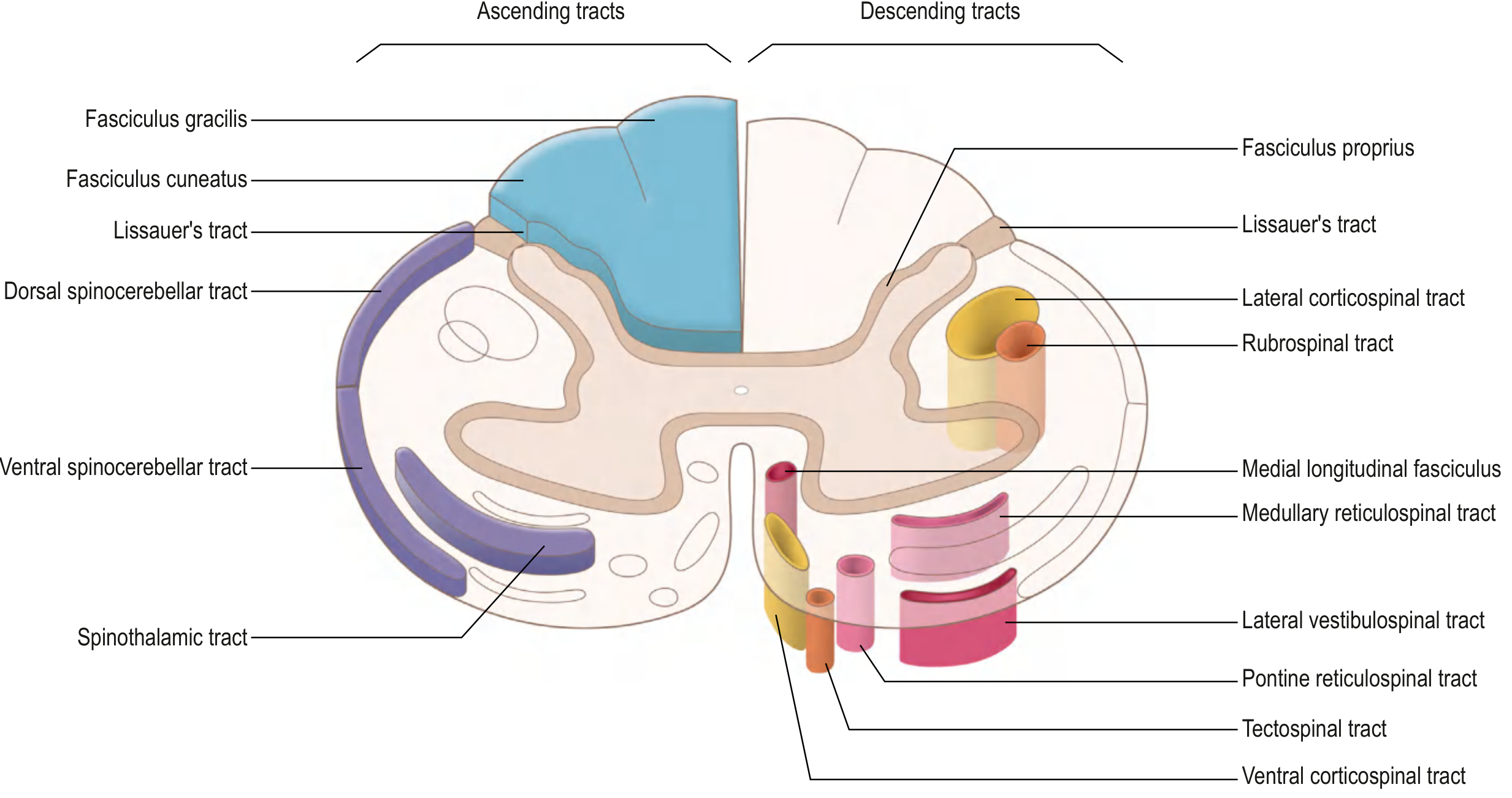
Gray's Anatomy for Students - Tracts of the Spinal Cord. Lateral corticospinal (yellow) and rubrospinal (orange) are in the lateral column. Vestibulospinal (pink), reticulospinal, tectospinal, and anterior corticospinal occupy the anterior column.
1. LATERAL CORTICOSPINAL TRACT (Pyramidal Tract)
The most clinically significant of all descending tracts.
Origin:
- ~30% from primary motor cortex (Area 4)
- ~30% from premotor and supplementary motor cortex (Area 6)
- ~40% from somatosensory cortex (Areas 3, 1, 2) - these modulate sensory input
Key fibers: Giant Betz cells (60 µm diameter, only in Area 4) - ~34,000 per tract - conduct at 70 m/sec; represent only 3% of total fibers. Over 1 million fibers total per tract.
Flowchart - Lateral Corticospinal Tract
Motor Cortex (Area 4, 6, 3, 1, 2)
↓
Corona Radiata
↓
Posterior Limb of Internal Capsule
[face fibers anterior → leg fibers posterior]
↓
Crus Cerebri (Midbrain)
[middle 1/3 = corticobulbar + corticospinal]
↓
Longitudinal Fascicles of Pons
[scattered as pontine nuclei separate them]
↓
Medullary Pyramid (ventral medulla bulge)
↓
PYRAMIDAL DECUSSATION (caudal medulla)
←85% cross to CONTRALATERAL side
↓
Lateral Corticospinal Tract
[lateral white column - all spinal levels]
[somatotopy: arm medial, leg lateral]
↓
Synapse on LMNs in LATERAL anterior horn
→ α-motor neurons → peripheral nerve → muscle
(predominantly interneurons, some direct)
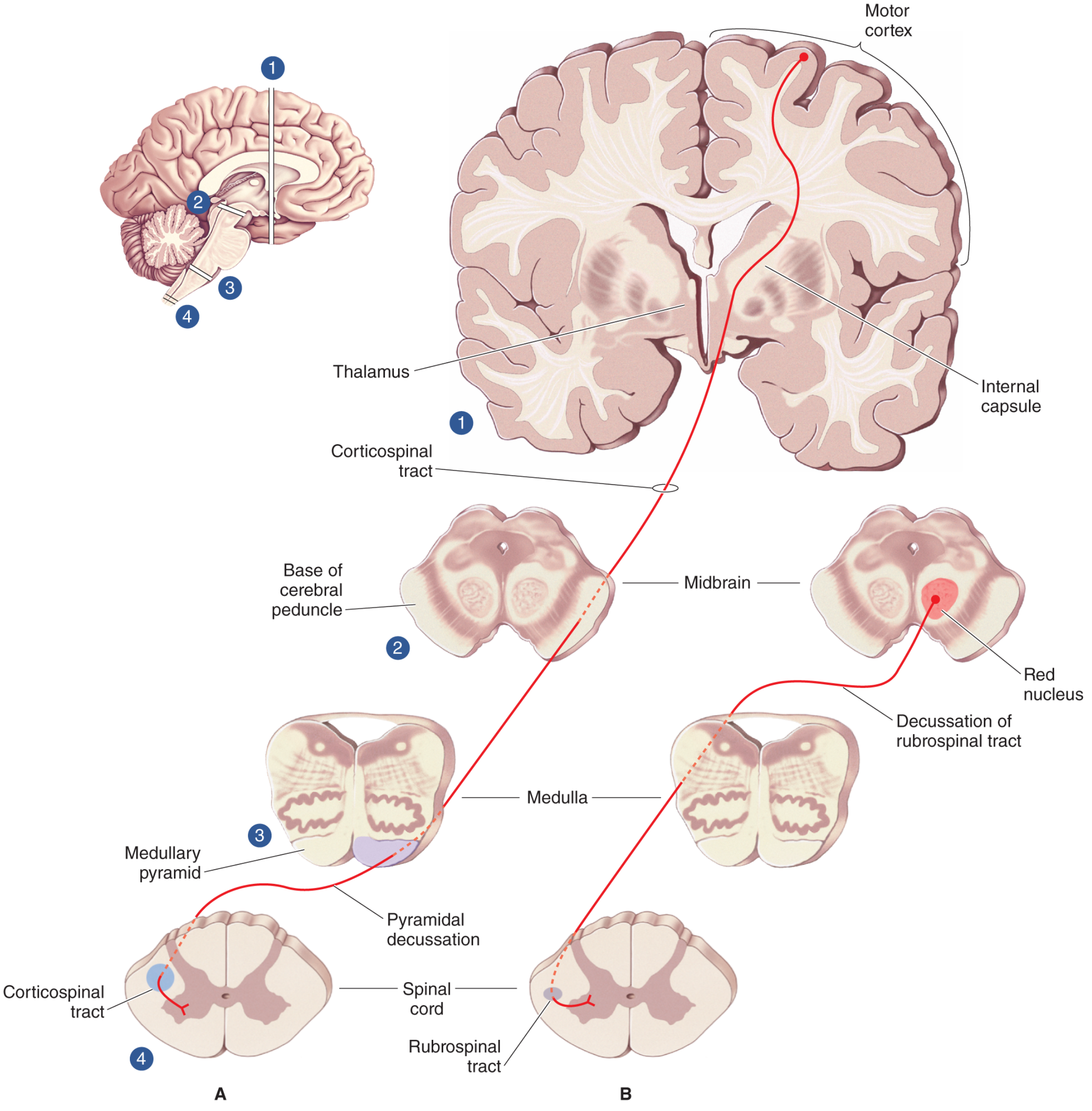
Lateral pathways: (A) Corticospinal tract from motor cortex through internal capsule, cerebral peduncle, medullary pyramid, pyramidal decussation → lateral corticospinal tract. (B) Rubrospinal tract - from red nucleus in midbrain, crosses in pons → lateral column.
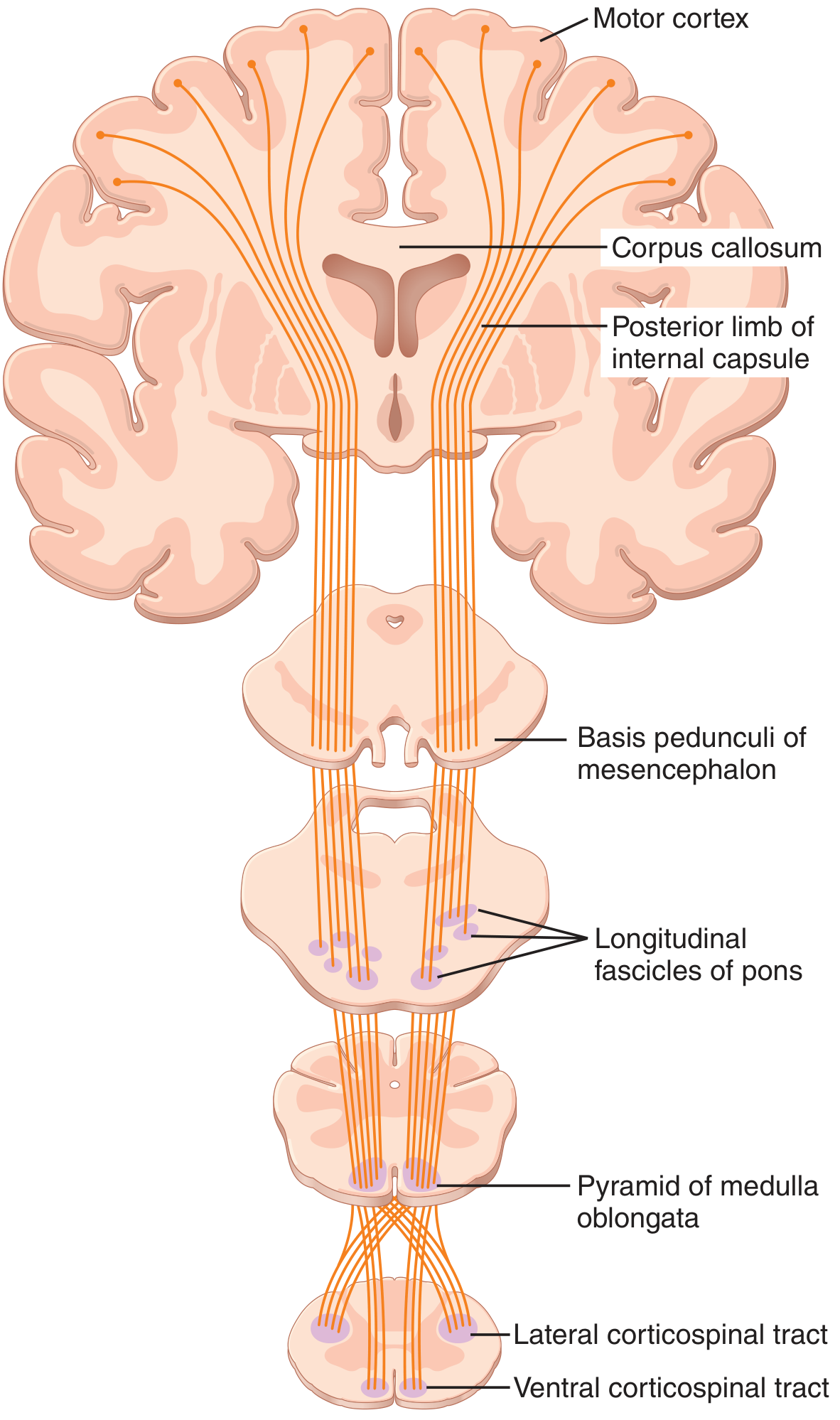
Guyton & Hall Medical Physiology - Corticospinal (pyramidal) tract: motor cortex → posterior limb of internal capsule → basis pedunculi → longitudinal fascicles of pons → medullary pyramids → lateral and ventral corticospinal tracts.
Function: Controls voluntary, skilled, fine movements of the distal extremities (especially hands and fingers). Contralateral control.
2. ANTERIOR (VENTRAL) CORTICOSPINAL TRACT
Flowchart
Motor Cortex (supplementary motor area)
↓
Same route as lateral CST to medullary pyramid
↓
~15% of fibers do NOT decussate at pyramidal decussation
↓
Anterior Corticospinal Tract
[anterior white column - ipsilateral]
[descends to CERVICAL and UPPER THORACIC only]
↓
Fibers cross at SPINAL CORD LEVEL (via anterior white commissure)
↓
Synapse on LMNs in MEDIAL anterior horn
→ controls bilateral AXIAL and PROXIMAL muscles
Function: Postural control of axial and proximal limb muscles (neck, back, shoulder). Works bilaterally.
3. RUBROSPINAL TRACT
Flowchart
Red Nucleus (magnocellular part) - MIDBRAIN tegmentum
↓
Ventral Tegmental Decussation (immediately crosses)
↓
Rubrospinal Tract - LATERAL column of cord
[only to CERVICAL spinal cord in humans]
↓
Synapse on interneurons in LATERAL anterior horn
→ excites FLEXOR motor neurons
→ inhibits EXTENSOR motor neurons (upper limb)
Function: Facilitates flexor activity and inhibits extensor activity in the upper limb. Supplementary to the lateral corticospinal tract. Relatively minor in humans; important in decorticate posturing.
4. VESTIBULOSPINAL TRACTS (Medial system)
Two components:
4a. Lateral Vestibulospinal Tract
Lateral Vestibular Nucleus (Deiter's nucleus)
↓
Uncrossed (IPSILATERAL) - descends to ALL spinal levels
↓
Anterior white column
↓
Synapse on medial anterior horn LMNs
→ EXCITES extensor (antigravity) motor neurons
→ Inhibits flexor motor neurons
→ Controls PROXIMAL limb and axial muscles
Function: Maintains posture and balance via excitation of ipsilateral extensor muscles. Responds to vestibular (gravitational) input.
4b. Medial Vestibulospinal Tract
Medial + Inferior Vestibular Nuclei
↓
Projects BILATERALLY
↓
Cervical spinal cord only
↓
Controls NECK musculature (head position reflexes)
5. RETICULOSPINAL TRACTS (Medial system)
Two components with opposing actions:
5a. Pontine (Medial) Reticulospinal Tract
Pontine Reticular Formation (Nucleus reticularis pontis)
↓
IPSILATERAL descent (some bilateral)
↓
Anterior white column → all spinal levels
↓
EXCITES extensor motor neurons
→ Increases muscle tone
→ Facilitates antigravity muscles
5b. Medullary (Lateral) Reticulospinal Tract
Medullary Reticular Formation (Nucleus reticularis gigantocellularis)
↓
Bilaterally (predominantly ipsilateral)
↓
Lateral and anterior white columns → all spinal levels
↓
INHIBITS extensor motor neurons
→ Decreases muscle tone
→ Also modulates pain (gate control via raphespinal fibers)
Combined reticulospinal function: Modulates muscle tone, spinal reflexes, and pain transmission. γ-motor neuron regulation (muscle spindle sensitivity). The balance between pontine (excitatory) and medullary (inhibitory) reticulospinal tracts determines overall muscle tone.
6. TECTOSPINAL TRACT (Medial system)
Superior Colliculus (MIDBRAIN tectum)
↓
Crosses in DORSAL tegmental decussation (Meynert's decussation)
↓
CONTRALATERAL descent - CERVICAL cord only
↓
Anterior white column
↓
Synapse on anterior horn cells (neck muscles)
→ Reflex turning of HEAD and EYES
→ Coordinates visual-guided head movements
Function: Coordinates head and eye movements in response to visual (and auditory) stimuli. Limited to cervical spinal cord.
Summary Comparison Table
| Tract | Origin | Column | Decussation | Level | Function |
|---|---|---|---|---|---|
| Lateral Corticospinal | Motor cortex | Lateral | Pyramidal decussation (medulla) - 85% | All levels | Fine voluntary distal limb movement |
| Anterior Corticospinal | Motor cortex | Anterior | Spinal cord commissure | Cervical/upper thoracic | Axial/proximal bilateral postural control |
| Rubrospinal | Red nucleus (midbrain) | Lateral | Ventral tegmental decussation (midbrain) | Cervical | Flexor facilitation, extensor inhibition (UL) |
| Lateral Vestibulospinal | Lateral vestibular nucleus | Anterior | None (ipsilateral) | All levels | Extensor facilitation, antigravity posture |
| Medial Vestibulospinal | Medial/inferior vestibular nucleus | Anterior | Bilateral | Cervical only | Neck muscle control, head stabilization |
| Pontine Reticulospinal | Pontine reticular formation | Anterior | Ipsilateral | All levels | Extensor facilitation, increased tone |
| Medullary Reticulospinal | Medullary reticular formation | Anterior/lateral | Bilateral | All levels | Extensor inhibition, decreased tone |
| Tectospinal | Superior colliculus (midbrain) | Anterior | Dorsal tegmental decussation (midbrain) | Cervical only | Head/eye reflexes to visual stimuli |
Medial vs. Lateral Brainstem Pathway Diagrams
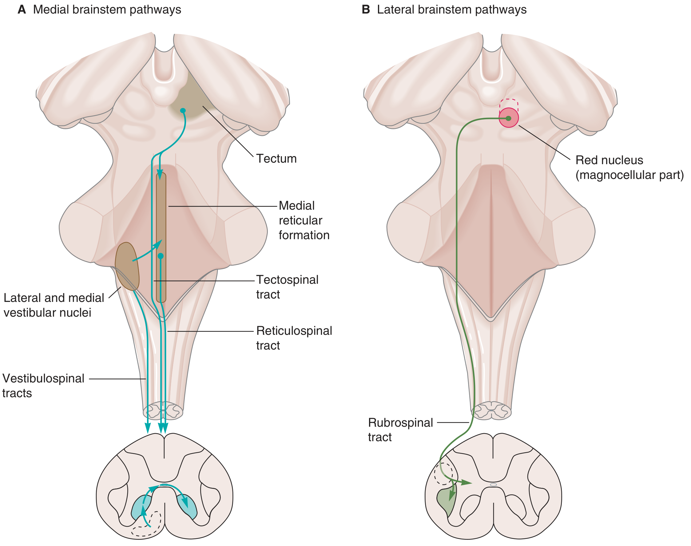
Ganong's Review of Medical Physiology - A) Medial brainstem pathways (reticulospinal, vestibulospinal, tectospinal) terminate in ventromedial anterior horn - control axial and proximal muscles. B) Lateral pathway (rubrospinal) terminates in dorsolateral anterior horn - controls distal muscles.
CLINICAL CORRELATIONS
1. Upper Motor Neuron (UMN) vs. Lower Motor Neuron (LMN) Lesions
The most critical clinical distinction in neurology:
| Sign | UMN Lesion | LMN Lesion |
|---|---|---|
| Weakness | Yes | Yes |
| Atrophy | No (mild disuse atrophy possible) | Yes (neurogenic, significant) |
| Fasciculations | No | Yes (spontaneous motor unit firing) |
| Reflexes | Increased (hyperreflexia) | Decreased (hyporeflexia) |
| Tone | Increased (spasticity) | Decreased (flaccidity) |
| Babinski sign | Present | Absent |
| Clonus | Present | Absent |
Important nuance: In ACUTE UMN lesions (e.g., acute stroke, acute spinal cord injury), there may be initial flaccid paralysis and decreased reflexes (spinal shock). Spasticity and hyperreflexia develop over hours to months. - Neuroanatomy through Clinical Cases, 3rd Ed.
Why spasticity with UMN lesions? Spasticity is NOT caused by damage to the corticospinal tract alone. It results from loss of descending inhibitory pathways (particularly the medullary reticulospinal tract) that travel alongside the corticospinal tract. This loss increases excitability of anterior horn motor neurons → brisk reflexes + increased tone. - Neuroanatomy through Clinical Cases, 3rd Ed.
2. Decorticate vs. Decerebrate Posturing
These reflect interruption of descending tracts at different levels:
DECORTICATE POSTURING (upper midbrain lesion)
↓
Corticospinal + rubrospinal tracts interrupted
(cortical and red nucleus input cut off)
Reticulospinal + vestibulospinal tracts INTACT
↓
Result:
Upper limbs: FLEXED (elbows, wrists, fingers)
Lower limbs: EXTENDED (legs, feet)
Rubrospinal inhibition on extensors lost → upper limb flexion dominant
DECEREBRATE POSTURING (lower midbrain / upper pontine lesion)
↓
Corticospinal + rubrospinal + cortical reticulospinal tracts interrupted
Vestibulospinal (excitatory to extensors) + pontine reticulospinal INTACT
↓
Result:
ALL limbs: EXTENDED
Upper limbs: extended + pronated + wrists flexed
Head: extended (neck retroflexed)
Indicates more severe/deeper brainstem involvement
WORSE prognosis than decorticate
Mnemonic:
- Decorticate = Cortex lesion = arms curled toward core (flexed)
- Decerebrate = Cerebellum/brainstem level = all extended (DE-cerebrate = DE-extensed... all straight)
3. Hemisection of Spinal Cord - Brown-Séquard Syndrome
A classic clinical scenario showing tract localization:
Hemisection at, e.g., T10 (right side)
IPSILATERAL (right) below lesion:
→ Lateral corticospinal tract cut
→ UMN signs (weakness, hyperreflexia, spasticity)
→ Dorsal column cut
→ Loss of proprioception, vibration, fine touch
CONTRALATERAL (left) below lesion:
→ Spinothalamic tract cut (crosses ~2 levels above entry)
→ Loss of pain and temperature
4. Internal Capsule Lesion (e.g., Lacunar Stroke)
Because the internal capsule compacts ALL corticospinal fibers (face, arm, leg) into a small area:
- A small lesion (as small as a lacunar infarct) produces contralateral hemiplegia - face + arm + leg
- The posterior limb of the internal capsule is most commonly affected (where the corticospinal tract runs)
- Somatotopy: face fibers most anterior → arm → leg most posterior
5. Amyotrophic Lateral Sclerosis (ALS)
- Degenerative disease affecting BOTH lateral corticospinal tract (UMN) AND anterior horn cells (LMN)
- Result: Combined UMN + LMN signs simultaneously
- UMN: spasticity, hyperreflexia, Babinski
- LMN: wasting, fasciculations, weakness
- This combination is essentially diagnostic of ALS
6. Anterior Cord Syndrome
Infarction of anterior spinal artery affects:
- Lateral corticospinal tract → bilateral motor paralysis below lesion
- Lateral spinothalamic tract → bilateral loss of pain and temperature
- Sparing: Dorsal columns (posterior columns) → proprioception and vibration INTACT
7. Spinal Cord Injury - Spinal Shock
Following acute complete cord transection:
- All descending tracts interrupted
- Initially: Flaccid paralysis, areflexia, autonomic dysfunction (neurogenic shock)
- Recovery phase: Gradual return of reflexes, development of spastic paraplegia
- This progression reflects re-establishment of local reflex arcs independent of supraspinal control
Quick Reference Flowchart: Descending System Organization
CEREBRAL CORTEX
(Areas 4, 6, 3,1,2)
|
├──────────────────────────────────────────┐
↓ ↓
CORTICOSPINAL TRACT CORTICAL MODULATION
(Lateral + Anterior) of Brainstem nuclei
| |
| ┌────────────────┼─────────────────┐
| ↓ ↓ ↓
| RED NUCLEUS RETICULAR FORMATION VESTIBULAR NUCLEI
| (Midbrain) (Pons + Medulla) (4 nuclei)
| | | |
| RUBROSPINAL RETICULOSPINAL VESTIBULOSPINAL
| TRACT TRACTS TRACTS
| (lateral col) (ant column) (ant column)
| | | |
└──────────────┬──────────┘ | |
↓ ↓ ↓
LATERAL ANTERIOR HORN MEDIAL ANTERIOR HORN (all levels)
(distal limb LMNs) (axial + proximal LMNs)
| |
└───────────┬───────────────┘
↓
α-MOTOR NEURONS
↓
Peripheral Nerve → NMJ → Muscle
Sources
- Gray's Anatomy for Students, Descending Tracts, pp. 1328-1332
- Guyton and Hall Textbook of Medical Physiology, Corticospinal (Pyramidal) Tract, p. 689
- Neuroanatomy through Clinical Cases, 3rd Edition, KCC 6.1 UMN vs LMN Lesions
- Neuroscience: Exploring the Brain, 5th Edition, Descending Spinal Tracts, pp. 1323-1330
- Ganong's Review of Medical Physiology, 26th Edition, Medial Brainstem Pathways, p. 247
Give clinical correlations seperately
Finding Sources
Finding Sources
Finding Sources
Finding Sources
Finding Sources
Finding Sources
Now read the detailed clinical content from these sources in parallel:
Reading File
Reading File
Reading File
Reading File
Now let me get the ALS/motor neuron disease content and the decorticate/decerebrate/internal capsule lesion content:
Finding Sources
Finding Sources
Reading File
Now let me get the ALS pathology content and the Adams & Victor internal capsule content:
Reading File
Reading File
Excellent - I now have all the content I need. Let me also fetch the spinal cord syndrome cross-section images:
Loading Image
Loading Image
Loading Image
I now have everything needed. Let me compose the comprehensive clinical correlations answer.
Clinical Correlations of Descending Tracts - Detailed
CLINICAL CORRELATION 1: UMN vs. LMN Lesions - The Foundation
Every descending tract lesion above the anterior horn cell is, by definition, an Upper Motor Neuron (UMN) lesion. Understanding this distinction is the bedrock of neurological localization.
UMN vs. LMN: Signs Compared
| Sign | UMN Lesion | LMN Lesion | Why |
|---|---|---|---|
| Weakness | Yes | Yes | Both interrupt the motor pathway |
| Atrophy | No (mild disuse only) | Yes (significant, neurogenic) | LMN denervation → muscle wasting |
| Fasciculations | No | Yes | Spontaneous firing of dying motor units |
| Tone | Increased (spasticity) | Decreased (flaccidity) | Loss of descending inhibitory control vs. denervation |
| Reflexes | Hyperreflexia | Hyporeflexia / areflexia | Disinhibition of reflex arc vs. arc disruption |
| Babinski sign | Present (extensor plantar) | Absent | Release of primitive reflex |
| Clonus | Present | Absent | Hyperexcitable stretch reflex arc |
| Distribution | Groups of muscles (never single muscle) | Single muscles or muscle groups |
Critical point from Adams & Victor's Principles of Neurology: A pure corticospinal tract lesion alone (e.g., medullary pyramid) produces only flaccid weakness and NO spasticity. Spasticity arises from co-damage to the corticoreticulospinal pathway (descending inhibitory fibers from the cortex to medullary reticular formation). Loss of medullary reticulospinal inhibition → increased excitability of anterior horn cells → spasticity and hyperreflexia.
Acute vs. Chronic UMN Lesion
ACUTE UMN LESION (first hours to days)
↓
Flaccid paralysis
Hypotonia, areflexia
(Resembles LMN lesion!)
= "Spinal shock" or "cerebral shock"
↓ (days to weeks to months)
CHRONIC UMN LESION
↓
Spasticity, hyperreflexia
Babinski sign, clonus
Clasp-knife response
Upright posture preserved better than voluntary movement
CLINICAL CORRELATION 2: Spinal Shock
Following acute complete spinal cord transection, ALL descending tracts are severed.
Four Phases of Spinal Shock (Miller's Review of Orthopaedics)
PHASE 1 - Areflexic / Hyporeflexic Phase
├── Timing: First 24-48 hours
├── Features: Loss of ALL reflexes below lesion
└── Mechanism: Sudden loss of tonic excitatory input from brain
PHASE 2 - Initial Reflex Return
├── Timing: Next 1-2 days
├── Features: Return of POLYSYNAPTIC reflexes first
└── Key sign: Bulbocavernosus reflex (BCR) returns
(anal sphincter contraction on squeezing glans / tugging Foley)
BCR return = END of spinal shock
PHASE 3 - Initial Hyperreflexia
├── Timing: 1-4 weeks
├── Features: Abnormally brisk reflexes
└── Mechanism: Upregulation of neurotransmitter receptors
→ increased sensitivity with minimal stimulus
PHASE 4 - Final Hyperreflexia / Spasticity
├── Timing: 1-12 months
├── Features: Hypertonia + hyperreflexia + altered muscle performance
└── Mechanism: Permanent loss of descending inhibition
→ spastic paraplegia / tetraplegia
Clinical pearl: A cord lesion CANNOT be declared complete until spinal shock has resolved (BCR returns). Before that, flaccid areflexia does NOT confirm a complete injury.
Autonomic involvement: Bladder is flaccid and unresponsive during spinal shock → overflow incontinence. After resolution → automatic (reflex) bladder. - Ganong's Review of Medical Physiology, 26th Ed.
CLINICAL CORRELATION 3: Incomplete Spinal Cord Syndromes
These represent the most clinically testable scenarios - each involves a specific pattern of tract damage.
3A. Anterior Cord Syndrome
Tracts damaged: Lateral corticospinal + lateral spinothalamic tracts (anterior 2/3 of cord)
Tracts spared: Dorsal columns (posterior 1/3, supplied by posterior spinal artery)
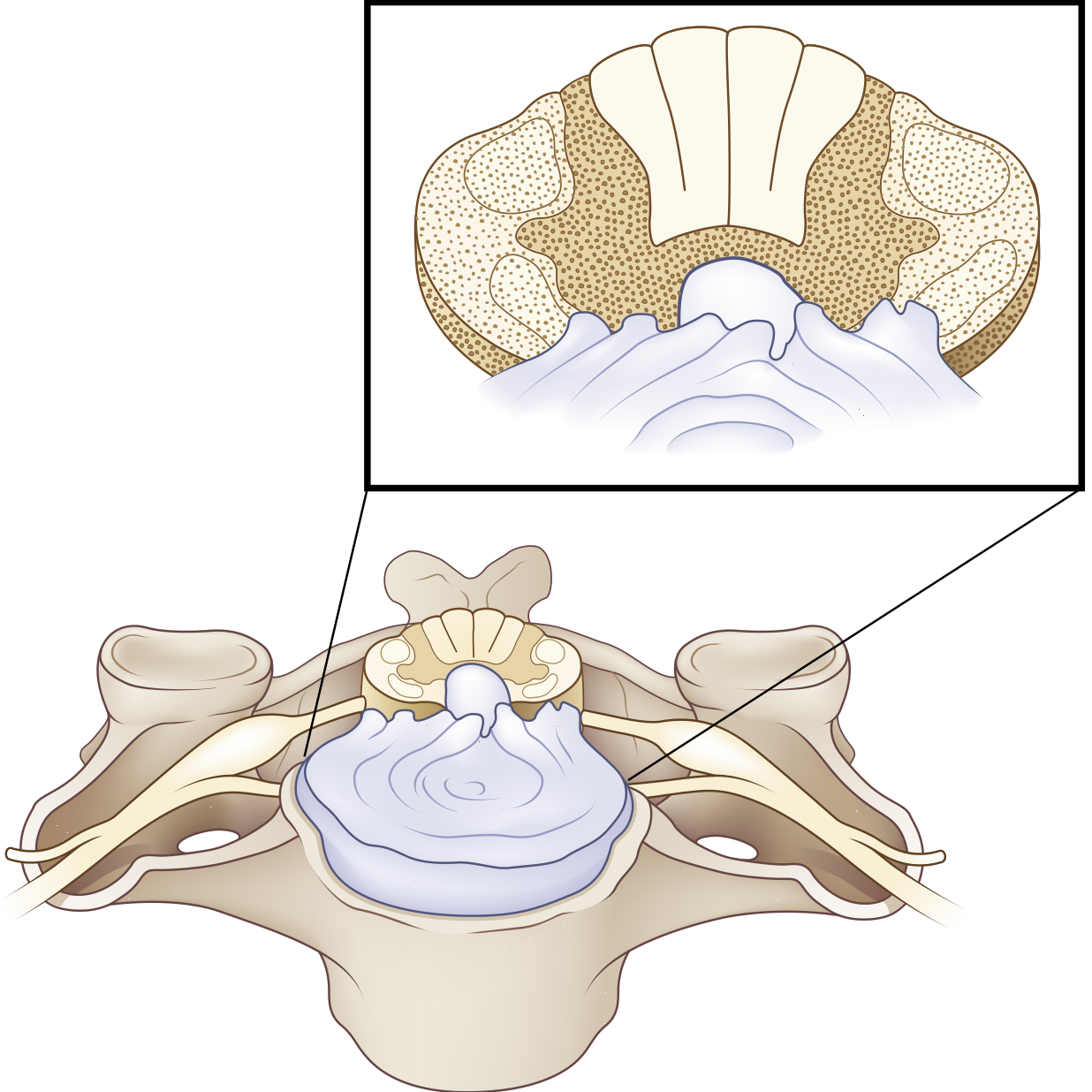
Bradley & Daroff's Neurology - Anterior cord damage: injury to corticospinal + spinothalamic tracts, dorsal columns preserved.
ANTERIOR CORD SYNDROME
↓
Corticospinal tract damaged
→ BILATERAL PARALYSIS below lesion (UMN signs)
→ Greater loss in LEGS than arms
Spinothalamic tract damaged
→ Bilateral loss of PAIN & TEMPERATURE below lesion
Dorsal columns SPARED
→ Proprioception, vibration, fine touch INTACT
Autonomic fibers damaged
→ Bladder, bowel, sexual dysfunction
Causes: Anterior spinal artery occlusion, flexion-compression injury, retropulsed disc/bone, aortic surgery (artery of Adamkiewicz compromise - arises between T9-L1, usually on left), cardiac arrest/hypotension.
Prognosis: WORST of all incomplete cord syndromes (only 10-20% recover useful motor function). - Miller's Review of Orthopaedics, 9th Ed.
3B. Central Cord Syndrome
Most common incomplete spinal cord injury (90% of incomplete injuries fall into central cord, Brown-Séquard, or anterior cord categories)
Tracts damaged: Central gray matter and surrounding white matter - disproportionately affects arm fibers (which run more centrally in the cord) vs. leg fibers (which run more peripherally)
Typical mechanism: Cervical hyperextension in elderly patients with pre-existing spondylosis - cord compressed anteriorly by osteophytes and posteriorly by infolded ligamentum flavum.
CENTRAL CORD SYNDROME
↓
Central gray matter + central white matter affected
MOTOR: Arms weaker than legs
(arm fibers run centrally in lateral CST → more damaged)
(leg fibers run peripherally → relatively spared)
SENSORY: Variable sensory loss
"Cape-like" distribution of pain/temperature loss
(cervical injury → affects arms and upper trunk)
Vibration + proprioception relatively preserved
AUTONOMIC: Bladder dysfunction (urinary retention most common)
Causes: Cervical hyperextension + spondylosis (elderly), syringomyelia, intrinsic tumors, myelitis.
Prognosis: Fair - most patients regain ambulation; upper extremity function less likely to return. - Bradley & Daroff's Neurology
3C. Brown-Séquard Syndrome (Hemisection)
Tracts damaged: ALL ipsilateral tracts - lateral CST, dorsal column + contralateral spinothalamic tract
Cause: Classically penetrating trauma (stab/bullet wound); also compressive tumors, epidural hematoma, disc herniation, MS plaque.
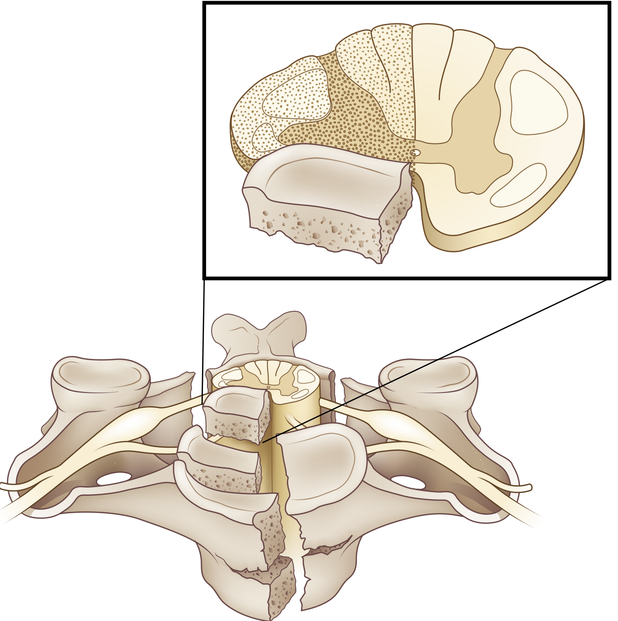
Bradley & Daroff's Neurology - Brown-Séquard: corticospinal, dorsal columns injured ipsilaterally; spinothalamic tract injured contralaterally (fibers cross before ascending).
BROWN-SÉQUARD SYNDROME (Right-side hemisection at T10)
IPSILATERAL (Right) below lesion:
├── Lateral CST cut → UMN weakness/paralysis (right leg)
├── Dorsal column cut → Loss of proprioception + vibration (right leg)
└── At level of lesion: LMN signs (anterior horn involved)
+ Ipsilateral loss of pain/temp at that dermatome
CONTRALATERAL (Left) below lesion:
└── Spinothalamic tract cut (fibers cross 1-2 levels above entry)
→ Loss of PAIN + TEMPERATURE (left leg)
→ Begins ~2 segments BELOW the lesion level
Key clinical pearl: The spinothalamic deficit starts 1-2 segments BELOW the actual lesion level because the fibers ascend 1-2 levels in Lissauer's tract before crossing.
Prognosis: BEST of all incomplete cord syndromes - ~90% recover the ability to walk. - Miller's Review of Orthopaedics, 9th Ed.
3D. Posterior Cord Syndrome
Tracts damaged: Dorsal columns only
Tracts spared: Corticospinal + spinothalamic tracts
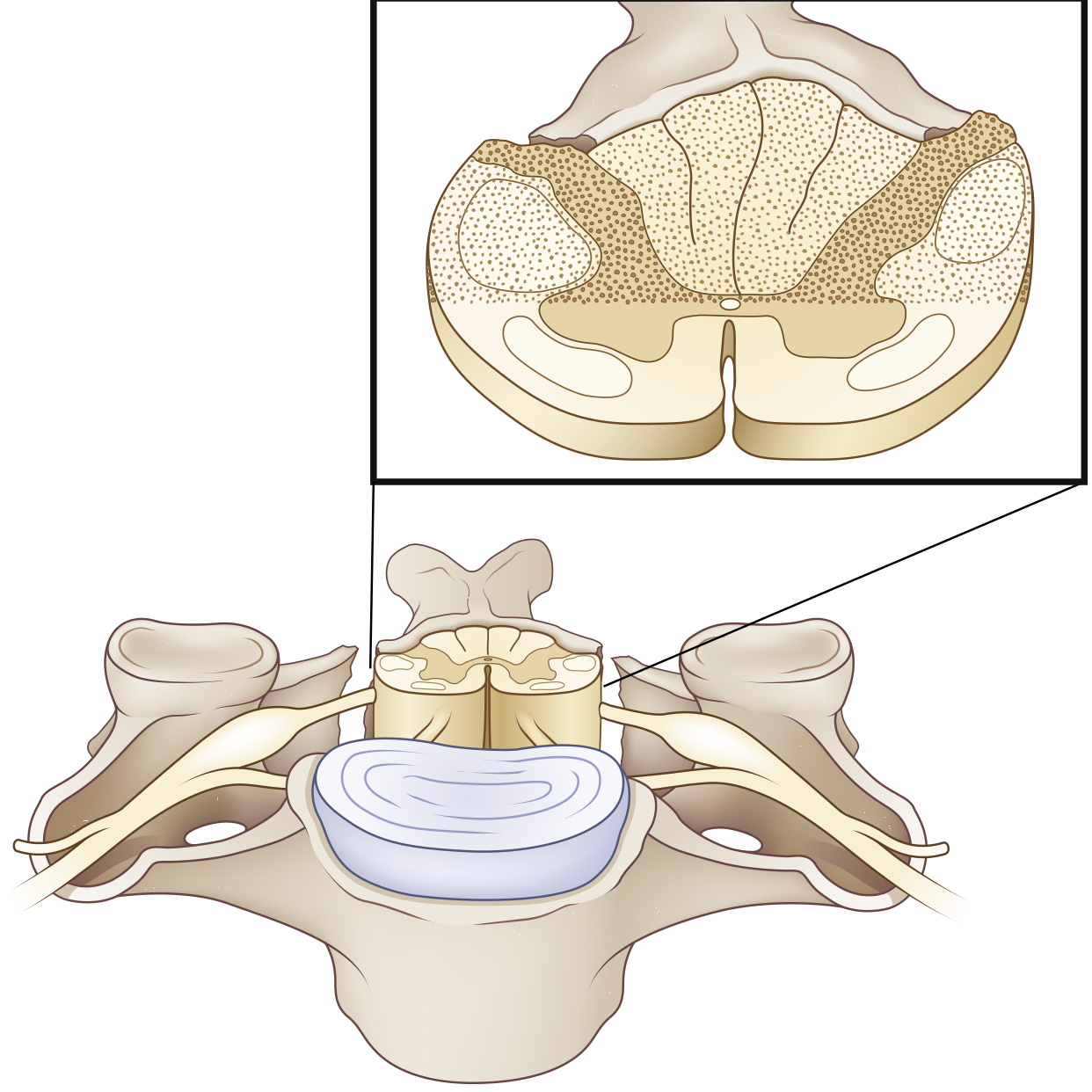
Bradley & Daroff's Neurology - Posterior cord: dorsal column injury, spinothalamic tracts preserved.
POSTERIOR CORD SYNDROME
↓
Dorsal columns damaged
→ Loss of proprioception, vibration, fine/discriminative touch
→ Sensory (proprioceptive) ATAXIA - wide-based, worsens eyes closed
→ Positive Romberg sign
→ Paresthesias
Motor function INTACT (CST spared)
Pain and temperature INTACT (spinothalamic spared)
Causes: Neck hyperextension, posterior spinal artery infarction (rare), subacute combined degeneration (B12 deficiency), tabes dorsalis (syphilis), MS. Less common than anterior cord syndrome.
Incomplete Cord Syndromes - Comparison Table
| Syndrome | Tracts Damaged | Motor | Pain/Temp | Proprioception/Vibration | Prognosis |
|---|---|---|---|---|---|
| Anterior Cord | Lateral CST + Spinothalamic | Bilateral paralysis (legs > arms) | Bilateral loss | INTACT | Poor (worst) |
| Central Cord | Central CST + central gray | Arms > Legs weakness | Variable, cape-like | Variable | Fair |
| Brown-Séquard | Ipsi CST + Ipsi dorsal column + Contra spinothalamic | Ipsi UMN weakness | Contra loss (1-2 levels below) | Ipsi loss | Best |
| Posterior Cord | Dorsal columns | INTACT | INTACT | Bilateral loss (ataxia) | Good |
CLINICAL CORRELATION 4: Internal Capsule Lesion
Because the internal capsule packs corticospinal fibers with corticobulbar, corticorubral, corticopontine, thalamocortical, and corticoreticular fibers into an extremely compact region:
INTERNAL CAPSULE LESION (e.g., lacunar infarct, hypertensive hemorrhage)
↓
POSTERIOR LIMB affected (most common)
↓
Contralateral HEMIPLEGIA
- Face + Arm + Leg affected (all fibers compressed together)
- Somatotopy: face (anterior) → arm → leg (most posterior)
Acute phase: Contralateral flaccid hemiplegia (capsular/cerebral shock)
Chronic phase: Spastic hemiplegia
- Arm: flexed (upper limb flexors > extensors)
- Leg: extended (lower limb extensors > flexors)
= Wernicke-Mann posture
Important from Adams & Victor: Spasticity after internal capsule lesion is NOT purely from corticospinal tract damage - it reflects damage to corticoreticular fibers (which modulate the medullary reticulospinal tract) running alongside the CST. A pure CST lesion produces flaccid weakness, not spasticity.
Anterior limb lesion: Frontal lobe disconnection, personality changes, less motor deficit
Genu lesion: Corticobulbar fibers → dysarthria, dysphagia, facial weakness (contralateral lower face)
CLINICAL CORRELATION 5: Decorticate vs. Decerebrate Posturing
These reflect release phenomena - when descending inhibitory tracts are cut, the unopposed activity of surviving tracts determines the posture.
DECORTICATE POSTURING
Lesion level: ABOVE midbrain (upper midbrain, hemispheric)
Tracts cut: Corticospinal + Rubrospinal
Tracts intact: Vestibulospinal (extensor) + Reticulospinal
↓
Upper limbs: FLEXED (elbows, wrists, fingers)
Lower limbs: EXTENDED (legs extended, feet plantar-flexed)
Head: Slight extension
Why: Rubrospinal (normally inhibits extensors + facilitates flexors in UL)
is cut → Upper limb extensors uninhibited by rubrospinal
But... actually, loss of corticospinal input leaves vestibulospinal
to dominate in lower limbs (extension)
In upper limbs: spinal flexor reflexes dominate without cortical control
DECEREBRATE POSTURING
Lesion level: LOWER midbrain / Upper pons (between superior and inferior colliculi)
Tracts cut: Corticospinal + Rubrospinal + Cortical influence on reticular formation
Tracts intact: Vestibulospinal (excitatory to extensors) + Pontine reticulospinal (excitatory)
↓
ALL FOUR LIMBS: EXTENDED
Upper limbs: Extended, forearms PRONATED, wrists flexed, fingers flexed
Lower limbs: Extended, toes pointed inward
Neck: Extended (opisthotonus in severe cases)
Worse prognosis than decorticate
COMPARISON SUMMARY:
DECORTICATE DECEREBRATE
Lesion level: Upper midbrain Lower midbrain/upper pons
UL posture: FLEXED EXTENDED
LL posture: EXTENDED EXTENDED
Prognosis: Less severe More severe
Tracts intact: Vestibulospinal Vestibulospinal + pontine reticulospinal
Mnemonic: "Toward core" (arms "Extends" (all limbs)
flex toward body)
CLINICAL CORRELATION 6: Amyotrophic Lateral Sclerosis (ALS)
The only condition that simultaneously destroys BOTH the UMN (lateral corticospinal tract) and LMN (anterior horn cells).
ALS = BOTH UMN + LMN signs TOGETHER
UMN signs (lateral CST degeneration):
→ Spasticity, hyperreflexia, Babinski
→ MRI: T2 hyperintensity along corticospinal tracts (Wallerian degeneration)
LMN signs (anterior horn cell degeneration):
→ Muscle wasting, atrophy
→ Fasciculations (pathognomonic)
→ Weakness (asymmetric onset, typically distal hands first)
→ Bulbar: dysarthria, dysphagia (if bulbar motor neurons involved)
SPARED (important for diagnosis):
→ Oculomotor neurons (eye movements intact)
→ Onuf's nucleus (bowel/bladder sphincters initially spared)
→ Sensory pathways (purely motor disease)
→ Cerebellar pathways (no ataxia)
Key point from Harrison's, 22nd Ed.: T2-weighted MRI shows bilateral high-signal intensity along the corticospinal tracts from the internal capsule downward into the medulla - this represents Wallerian degeneration from dying cortical motor neurons. This can also be seen in AIDS encephalopathy and other symmetric CST disorders.
CLINICAL CORRELATION 7: Subacute Combined Degeneration (Vitamin B12 Deficiency)
A clinical scenario that specifically targets two spinal cord systems:
SUBACUTE COMBINED DEGENERATION (B12 deficiency)
Dorsal columns → loss of proprioception, vibration
→ Sensory ataxia, positive Romberg sign
→ Lhermitte's sign (electric shock down spine on neck flexion)
Lateral corticospinal tract → UMN signs
→ Spastic paraparesis, brisk reflexes, Babinski
COMBINATION:
Brisk reflexes (UMN) + Absent ankle jerks (peripheral neuropathy also present)
Positive Romberg (dorsal column) + Spastic gait (CST)
This paradoxical combination is highly characteristic
Common clinical vignette: A 34-year-old woman with anorexia nervosa develops paresthesias, spastic paraparesis, brisk reflexes, and a positive Romberg sign. Cause: B12 deficiency from poor intake.
CLINICAL CORRELATION 8: Syringomyelia
Expansion of a fluid-filled cavity (syrinx) in the central cord - typically cervical.
SYRINGOMYELIA (central cavity expansion)
Central crossing fibers of spinothalamic tract disrupted first
→ BILATERAL loss of PAIN + TEMPERATURE in "cape" distribution
(arms, shoulders, upper chest)
→ Light touch and proprioception PRESERVED (dorsal columns intact)
= "Dissociated sensory loss"
As syrinx expands outward:
→ Lateral CST involved → UMN signs in legs (spastic paraparesis)
→ Anterior horn involved → LMN signs in arms (wasting, areflexia)
Late: Descending sympathetic fibers involved
→ Horner syndrome (ptosis, miosis, anhidrosis)
if syrinx in cervicothoracic cord
CLINICAL CORRELATION 9: Spinal Cord Compression (Myelopathy)
From disc herniation, epidural abscess/hematoma, tumor, spondylosis:
LEVEL-BASED LOCALIZATION:
At level of lesion:
→ LMN signs (anterior horn cells + nerve roots compressed)
→ Dermatomal sensory changes at that level
Below level of lesion:
→ UMN signs (CST compressed)
→ Spastic paresis + hyperreflexia below lesion
→ Sensory level (loss of all modalities below)
Bladder involvement:
→ Early: Urgency, frequency (UMN bladder)
→ Late: Retention → overflow incontinence
Presence of "sensory level" + UMN signs below it
= Strong localizer for cord compression
CLINICAL CORRELATION 10: Brainstem Lesions
Brainstem lesions affecting the corticospinal tract produce a specific pattern - crossed syndromes - because cranial nerve nuclei are at the level of the lesion, while the CST has not yet crossed:
BRAINSTEM CST LESION (before pyramidal decussation):
↓
Ipsilateral cranial nerve palsy (nucleus or fascicle at lesion level)
+
Contralateral hemiplegia (CST - already crossed fibers below lesion)
= "CROSSED HEMIPLEGIA" or "Alternating hemiplegia"
Examples:
Weber syndrome (midbrain): CN III palsy (ipsi) + hemiplegia (contra)
Millard-Gubler syndrome (pons): CN VI + VII palsy (ipsi) + hemiplegia (contra)
Medial medullary syndrome: CN XII palsy (ipsi) + hemiplegia (contra)
Master Summary Flowchart: Tract Damage → Clinical Syndrome
LEVEL OF LESION → CLINICAL PICTURE
Motor cortex (Area 4 only)
→ Distal limb weakness + hypotonia (minimal spasticity)
Cortex + Area 6 / subcortical white matter
→ Contralateral hemiplegia, spasticity, hyperreflexia
→ Broca's area (if left dominant) → expressive aphasia
Posterior limb, internal capsule
→ Dense contralateral hemiplegia (face + arm + leg)
→ Acute: flaccid; Chronic: spastic (Wernicke-Mann)
Brainstem (before decussation)
→ Crossed syndrome (ipsi CN + contra hemiplegia)
Medullary pyramid (pure CST)
→ Contralateral flaccid hemiplegia (NO spasticity)
→ Face spared (corticobulbar above)
Lateral CST (spinal cord, one side)
→ Ipsilateral UMN signs below lesion
Bilateral CST (cord transection)
→ Paraplegia / tetraplegia (UMN)
→ Spinal shock initially → spastic paraplegia
Anterior horn cells (LMN)
→ Ipsilateral LMN signs at that segment
Sources
- Bradley and Daroff's Neurology in Clinical Practice - Spinal Cord Injury Syndromes, pp. 931-933; Localization of Motor Deficits
- Adams and Victor's Principles of Neurology, 12th Ed. - Paralysis from UMN Lesions
- Miller's Review of Orthopaedics, 9th Ed. - Spinal Shock, Cord Injury Syndromes Table 8.9
- Harrison's Principles of Internal Medicine, 22nd Ed. - ALS Clinical Manifestations
- Neuroanatomy through Clinical Cases, 3rd Ed. - KCC 6.1 UMN vs. LMN; KCC 7.4 Spinal Cord Syndromes
- Ganong's Review of Medical Physiology, 26th Ed. - Effects of Spinal Cord Transection; Decerebrate/Decorticate Rigidity
- Frameworks for Internal Medicine - Weakness from UMN Spinal Cord Lesions
This is a shared conversation. Sign in to Orris to start your own chat.