


Answer these questions
uterus supports ligaments prolapse anatomy
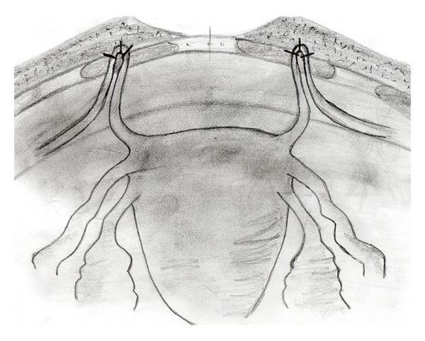
Anatomical Diagram: A black-and-white schematic illustration demonstrating the surgical fixation of the round ligaments to the anterior rectus fascia, likely for the management of pelvic organ prolapse. The diagram provides an anterior view of the uterus and its supporting structures. The uterine fundus is shown in the center, with the round ligaments extending bilaterally. These ligaments are depicted being pulled cranially and looped through small fenestrations in the rectus abdominis muscle layer. The apical 'ans' or loop of each round ligament is secured to the overlying fascia using three distinct sutures, represented by 'x' markers. Additional adnexal structures, including the fallopian tubes and utero-ovarian ligaments, are visible extending from the uterine cornua. The illustration emphasizes the anatomical relationship between the pelvic viscera and the abdominal wall fascia during a suspension procedure, highlighting the point of mechanical support and the method of non-absorbable suture fixation used to stabilize the uterine position.
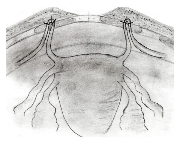
**Imaging Modality:** Medical schematic illustration (pencil/grayscale sketch). **Anatomical Region:** Female pelvic anatomy focusing on the uterus, adnexa, and anterior abdominal wall. **Observed Procedure/Landmarks:** The illustration depicts a surgical uterine suspension procedure. Key anatomical structures include the uterine fundus, fallopian tubes, and the round ligaments of the uterus. The round ligaments are shown extending bilaterally from the uterine cornua. **Characteristic Visual Features:** The schematic highlights the bilateral fixation of the round ligaments to the rectus fascia of the anterior abdominal wall. The distal portions of the round ligaments are looped through or anchored to the fascial layer, secured with visible sutures. This anatomical arrangement demonstrates the shortening or plication of the ligaments to provide superior and anterior support to the uterus. **Clinical Context:** This visual represents a surgical technique for the correction of uterine retroversion or pelvic organ prolapse. The procedure aims to reposition the uterus into an anteverted state by utilizing the native support of the round ligaments and anchoring them to the sturdy fascial layers of the abdominal wall.
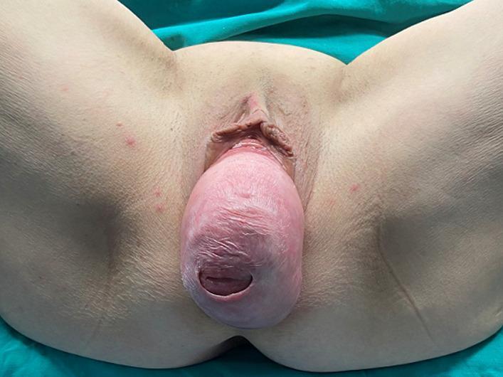
A clinical photograph showing a stage IV total uterine prolapse in a patient positioned for gynecological examination. The visual demonstrates a significant protrusion of the uterus and everted vaginal walls through the vaginal introitus, presenting as a large, rounded, fleshy mass. The prolapsed tissue exhibits a pale pink to reddish color with a glistening, moist surface and visible horizontal rugae or mucosal folds. At the most distal end of the mass, the external os of the cervix is clearly visible as a dark, oval orifice. The surrounding anatomy includes the labia minora and the vulvar region. The skin of the inner thighs and perineum is visible, showing some minor scattered erythematous macules or blemishes, but notably lacks extensive ulceration or necrosis of the vaginal mucosa. This image illustrates a severe case of pelvic organ prolapse (POP) which, as noted in the clinical context, can lead to secondary complications such as bilateral hydronephrosis due to ureteral compression.
appendix positions retrocecal pelvic iliac diagram
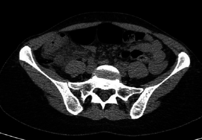
**Modality:** Computed Tomography (CT), axial section. **Anatomical Region:** Abdomino-pelvic region at the level of the iliac crests and sacrum. **Key Findings:** The image demonstrates a thickened, dilated vermiform appendix located in a retrocecal position. There is clear evidence of periappendiceal fat stranding (increased attenuation/infiltration of the surrounding mesenteric fat), indicating a localized inflammatory process. The cecum is visible anterior to the inflamed appendix. No gross evidence of abscess formation or free intraperitoneal air is noted in this specific slice. **Pathology:** Acute retrocecal appendicitis. **Diagnostic Features:** - **Increased appendiceal diameter:** Tubular structure showing wall thickening. - **Fat stranding:** Hallmark secondary sign of acute inflammation in the right lower quadrant. - **Anatomic variant:** Retrocecal orientation of the appendix. **Clinical Significance:** These radiologic features are diagnostic of acute appendicitis, used to guide surgical intervention or conservative management. The retrocecal position is a critical surgical landmark for operative planning.
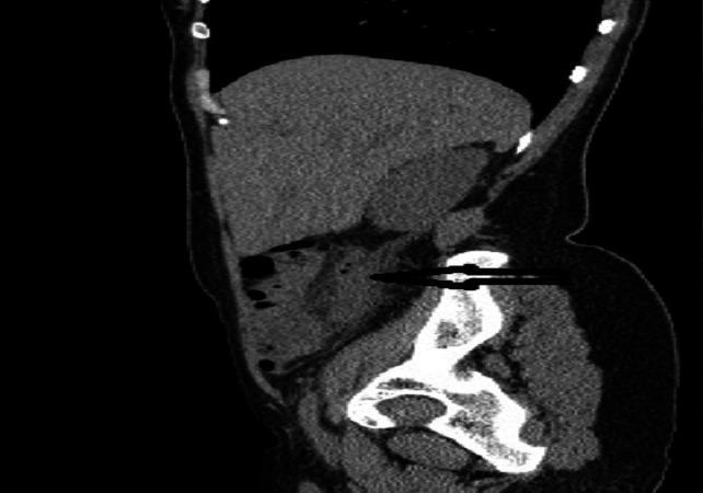
This diagnostic image is a sagittal view of an abdominal-pelvic CT scan (TDM). It demonstrates key radiological findings indicative of acute retrocecal appendicitis. A prominent feature is the distended and thickened vermiform appendix, indicated by a horizontal black arrow. The appendix measures approximately 12 mm in diameter, exceeding the standard threshold for inflammation. It is positioned posteriorly to the cecum, confirming a retrocecal anatomical variant. Furthermore, there is a visible loss of the normal low-density appearance of the surrounding mesenteric fat, characterized as periappendiceal fat stranding or infiltration. This increased attenuation in the fat neighboring the thickened appendix is a secondary sign of localized inflammatory response. The image serves as a clinical example for medical students and radiology residents to identify primary and secondary signs of appendicitis in unconventional anatomical positions using cross-sectional imaging.
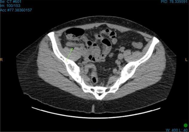
This diagnostic image is an axial computed tomography (CT) scan of the lower abdomen and pelvis, demonstrating diagnostic findings of acute appendicitis. The view captures the cross-sectional anatomy of the iliac bones (bright white, high-density structures) and pelvic musculature. A green arrow points specifically to a thickened, tubular structure in the right iliac fossa, located in a retrocecal position, which is consistent with an inflamed appendix. Pathological features include increased density and a hazy, smudged appearance of the adjacent mesenteric fat, known as fat stranding, which indicates local inflammatory infiltration. Several air-filled bowel loops are visible within the abdominal cavity. This imaging is characteristic of an emergency surgical presentation, used to differentiate between causes of right lower quadrant pain, such as acute appendicitis or appendiceal diverticulitis.
knee cruciate ligaments anatomy diagram ACL PCL attachment
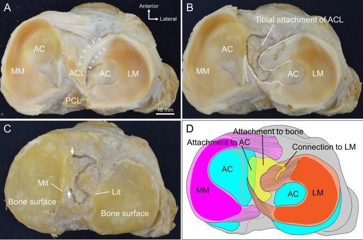
This composite educational graphic details the tibial attachment of the anterior cruciate ligament (ACL) through clinical specimen photographs and an anatomical diagram. Panel A shows a superior view of a human knee tibial plateau, illustrating the spatial relationship between the ACL, posterior cruciate ligament (PCL), medial meniscus (MM), and lateral meniscus (LM) with associated articular cartilage (AC). White arrowheads indicate fibers connecting the ACL to the LM's anterior horn. Panel B demonstrates the footprint after ACL removal, outlined with a white dashed line, showing its irregular shape abutting the AC medially and coinciding with LM horn margins. Panel C reveals the underlying bone surface after further dissection of the menisci and cartilage, highlighting the medial intercondylar tubercle (Mit) and lateral intercondylar tubercle (Lit). Panel D is a color-coded summary diagram categorizing the ACL attachment into three distinct zones: direct attachment to bone (yellow), attachment to articular cartilage (green), and connection to the lateral meniscus (orange). This material demonstrates that the ACL tibial footprint extends beyond bone to include fibrocartilaginous and meniscal tissues.
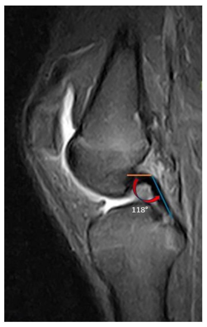
This diagnostic image is a T2-weighted sagittal MRI view of a human knee joint, illustrating the measurement of the Posterior Cruciate Ligament (PCL) buckling angle. The anatomical focus includes the distal femur, proximal tibia, patella, and the intra-articular ligaments. The PCL is highlighted as a low-signal (dark) band extending from its femoral attachment to its tibial insertion. Superimposed on the MRI are two geometric lines: an orange line following the proximal femoral portion of the PCL and a blue line following the distal tibial portion. A red curved arrow indicates the vertex where these lines intersect, with a displayed measurement of 118 degrees. This radiological measurement is used in orthopedic surgery and sports medicine to evaluate PCL morphology and ligamentous laxity, which can be secondary to anterior cruciate ligament (ACL) insufficiency or posterior tibial slope variations. The image serves as an educational reference for quantitative assessment of cruciate ligament orientation in the sagittal plane.
spleen ligaments anatomy gastrosplenic lienorenal
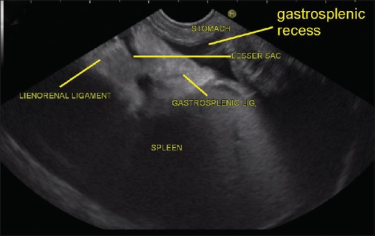
**Imaging Modality:** Endoscopic Ultrasound (EUS) **Anatomical Region:** Upper abdomen, specifically the left upper quadrant focusing on the perisplenic space and gastric anatomy. **Key Landmarks and Visual Features:** This ultrasound image demonstrates the anatomical relationships between the spleen, stomach, and their supporting ligamentous structures. The spleen is visible as a large, homogeneous, moderately echogenic organ in the lower portion of the sector. Superior and adjacent to the splenic hilum, the following structures are identified and labeled: * **Gastrosplenic Ligament:** Visible as an echogenic band connecting the splenic hilum to the stomach. * **Lienorenal (Splenorenal) Ligament:** An echogenic ligamentous attachment extending toward the retroperitoneum. * **Stomach:** Positioned at the top of the image near the transducer interface. * **Gastrosplenic Recess and Lesser Sac:** These potential spaces are identified as hypoechoic (fluid-filled) areas adjacent to the ligamentous attachments. **Clinical Context:** This image serves as a reference for EUS-based identification of perisplenic ligaments and the boundaries of the lesser sac, essential for navigating the omental bursa and identifying regional vascular or lymph node pathology during diagnostic procedures.
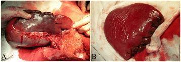
This clinical photograph displays two stages of a subtotal splenectomy during a surgical procedure for portal hypertension. Image A shows the intraoperative identification of the ischemic separatrix (indicated by an arrow) following the ligation of the splenic artery stem and the lienorenal, splenophrenic, and gastrosplenic ligaments. A clear demarcation is visible between the well-perfused red splenic tissue and the darker, congested ischemic portion designated for resection. Image B illustrates the surgical reconstruction of the residual spleen. The cut surface is managed using a horizontal mattress cross-suture technique (indicated by an arrow), which provides effective hemostasis and closure of the splenic parenchyma. The image demonstrates a specialized surgical approach aimed at preserving splenic function (immunological and phagocytic) while addressing complications of portal hypertension and splenomegaly. Key educational concepts include surgical anatomy of splenic ligaments, vascular ligation patterns, and techniques for parenchymal suturing in organ-preserving surgery.
Complete Answers
Question C - Uterine Prolapse (Female with perineal protrusion while standing)
C.i. What organ descends down?
C.ii. Supports of the Uterus
- Transverse cervical (Cardinal / Mackenrodt's) ligaments - the most important; extend from the cervix and lateral vaginal fornix to the lateral pelvic wall
- Uterosacral ligaments - extend from the posterior cervix to the sacrum (S2-S4); provide the critical level I support that prevents uterine prolapse. These are the key ligaments lost in prolapse.
- Pubocervical ligaments - anterior ligaments to the pubic bone
Repeated childbirth damages especially the levator ani and stretches the cardinal/uterosacral ligaments, causing prolapse.
C.iii. Arterial Supply of the Uterus
| Artery | Origin | Supply |
|---|---|---|
| Uterine artery (main) | Internal iliac artery (anterior division) | Body, cervix, upper vagina, tubes |
| Ovarian artery | Directly from abdominal aorta (L1-L2) | Fundus and upper body via anastomosis |
| Vaginal artery | Internal iliac artery | Cervix and upper vagina |
Question D - Appendicitis (18-year-old male with pain shift from umbilicus to RLQ)
D.i. Diagnosis
- Age 18, male (peak incidence)
- Pain starting periumbilically then shifting to right iliac fossa (McBurney's point / RLQ)
- Nausea over several hours
- Low-grade fever (99.4°F / 37.4°C)
- RLQ tenderness on examination
- Normal urine analysis (rules out ureteric colic/UTI)
D.ii. Causes for the Shift in Location of Pain
- The appendix is a midgut derivative (embryologically derived from the midgut)
- Inflammation initially stimulates visceral afferent (autonomic) nerves in the appendicular wall
- Visceral pain from the midgut is referred to the periumbilical region (T10 dermatome), which is imprecise and dull/colicky
- The pain is felt around the umbilicus because midgut visceral afferents enter the spinal cord at T10
- As inflammation progresses, the inflamed appendix irritates the parietal peritoneum overlying it
- Parietal peritoneum is supplied by somatic (spinal) nerves (ilioinguinal nerve, branches of L1)
- Somatic pain is sharp, well-localized and points to the exact site of the appendix in the right iliac fossa
- This is why the pain "shifts" from central to RLQ as appendicitis progresses
D.iii. Different Positions of the Appendix (with diagram)
| Position | Frequency | Clinical Importance |
|---|---|---|
| Retrocecal | ~65-75% (most common) | Pain may be posterior/flank; psoas sign positive; diagnosis may be delayed |
| Pelvic (descending) | ~20-30% | Pain in suprapubic area or pelvis; mimics ovarian/bladder pathology; obturator sign positive |
| Pre-ileal / Post-ileal | Rare | Central abdominal pain; can obstruct ileum |
| Subcecal | Rare | RLQ pain but lower |
| Paracolic (right) | Rare | Lateral pain |
| Splenic (left-sided) | Very rare | In situs inversus only |
         [Ileum]
              \
    [Cecum]----[Ileocecal valve]
         |
    [Base of appendix] (constant position)
         |
         ├─── Retrocecal (behind cecum) ← MOST COMMON ~70%
         ├─── Pelvic/Descending (into true pelvis) ← 2nd most ~25%
         ├─── Pre-ileal (in front of ileum)
         ├─── Post-ileal (behind ileum)
         └─── Subcecal (below cecum)
Question D (second set) - Spleen Rupture (boy kicked in upper abdomen)
D.i. What organ is likely to rupture?
D.ii. Ligaments Associated with the Spleen
| Ligament | Connects | Contents |
|---|---|---|
| Gastrosplenic (gastrolienal) ligament | Spleen to greater curvature of stomach | Short gastric arteries & veins, left gastroepiploic vessels |
| Splenorenal (lienorenal) ligament | Spleen to left kidney | Splenic artery & vein, tail of pancreas |
| Phrenicocolic ligament | Left colic flexure to diaphragm | Acts as a "shelf" supporting the spleen inferiorly |
| Phrenicosplenic ligament | Spleen to diaphragm | (sometimes described) |
| Splenocolic ligament | Spleen to transverse colon | Variable |
D.iii. Precautions Necessary During Removal (Splenectomy)
-
Identify and protect the tail of the pancreas - it lies within the splenorenal ligament, very close to the splenic hilum. Injury causes post-operative pancreatic fistula/pancreatitis.
-
Ligate the splenic artery first (before the vein) in the lesser sac - this reduces blood loss and allows autotransfusion of splenic blood back into the circulation.
-
Protect the stomach - the short gastric vessels in the gastrosplenic ligament must be carefully ligated to avoid gastric perforation.
-
Protect the left colon/splenic flexure - avoid injury to colon during mobilization.
-
Search for accessory spleens (splenunculi) - present in ~10-30% of people, commonly at the splenic hilum, gastrosplenic ligament, or gonadal area. Must be removed to prevent recurrence of conditions like ITP.
-
Post-splenectomy sepsis (OPSI) prevention - vaccinate against encapsulated organisms (Streptococcus pneumoniae, Haemophilus influenzae, Neisseria meningitidis) before or immediately after splenectomy.
-
In trauma, splenic conservation (splenorrhaphy) is preferred over total splenectomy especially in children, to preserve immune function.
Question E - Knee Injury (football player, knee at right angle, tibia driven forward)
E.i. Which Ligament is Injured?
Note: If the tibia were driven posteriorly, it would be the PCL. Since the tibia is driven forwards, this is an ACL injury.
E.ii. Attachments of the ACL
| End | Attachment |
|---|---|
| Proximal (Femoral) | Posterior part of the medial surface of the lateral femoral condyle, in the intercondylar notch |
| Distal (Tibial) | Anterior intercondylar area of the tibia, anterior and lateral to the anterior tibial spine; partially blends with the anterior horn of the lateral meniscus |
E.iii. Other Ligaments That Can Be Injured (Associated Injuries)
- ACL (primary injury)
- Medial Collateral Ligament (MCL) - resists valgus stress; commonly torn with ACL
- Medial Meniscus - attached to MCL; torn with valgus-rotation mechanism
- Lateral Collateral Ligament (LCL) - with varus force
- Posterior Cruciate Ligament (PCL) - in severe combined injuries
- Posterolateral Corner (PLC) - popliteus-arcuate complex, in high-energy trauma
- Medial and/or lateral menisci - frequently co-injured (hemarthrosis within hours of injury is common)
- ACL tears cause hemarthrosis within 1-2 hours (ACL is vascular)
- A Segond fracture (avulsion of lateral tibial condyle) on X-ray is pathognomonic of ACL rupture
- MRI has ~90-95% accuracy in identifying cruciate and meniscal disruption
What is mc berny point
McBurney's point appendix surface marking anatomy diagram
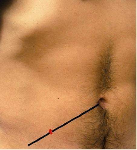
A clinical photograph of the human lower abdomen illustrating surface anatomy landmarks used in surgical evaluation. A black line represents the spino-umbilical line, extending from the umbilicus to the right anterior superior iliac spine (ASIS). A red mark is placed on this line at the junction of the lateral one-third and the medial two-thirds, identifying McBurney's point. This landmark is clinically significant as it traditionally corresponds to the base of the vermiform appendix and serves as a primary site for tenderness assessment in cases of suspected acute appendicitis. The photograph provides a clear visual guide for physical examination techniques in emergency medicine and general surgery.
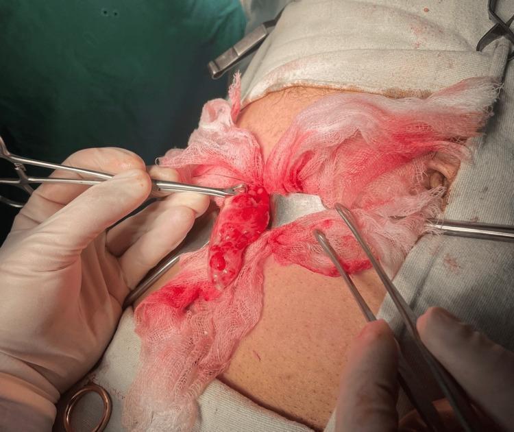
This clinical photograph captures an intraoperative view of an open appendectomy. An inflamed, erythematous, and enlarged appendix is being exteriorized through a gridiron incision at McBurney's point. The appendix exhibits signs of acute inflammation, including significant hypervascularity and a thickened, irregular surface texture. Surgical instruments, including a Babcock forcep and other clamps, are visible as they manipulate the tissue. The operative field is packed with blood-stained sterile gauze to maintain visibility and control minor bleeding from the surrounding subcutaneous tissue and skin. The surrounding area is draped with sterile surgical towels. This image serves as an educational example of the surgical presentation of acute appendicitis, secondary to an obstructing appendicolith, highlighting the gross pathological changes of the organ and the standard surgical environment for an open abdominal procedure.
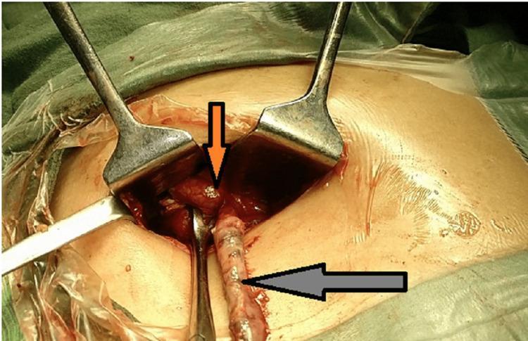
This clinical photograph captures an intraoperative view of an emergency appendicectomy through a McBurney's point incision, revealing a rare anatomical variation: a duplicated vermiform appendix. Two distinct appendices are visible within the surgical field, stabilized by metal retractors. An inferiorly positioned, elongated appendix is indicated by a gray arrow. Located superiorly, an orange arrow points to a second appendix that appears acutely inflamed and gangrenous; a small, pale, firm fecolith is visible near its base. The surrounding tissues exhibit signs of severe inflammation, including marked vascular congestion and erythema. There is visible pooling of glistening seropurulent fluid and pus in the peritoneal cavity, consistent with appendiceal perforation and localized peritonitis. This image serves as a high-level educational resource for surgical residents and medical students, illustrating the clinical presentation of appendiceal duplication and the surgical anatomy of the cecal base during complicated appendicitis.
McBurney's Point
Location / Definition
         UMBILICUS ●─────────────────────────● ASIS (right)
                     ←── 2/3 ──→←── 1/3 ──→
                                    ↑
                             McBurney's Point
Clinical Significance
| Feature | Detail |
|---|---|
| Named after | Charles McBurney (American surgeon, 1845-1913) |
| Marks | Base of the appendix (where it arises from the cecum) |
| Key clinical sign | Point of maximum tenderness in acute appendicitis |
| Surgical incision | The gridiron (McBurney's) incision for open appendectomy is centered over this point |
| Rebound tenderness | Releasing pressure at this point causes sharp pain (Blumberg's sign) in appendicitis |
Important Caveat
- Tenderness at McBurney's point strongly suggests appendicitis
- Absence of tenderness there does NOT rule it out (especially in retrocecal or pelvic appendix positions)
Associated Signs in Appendicitis
- Rovsing's sign - pressure in the LLQ causes pain in the RLQ (referred rebound)
- Psoas sign - pain on right hip extension (retrocecal appendix)
- Obturator sign - pain on internal rotation of right hip (pelvic appendix)
- Blumberg's sign - rebound tenderness at McBurney's point