Uh, write the, all the, um, I mean, all the veins and... I tell you, and where they arises and where they goes and which type of, um... I mean, which type of... Um... Gaseous form de, é... Quantidade, I mean, oxygenated or deoxygenated.
major veins of the human body diagram systemic venous system
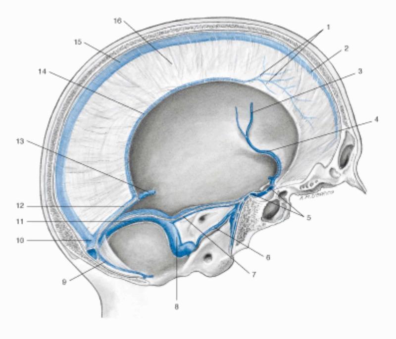
Anatomical illustration of a sagittal section of the human skull showcasing the intracranial venous drainage system and dural sinuses. The diagram highlights the major dural venous sinuses, including the superior sagittal sinus running along the superior margin of the falx cerebri, the inferior sagittal sinus at the inferior margin, and the straight sinus formed by the junction of the inferior sagittal sinus and the great cerebral vein (of Galen). These converge posteriorly at the confluence of sinuses. The image also depicts the drainage pathways at the base of the skull, illustrating the sphenoparietal sinus, intercavernous sinus, and the petrosal sinuses (superior and inferior). The transverse sinus and sigmoid sinus are shown providing the outflow pathway toward the jugular foramen. Key dural structures like the falx cerebri and dural veins are labeled numerically. This illustration serves as an educational resource for neuroanatomy and clinical radiology to understand cerebral venous circulation and potential sites for dural venous sinus thrombosis.
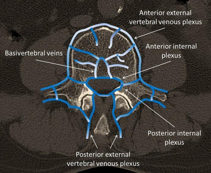
Anatomical Diagram overlaid on a computerized tomography (CT) cross-section of a human vertebra, illustrating the vertebral venous plexuses. The axial view displays four primary venous networks labeled with blue schematics: the anterior external vertebral venous plexus, which lies on the anterolateral surface of the vertebral body; the posterior external vertebral venous plexus, located around the vertebral arches and spinous process; and the internal plexuses (anterior and posterior) situated within the spinal canal. Centrally, the basivertebral veins are shown originating within the vertebral body and draining into the internal vertebral venous plexuses. The diagram highlights the complex, interconnected nature of the spinal venous system, which is clinically significant in understanding the potential pathways for Batson's plexus and the risk of cement embolism during spinal procedures like balloon kyphoplasty (BKP). This content is intended for intermediate medical education, focusing on spinal anatomy and perioperative complications.
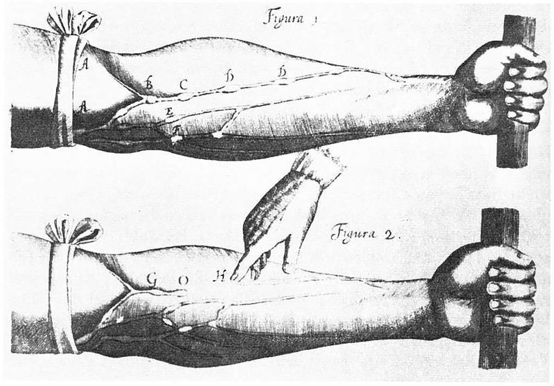
This historical anatomical diagram, famously from William Harvey's 1628 'De Motu Cordis', illustrates the physiological principles of blood circulation and venous valve function. The image consists of two comparative panels, Figura 1 and Figura 2, showing a human forearm with a tourniquet applied proximally. In Figura 1, the superficial veins (labeled B, C, D, E) appear distended and engorged between the tourniquet (A) and the distal hand, which is gripping a rod to further increase venous pressure. Nodes along the veins represent the locations of venous valves, where blood is pooling due to the proximal occlusion. In Figura 2, an external finger (H) is shown depressing a specific segment of the vein to demonstrate that blood flow is unidirectional; the vessel remains collapsed distal to the pressure point, proving that valves prevent retrograde flow toward the hand. This illustration serves as a foundational pedagogical tool in cardiovascular physiology, demonstrating the transition from Galenic theories to the modern understanding of systemic circulation and the function of one-way valves in the venous system.
All Major Veins of the Human Body
- Pulmonary veins - carry oxygenated blood (from lungs to left atrium)
- Umbilical vein (fetal only) - carries oxygenated blood from placenta to fetus
1. PULMONARY VEINS (Oxygenated)
| Vein | Arises From | Drains Into | Blood |
|---|---|---|---|
| Right superior pulmonary vein | Right upper & middle lobes of lung | Left atrium | ✅ Oxygenated |
| Right inferior pulmonary vein | Right lower lobe of lung | Left atrium | ✅ Oxygenated |
| Left superior pulmonary vein | Left upper lobe of lung | Left atrium | ✅ Oxygenated |
| Left inferior pulmonary vein | Left lower lobe of lung | Left atrium | ✅ Oxygenated |
2. VEINS OF THE HEAD & NECK (Deoxygenated)
| Vein | Arises From | Drains Into |
|---|---|---|
| Internal jugular vein | Sigmoid sinus (base of skull, jugular foramen) | Brachiocephalic vein |
| External jugular vein | Retromandibular vein + posterior auricular vein (behind the ear, parotid region) | Subclavian vein |
| Anterior jugular vein | Submental region (chin area) | External jugular vein or subclavian vein |
| Facial vein | Angular vein (near inner corner of eye) | Internal jugular vein |
| Retromandibular vein | Superficial temporal vein + maxillary vein (behind mandible) | External jugular or facial vein |
| Superficial temporal vein | Scalp, temporal region | Retromandibular vein |
| Maxillary vein | Pterygoid plexus (infratemporal fossa) | Retromandibular vein |
| Occipital vein | Posterior scalp | Internal jugular or vertebral vein |
| Vertebral vein | Suboccipital venous plexus (around vertebral artery, C1 level) | Brachiocephalic vein |
| Transverse facial vein | Lateral face | Superficial temporal vein |
| Sinus | Arises From | Drains Into |
|---|---|---|
| Superior sagittal sinus | Anterior cranial fossa, superior cerebral veins | Confluence of sinuses |
| Inferior sagittal sinus | Medial cerebral hemispheres | Straight sinus |
| Straight sinus | Inferior sagittal sinus + great cerebral vein (of Galen) | Confluence of sinuses |
| Confluence of sinuses | Superior sagittal, straight, occipital sinuses | Transverse sinuses |
| Transverse sinus | Confluence of sinuses | Sigmoid sinus |
| Sigmoid sinus | Transverse sinus | Internal jugular vein |
| Cavernous sinus | Ophthalmic veins, sphenoparietal sinus | Superior/inferior petrosal sinuses |
| Superior petrosal sinus | Cavernous sinus | Sigmoid/transverse sinus |
| Inferior petrosal sinus | Cavernous sinus | Internal jugular vein |
3. VEINS OF THE UPPER LIMB (Deoxygenated)
Deep Veins
| Vein | Arises From | Drains Into |
|---|---|---|
| Radial veins | Deep palmar arch, radial side of forearm | Brachial veins (join at elbow) |
| Ulnar veins | Deep palmar arch, ulnar side of forearm | Brachial veins (join at elbow) |
| Brachial veins (paired) | Radial + ulnar veins (at elbow) | Axillary vein |
| Axillary vein | Continuation of basilic vein + brachial veins (at lower border of teres major) | Subclavian vein (at lateral border of rib I) |
| Subclavian vein | Continuation of axillary vein | Brachiocephalic vein |
Superficial Veins
| Vein | Arises From | Drains Into |
|---|---|---|
| Cephalic vein | Dorsal venous network of hand (lateral/posterior hand) | Axillary vein (via deltopectoral groove) |
| Basilic vein | Dorsal venous network (medial/posterior hand and forearm) | Axillary vein (pierces deep fascia mid-arm) |
| Median cubital vein | Cephalic vein | Basilic vein (crosses the cubital fossa - classic IV/blood draw site) |
| Median antebrachial vein | Palm/anterior forearm | Basilic or median cubital vein |
4. VEINS OF THE THORAX (Deoxygenated)
| Vein | Arises From | Drains Into |
|---|---|---|
| Right brachiocephalic vein | Right subclavian + right internal jugular veins | Superior vena cava |
| Left brachiocephalic vein | Left subclavian + left internal jugular veins | Superior vena cava |
| Superior vena cava (SVC) | Union of two brachiocephalic veins | Right atrium |
| Azygos vein | Junction of right ascending lumbar vein + right subcostal vein (at L1-L2) | Superior vena cava (at vertebral level T4) |
| Hemiazygos vein | Junction of left ascending lumbar vein + left subcostal vein | Azygos vein (crosses at T9) |
| Accessory hemiazygos vein | Upper left posterior intercostal veins (T4-T8) | Azygos vein (crosses at T7-T8) |
| Left superior intercostal vein | 2nd, 3rd, 4th left intercostal veins | Left brachiocephalic vein |
| Posterior intercostal veins (right, 2nd-4th) | Intercostal spaces | Right superior intercostal vein → azygos |
| Posterior intercostal veins (right, 5th-11th) | Intercostal spaces | Azygos vein |
| Posterior intercostal veins (left, lower 4-5) | Intercostal spaces | Hemiazygos vein |
| Internal thoracic veins | Anterior intercostal spaces, anterior chest wall | Brachiocephalic vein |
| Coronary sinus | Great cardiac vein, middle cardiac vein, small cardiac vein (heart wall) | Right atrium |
| Great cardiac vein | Apex of heart, anterior surface | Coronary sinus |
| Middle cardiac vein | Posterior interventricular groove | Coronary sinus |
| Small cardiac vein | Right atrium/ventricle margin | Coronary sinus |
5. VEINS OF THE ABDOMEN (Deoxygenated unless noted)
Inferior Vena Cava (IVC) system
| Vein | Arises From | Drains Into |
|---|---|---|
| Inferior vena cava (IVC) | Union of two common iliac veins (at L5) | Right atrium |
| Common iliac veins | Union of internal + external iliac veins | IVC |
| External iliac vein | Continuation of femoral vein (at inguinal ligament) | Common iliac vein |
| Internal iliac vein | Pelvic viscera and walls | Common iliac vein |
| Renal veins (left and right) | Renal hilum (kidney) | IVC |
| Suprarenal (adrenal) veins | Adrenal glands | Right → IVC; Left → left renal vein |
| Gonadal veins (testicular/ovarian) | Testes or ovaries | Right → IVC; Left → left renal vein |
| Hepatic veins (right, middle, left) | Liver parenchyma | IVC (just below diaphragm) |
| Lumbar veins | Posterior abdominal wall muscles | IVC |
| Ascending lumbar veins | Lumbar veins | Azygos (right) / Hemiazygos (left) |
| Phrenic veins | Diaphragm | IVC |
Portal Venous System (Deoxygenated - carries nutrient-rich blood from gut)
| Vein | Arises From | Drains Into |
|---|---|---|
| Portal vein | Junction of superior mesenteric vein + splenic vein (behind neck of pancreas) | Liver (right and left branches) |
| Superior mesenteric vein | Small intestine, cecum, ascending & transverse colon, stomach | Portal vein |
| Splenic vein | Spleen, pancreas, short gastric veins | Portal vein |
| Inferior mesenteric vein | Descending colon, sigmoid colon, rectum | Splenic vein (or junction) |
| Left gastric (coronary) vein | Esophagus, lesser curvature of stomach | Portal vein |
| Right gastric vein | Lesser curvature of stomach | Portal vein |
| Cystic vein | Gallbladder | Portal vein (right branch) |
6. VEINS OF THE LOWER LIMB (Deoxygenated)
Deep Veins
| Vein | Arises From | Drains Into |
|---|---|---|
| Plantar veins (medial & lateral) | Plantar surface of foot | Posterior tibial veins |
| Posterior tibial veins | Plantar veins, deep muscles of posterior leg | Popliteal vein |
| Anterior tibial veins | Dorsal venous arch, anterior compartment of leg | Popliteal vein |
| Peroneal (fibular) veins | Lateral compartment of leg | Popliteal vein |
| Popliteal vein | Anterior + posterior tibial + peroneal veins (at knee) | Femoral vein |
| Femoral vein | Popliteal vein (at adductor hiatus) | External iliac vein (at inguinal ligament) |
| Deep femoral (profunda femoris) vein | Deep thigh muscles | Femoral vein |
Superficial Veins
| Vein | Arises From | Drains Into |
|---|---|---|
| Great (long) saphenous vein | Dorsal venous arch of foot (medial side) | Femoral vein (at saphenofemoral junction, femoral triangle) |
| Small (short) saphenous vein | Dorsal venous arch (lateral side) | Popliteal vein (at popliteal fossa) |
Summary: Oxygenated vs. Deoxygenated
| Category | Blood Type | Why |
|---|---|---|
| Pulmonary veins (x4) | ✅ Oxygenated | Carry freshly oxygenated blood from lungs to left atrium |
| Umbilical vein (fetal) | ✅ Oxygenated | Carries oxygenated blood from placenta to fetus |
| ALL other veins (systemic veins) | ❌ Deoxygenated | Return used blood from tissues to the right side of the heart |
Note: The portal vein is sometimes considered separately - it carries deoxygenated but nutrient-rich blood from the gut to the liver. It is still deoxygenated.
Just simplifily-- uh, just simplify the whole thing and then tell that, uh, just like left pulmonary artery, left pulmonary vein, left, uh, aortic, left artery, I mean, the-- then right pulmonary artery, the right pulmonary vein, uh, then inferior vena cava, uh, and all the right, right vena cava, left, uh, basically ventricle, right ventricle, basically aorta, the arch of aorta, uh, then... And superior vena cava, what's the inferior vena cava, all, uh, from-- arises from and drains into, and which type of, uh, o- uh, I mean, if they, uh, goes by the oxygenated or deoxygenated, describe, uh, as a graphical presentation and also, uh, in a table chart, so that, so that we can simplify the whole thing and just tell the main thing. Just not go thoroughly into the, uh, matter. So...
🫀 Blood Flow Diagram (Simplified)
BODY TISSUES
     |
     | (deoxygenated blood)
     ↓
┌─────────────────────────────────┐
│  SUPERIOR VENA CAVA             │  ← from upper body (head, arms, chest)
│  INFERIOR VENA CAVA             │  ← from lower body (abdomen, legs)
└────────────┬────────────────────┘
             ↓
     RIGHT ATRIUM
             ↓
     RIGHT VENTRICLE
             ↓
┌────────────────────────────────┐
│  RIGHT PULMONARY ARTERY        │ → right lung
│  LEFT PULMONARY ARTERY         │ → left lung
└────────────────────────────────┘
             ↓
         (in lungs: CO₂ out, O₂ in)
             ↓
┌────────────────────────────────┐
│  RIGHT PULMONARY VEIN          │ ← right lung
│  LEFT PULMONARY VEIN           │ ← left lung
└────────────┬───────────────────┘
             ↓
      LEFT ATRIUM
             ↓
      LEFT VENTRICLE
             ↓
          AORTA
             ↓
       ARCH OF AORTA
             ↓
    (branches to whole body)
             ↓
       BODY TISSUES
📊 Main Vessels - Simple Table
| Vessel | Arises From | Drains / Goes Into | Blood Type |
|---|---|---|---|
| Superior Vena Cava (SVC) | Upper body (head, arms, chest) - union of two brachiocephalic veins | Right atrium | ❌ Deoxygenated |
| Inferior Vena Cava (IVC) | Lower body (abdomen, legs) - union of two common iliac veins at L5 | Right atrium | ❌ Deoxygenated |
| Right Ventricle | Receives from right atrium | Pumps into right + left pulmonary arteries | ❌ Deoxygenated |
| Right Pulmonary Artery | Right ventricle (via pulmonary trunk) | Right lung | ❌ Deoxygenated |
| Left Pulmonary Artery | Right ventricle (via pulmonary trunk) | Left lung | ❌ Deoxygenated |
| Right Pulmonary Vein | Right lung | Left atrium | ✅ Oxygenated |
| Left Pulmonary Vein | Left lung | Left atrium | ✅ Oxygenated |
| Left Ventricle | Receives from left atrium | Pumps into aorta | ✅ Oxygenated |
| Aorta | Left ventricle | Arch of aorta → whole body | ✅ Oxygenated |
| Arch of Aorta | Ascending aorta (from left ventricle) | Descending aorta + branches to head/arms | ✅ Oxygenated |
🔑 One Rule to Remember
Arteries away from heart, Veins return to heart. Only the pulmonary vessels are "backwards" - pulmonary artery carries deoxygenated, pulmonary vein carries oxygenated.
| Carries | Goes to | |
|---|---|---|
| All pulmonary arteries | ❌ Deoxygenated | Lungs (to pick up O₂) |
| All pulmonary veins | ✅ Oxygenated | Heart (left atrium) |
| Aorta + all systemic arteries | ✅ Oxygenated | Body |
| SVC + IVC + all systemic veins | ❌ Deoxygenated | Heart (right atrium) |
And also tell me about the arts of Ayodhya, all the, um...
Also, tell me about the Arch of Arta.
You mentioned 'arts of Ayodhya' and 'Arch of Arta' - which of these did you mean?
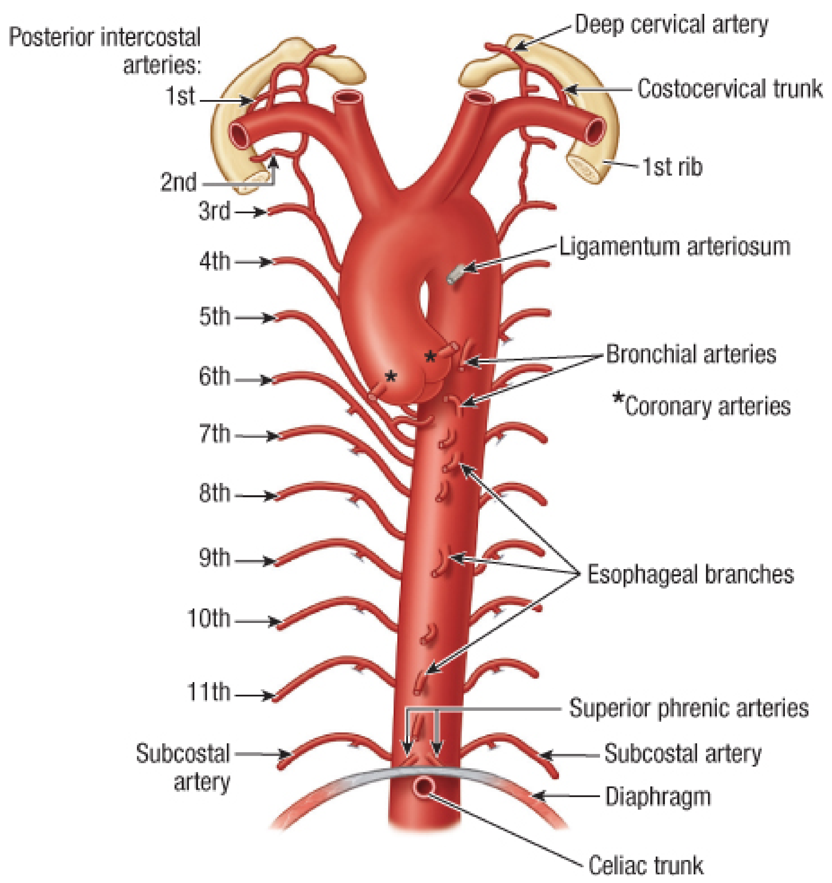
Arch of Aorta - Simplified
What & Where
- It is the curved part of the aorta that sits in the superior mediastinum (upper chest), behind the manubrium of the sternum
- It begins where the ascending aorta exits the heart's pericardial sac
- It curves upward, backward, and to the left
- It ends at the level of vertebra T4/T5 on the left side, where it becomes the descending (thoracic) aorta
- Blood type: ✅ Oxygenated (all parts of the aorta carry oxygenated blood)
Flow Diagram
LEFT VENTRICLE
      ↓
ASCENDING AORTA
      ↓
ARCH OF AORTA  ──────────────────────┐
  (curves left, at level of T4/T5)   │
      ↓                              │
DESCENDING AORTA          3 Branches off the top:
                          1. Brachiocephalic Trunk
                          2. Left Common Carotid Artery
                          3. Left Subclavian Artery
3 Branches of the Arch - Table
| Branch | Arises From | Supplies | Blood |
|---|---|---|---|
| 1. Brachiocephalic Trunk (largest, first/rightmost) | Arch of aorta | Splits into → Right common carotid (right head & neck) + Right subclavian (right arm) | ✅ Oxygenated |
| 2. Left Common Carotid Artery | Arch of aorta | Left side of head & neck | ✅ Oxygenated |
| 3. Left Subclavian Artery | Arch of aorta | Left upper limb | ✅ Oxygenated |
All three branches are crossed in front by the left brachiocephalic vein.
One Extra Structure - Ligamentum Arteriosum
- A small fibrous cord connecting the arch of aorta to the left pulmonary artery
- In the fetus it was a real open vessel called the ductus arteriosus - it allowed blood to bypass the lungs (since the fetus doesn't breathe)
- After birth, it closes and becomes just a ligament
Aorta Summary (all 3 parts together)
LEFT VENTRICLE
      ↓
ASCENDING AORTA   → gives off: Left & Right Coronary arteries (heart supply)
      ↓
ARCH OF AORTA     → gives off: Brachiocephalic trunk, Left carotid, Left subclavian
      ↓
DESCENDING AORTA  → gives off: Intercostal arteries, Bronchial arteries, then
      ↓             passes through diaphragm → becomes Abdominal Aorta
ABDOMINAL AORTA   → gives off: Celiac trunk, Mesenteric arteries, Renal arteries, etc.