Bladder outflow obstruction
"bladder outlet obstruction" management
Bladder Outflow Obstruction (BOO)
Definition
- Bailey and Love's Short Practice of Surgery, 28th Ed., p. 1545
Causes
- Benign prostatic hyperplasia (BPH) - the most frequent cause
- Bladder neck stenosis
- Bladder neck dyssynergia / functional bladder neck obstruction
- Bladder neck hypertrophy
- Prostate cancer
- Urethral stricture
- Functional obstruction from neuropathic conditions
-
Post anti-incontinence surgery (e.g. sling procedures)
-
Pelvic organ prolapse / cystocele
-
Primary bladder neck obstruction
-
Dysfunctional voiding
-
Urethral stricture
-
Bailey and Love's, p. 1545; Campbell-Walsh-Wein Urology, p. 3381
Urodynamic Parameters
Normal flow rate
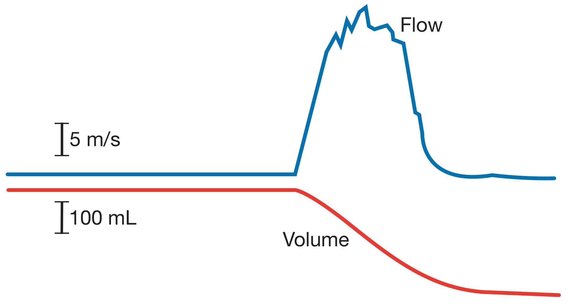
| Flow Rate | Interpretation |
|---|---|
| >15 mL/s (voided vol >200 mL) | Normal |
| 10-15 mL/s | Equivocal |
| <10 mL/s | Low (suggestive of BOO) |
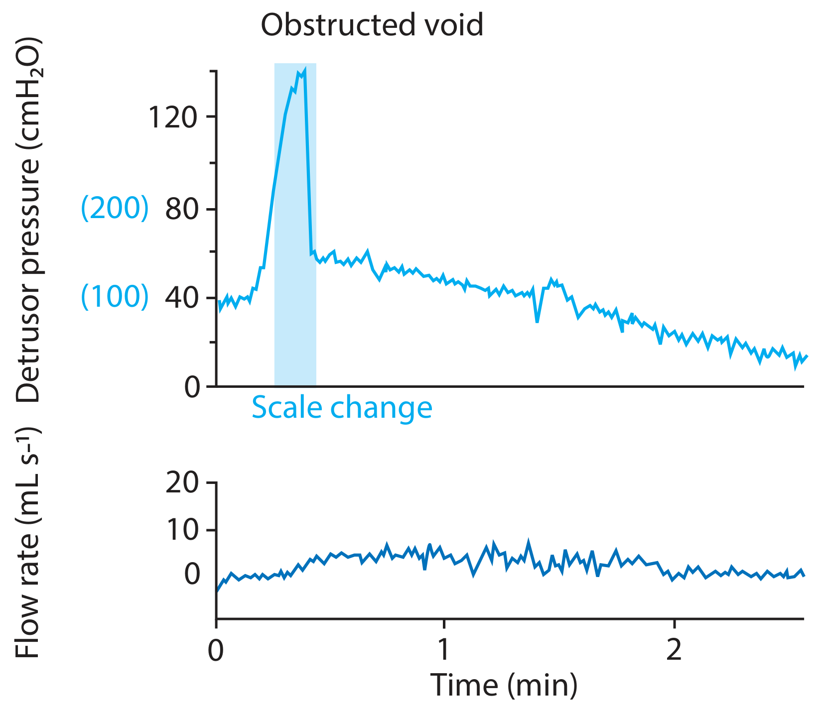
Voiding pressures (detrusor pressure at max flow)
| Pressure | Interpretation |
|---|---|
| >80 cmH₂O | High (obstructed) |
| 60-80 cmH₂O | Equivocal |
| <60 cmH₂O | Normal |
Obstructed void trace
BOO in women
-
Qmax ≤ 11-15 mL/s plus pdetQmax ≥ 20-25 cmH₂O
-
Videourodynamics (fluoroscopic imaging) adds precision by localizing the site of obstruction
-
Campbell-Walsh-Wein, p. 3381
Pathophysiology - Effects on the Bladder
Immediate / functional changes
- Detrusor hypertrophy - the bladder wall hypertrophies in response to chronic high-pressure voiding; muscle cell size increases massively
- Detrusor overactivity (DO) - obstruction-induced DO with irritative symptoms is attributed to:
- Denervation supersensitivity (increased cholinergic contractile responses)
- Altered contractile proteins (SM-A isoform expression)
- Upregulation of Cx43 (gap-junction protein) leading to enhanced smooth muscle coupling and coordinated myogenic contractions
- Increased NGF (nerve growth factor) in bladder tissue and urine
- Spinal reflex enhancement - a spinal micturition reflex is enhanced in BOO
Long-term effects
-
Bladder decompensation - detrusor contraction becomes progressively less efficient; residual urine accumulates - chronic retention
-
Increased bladder irritability - decreased functional capacity, detrusor overactivity, urgency/frequency
-
High-pressure chronic retention - residual volume >250 mL raises intramural wall tension, causing functional obstruction of the upper tracts and bilateral hydronephrosis, upper tract infection, and renal impairment
-
Bailey and Love's, p. 1545-1546; Campbell-Walsh-Wein, p. 3291
Complications
| Complication | Notes |
|---|---|
| Acute urinary retention | Sometimes the first presentation; painful |
| Chronic retention | Low-pressure (no upper tract damage) or high-pressure (hydronephrosis, renal failure) |
| Overflow incontinence / enuresis | Clue to high-pressure chronic retention |
| Urinary tract infection | From stasis and residual urine |
| Bladder calculi | Stasis promotes stone formation |
| Bladder diverticula | Herniation through areas of muscular weakness |
| Haematuria | May relate to prostatic vascularity; other causes must be excluded |
| Renal failure | From chronic high-pressure back-pressure |
Investigation
- Uroflowmetry - screening; >200 mL voided for reliability
- Post-void residual (PVR) ultrasound - >300 mL warrants further workup; >250 mL suggests chronic retention
- Pressure-flow urodynamics - gold standard for diagnosis
- Videourodynamics - especially valuable in women to localize site of obstruction
- Renal ultrasound - to detect hydronephrosis / upper tract damage
- PSA + prostate size - to guide BPH management
- CT urography + cystoscopy + urine cytology - when haematuria is present
Management
Conservative (watchful waiting)
- Fluid manipulation: avoid fluid binges, limit late-night intake
- Reduce caffeinated and alcoholic drinks
- Appropriate for mild-moderate symptoms
Medical therapy
| Drug class | Agents | Mechanism |
|---|---|---|
| α-blockers | Tamsulosin, alfuzosin, silodosin | Relax smooth muscle of bladder neck and prostate |
| 5α-reductase inhibitors | Finasteride, dutasteride | Reduce prostatic volume (best for large glands) |
| Combination | α-blocker + 5ARI | Superior outcome for glands >35 g |
Note: Medical therapy is not safe in high-pressure chronic retention given the risk of worsening renal failure - these patients need surgical or catheter management.
Strong indications for surgery
- Acute retention in fit men without reversible cause (accounts for ~25% of prostatectomies)
- Chronic retention + renal impairment - residual >200 mL, hydro(ureter)nephrosis, uraemia (~15% of prostatectomies)
- Complications - stone, infection, diverticulum, recurrent haematuria
- Severe symptoms not responding to drugs, Qmax <10 mL/s, residual 100-250 mL (~60% of prostatectomies)
Surgical options
| Procedure | Notes |
|---|---|
| TURP (transurethral resection) | Gold standard for decades |
| HoLEP (holmium laser enucleation) | Preferred for large glands (>100 g), patients on anticoagulation; 7-year data shows equivalent QoL to TURP with fewer reoperations |
| Greenlight laser vaporization | Reduced bleeding and catheter time |
| Open / robotic simple prostatectomy | Large glands |
| PAE (prostate artery embolisation) | Minimally invasive |
| Rezūm (water vapour therapy) | Minimally invasive, preserves sexual function |
| UroLift (prostatic urethral lift) | No thermal energy, day-case procedure |
| Aquablation (water jet) | Robotic-assisted, anatomy-guided |
Patients with very large PVR (>1 L) often benefit more from intermittent self-catheterization or a permanent catheter, because detrusor weakness from chronicity reduces the chance of surgical success.
- Bailey and Love's, p. 1546-1548; Comprehensive Clinical Nephrology, 7th Ed., p. 2502-2504
Recent Evidence (2024-2026)
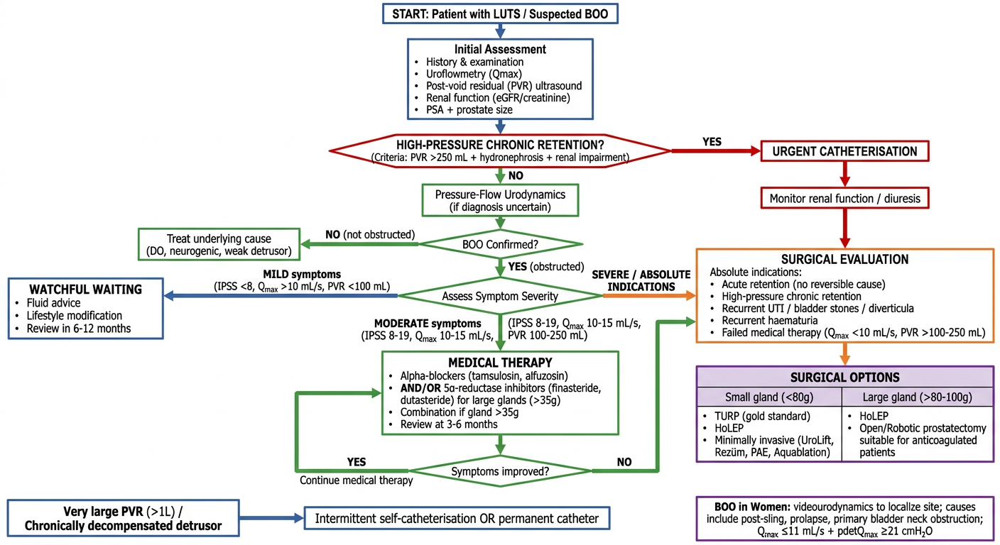
Generate a clinical decision-making flowchart for BOO management (medical vs surgical)
Clinical decision-making flowchart for Bladder Outflow Obstruction (BOO) management - medical vs surgical pathway. Clean professional medical flowchart with the following structure: START: "Patient with LUTS / Suspected BOO" at top First box: "Initial Assessment" with bullet points: History & examination, Uroflowmetry (Qmax), Post-void residual (PVR) ultrasound, Renal function (eGFR/creatinine), PSA + prostate size Diamond decision: "HIGH-PRESSURE CHRONIC RETENTION?" criteria: PVR >250 mL + hydronephrosis + renal impairment YES branch (red arrow) → "URGENT CATHETERISATION" box (red) → "Monitor renal function / diuresis" → then connects to Surgical Evaluation NO branch (green arrow) → "Pressure-Flow Urodynamics (if diagnosis uncertain)" → "BOO Confirmed?" diamond NO (not obstructed) → "Treat underlying cause (DO, neurogenic, weak detrusor)" YES (obstructed) → "Assess Symptom Severity" diamond with three branches: Branch 1 - MILD symptoms (IPSS <8, Qmax >10 mL/s, PVR <100 mL): "WATCHFUL WAITING" box (blue) with: Fluid advice, Lifestyle modification, Review in 6-12 months Branch 2 - MODERATE symptoms (IPSS 8-19, Qmax 10-15 mL/s, PVR 100-250 mL): "MEDICAL THERAPY" box (green) with: Alpha-blockers (tamsulosin, alfuzosin) AND/OR 5α-reductase inhibitors (finasteride, dutasteride) for large glands (>35g), Combination if gland >35g, Review at 3-6 months; then "Symptoms improved?" diamond → YES: Continue medical therapy / YES: Proceed to surgical evaluation Branch 3 - SEVERE / ABSOLUTE INDICATIONS: "SURGICAL EVALUATION" box (orange) listing absolute indications: Acute retention (no reversible cause), High-pressure chronic retention, Recurrent UTI / bladder stones / diverticula, Recurrent haematuria, Failed medical therapy (Qmax <10 mL/s, PVR >100-250 mL) SURGICAL OPTIONS box (purple) with table: Small gland (<80g): TURP (gold standard), HoLEP, Minimally invasive (UroLift, Rezūm, PAE, Aquablation); Large gland (>80-100g): HoLEP, Open/Robotic prostatectomy, suitable for anticoagulated patients Special box at bottom: "Very large PVR (>1L) / Chronically decompensated detrusor" → "Intermittent self-catheterisation OR permanent catheter" Women-specific note box: "BOO in Women: videourodynamics to localize site; causes include post-sling, prolapse, primary bladder neck obstruction; Qmax ≤11 mL/s + pdetQmax ≥21 cmH₂O" Color scheme: red for urgent/emergency, green for medical/conservative, orange for surgical indications, blue for watchful waiting, purple for surgical options. White background, dark text, clear arrows with labels, professional clinical style.