https://orris.care/shared/chat/1a98ec4c-e14d-4283-afd0-b57a7d4b8abf
Reading Web Page
This is a shared Orris chat about the Radiological Approach to the Solitary Pulmonary Nodule (based on Grainger & Allison's textbook). Here's the full content from that conversation:
Radiological Approach to the Solitary Pulmonary Nodule
(Grainger & Allison)
Definition
A solitary pulmonary nodule (SPN) is a single, well-defined, rounded opacity ≤3 cm in diameter, surrounded by lung parenchyma, with no associated lymphadenopathy or atelectasis.
Clinical Significance
SPNs are common incidental findings on chest CT. The key clinical question is distinguishing benign from malignant lesions, as ~40% of SPNs in high-risk populations may be malignant.
Step 1: Assess Nodule Size and Growth Rate
| Size | Interpretation |
|---|---|
| <5 mm (<80 mm³) | Malignancy risk so low that follow-up not generally recommended in average-risk patients |
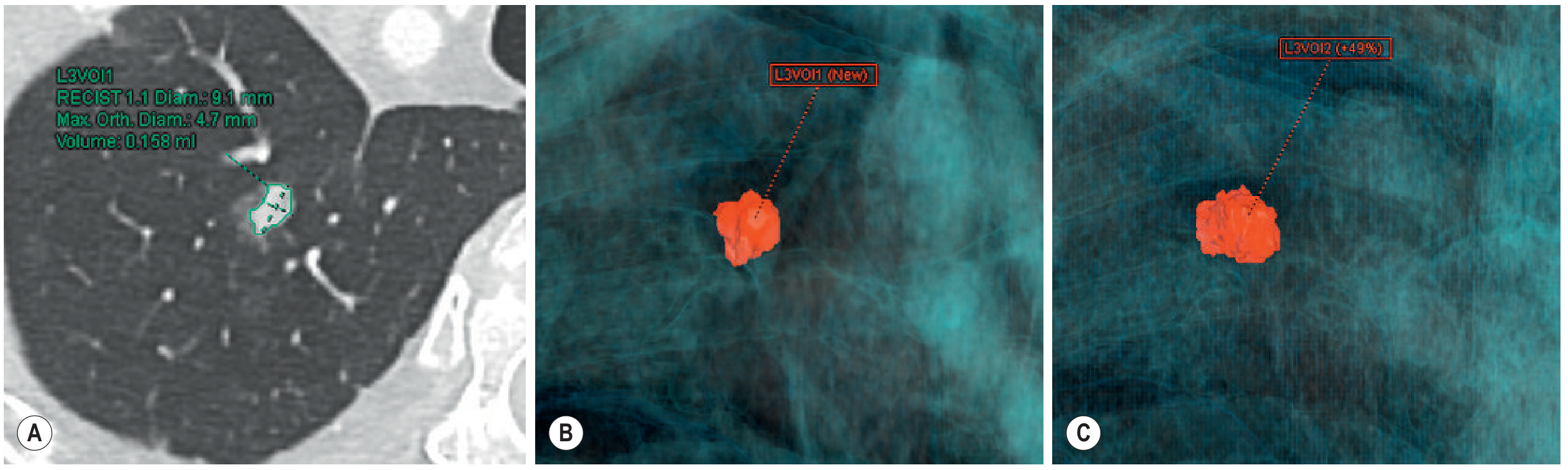
| 5-8 mm | Intermediate risk - CT surveillance guided by volumetry/VDT |
| >8 mm | Significant risk - further assessment with PET/CT or tissue sampling |
Step 2: Assess Location, Shape, and Morphology
Perifissural Nodules (Benign Features)
Nodules attached to or within 1 cm of a fissure, with a polygonal, lenticular, or triangular shape and smooth margins - these are almost always benign intrapulmonary lymph nodes and can be discharged.
Nodule Outline
- Spiculated/corona radiata: Highly suspicious for malignancy
- Lobulated: Intermediate suspicion
- Smooth: More likely benign, but not excluded
Step 3: Assess Calcification Pattern
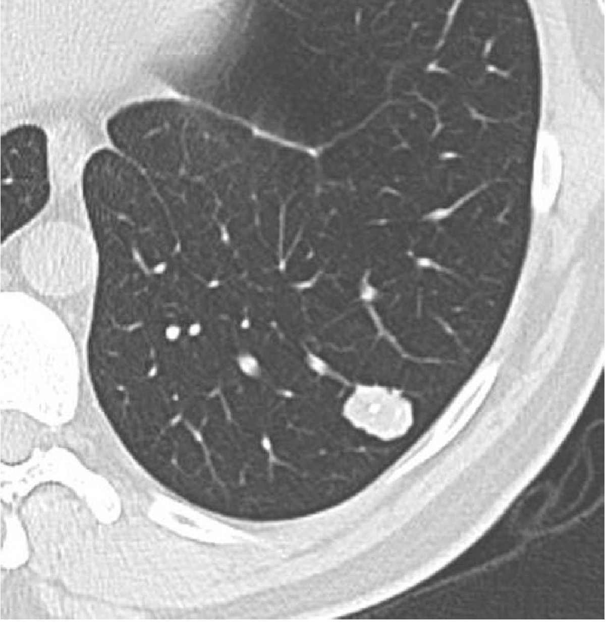
Calcification patterns:
- Benign: Central, diffuse, laminated ("eggshell"), or popcorn (hamartoma)
- Indeterminate/malignant: Eccentric, amorphous, or stippled calcification
Step 4: Classify as Solid vs. Subsolid
| Type | Definition | CT Appearance | Histological Correlate |
|---|---|---|---|
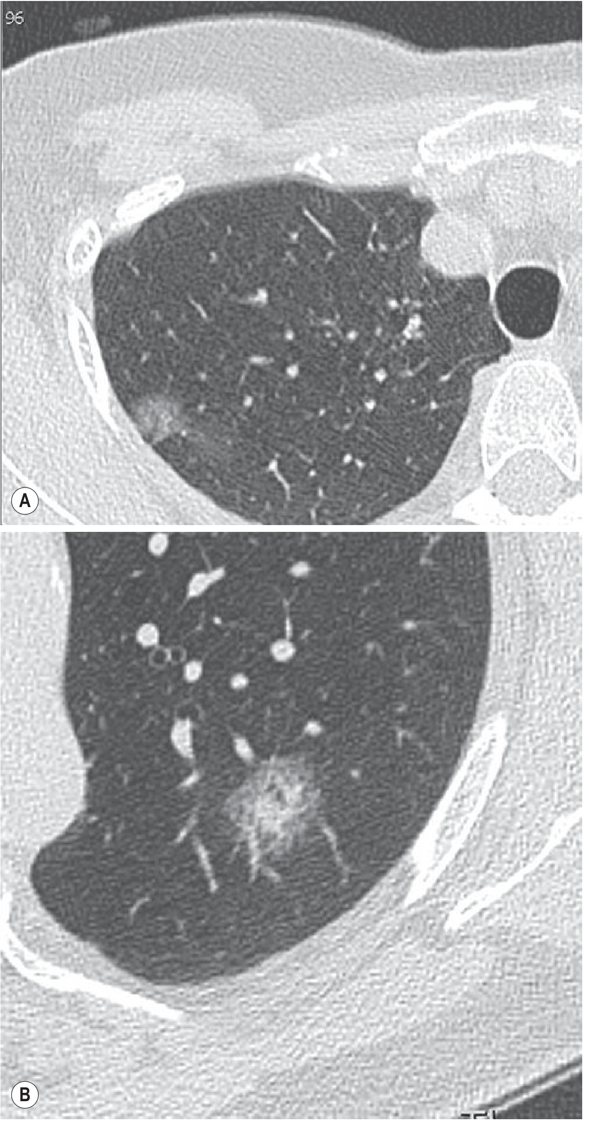
| Pure Ground-Glass Nodule (GGN) | Focal increased attenuation, lung architecture visible | Hazy, no solid component | Atypical adenomatous hyperplasia, adenocarcinoma in situ |
| Part-Solid Nodule | GGN with a central solid component that obscures architecture | Mixed density | Minimally invasive adenocarcinoma, invasive adenocarcinoma |
Part-solid nodules with a solid component >5 mm carry the highest malignancy risk of all SPN subtypes.
Step 5: Risk Stratification and Management Algorithms
Fleischner Society 2017 vs. BTS 2015 Guidelines
| Parameter | Fleischner 2017 | BTS 2015 |
|---|---|---|
| Population | Age ≥35, incidentally detected | Age ≥18, all presentations |
| Sizing method | Mean diameter (long + short axis) | Volumetry preferred |
| Follow-up threshold | 6 mm | 80 mm³ (or 5 mm) |
| Growth definition | ≥2 mm diameter increase | >25% volume increase |
| Small nodule (<6 mm) | No follow-up in low risk | Discharge; follow if definitely new vs. prior 1-2y CT |
| Medium nodule (6-8 mm) | CT at 6-12 months, then 18-24 months | CT at 3 months; further follow-up based on VDT |
| Large nodule (>8 mm) | CT at 3 months, PET/CT or biopsy | Brock model risk estimation |
BTS Brock Model (>8 mm nodules)
A validated malignancy prediction tool incorporating: family history of lung cancer, emphysema on CT, nodule size, spiculation, upper lobe location, nodule type (part-solid > solid > GGN), and count.
Step 6: Advanced Imaging Modalities
FDG-PET / PET-CT
- Best for nodules >8 mm
- Sensitivity ~87%, specificity ~83% for malignancy
- False negatives: carcinoid, BAC/AIS, mucinous adenocarcinoma, small nodules
- False positives: active infection/inflammation (TB, fungal, rheumatoid nodule)
CT Enhancement
- Benign: <15 HU enhancement
- Malignant: >15 HU enhancement (sensitivity ~98%, but low specificity)
MRI
- Emerging role; DWI useful for characterizing nodules, avoids radiation
Step 7: Tissue Sampling
- CT-guided percutaneous biopsy: For peripheral nodules >8 mm; risk of pneumothorax ~15-20%
- Bronchoscopy/EBUS: For central or peribronchial nodules; radial EBUS + fluoroscopy improves yield for peripheral lesions
- VATS: Diagnostic and potentially curative for peripheral nodules; preferred if high malignancy suspicion
Summary: Radiological Features Favouring Benign vs. Malignant
| Feature | Favours Benign | Favours Malignant |
|---|---|---|
| Size | <5 mm | >8 mm |
| Shape | Polygonal, flat, tubular, low sphericity | Round, spherical |
| Margins | Smooth, well-defined, concave surfaces | Spiculated (corona radiata), lobulated |
| Location | Subpleural, perifissural, lower lobe | Upper lobe (especially right) |
| Calcification | Central, diffuse, laminated, popcorn | Absent, eccentric, amorphous |
| Density | Solid with fat density (hamartoma) | Part-solid (solid component >5 mm) |
| VDT | >600 days or stable >2 years | <400 days |
| FDG-PET | No uptake | Uptake > mediastinal blood pool |
| CT enhancement | <15 HU | >15 HU |
That's the full shared chat. It covers the complete 7-step radiological approach to SPNs from Grainger & Allison. Would you like me to expand on any particular step, compare additional guidelines (e.g. Lung-RADS, ACCP), or discuss a specific clinical scenario?
This is a shared conversation. Sign in to Orris to start your own chat.