
Morphological spectrum of Nash/ nafld for md pathology exam
NASH nonalcoholic steatohepatitis histology hepatocyte ballooning steatosis
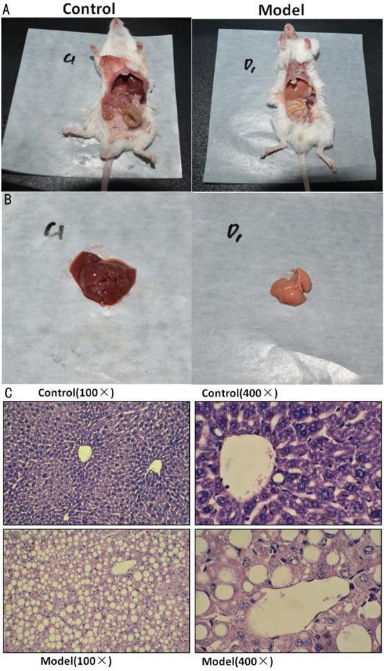
Educational panel illustrating the macroscopic and microscopic pathology of Nonalcoholic Steatohepatitis (NASH) in a mouse model. Panel A presents the gross anatomy of dissected control and model mice. Panel B provides a high-resolution clinical photograph comparing a healthy control liver (dark red/brown, smooth surface) with a NASH model liver (pale yellow-tan, suggestive of steatosis and atrophy). Panel C displays diagnostic histopathology using Hematoxylin and Eosin (H&E) staining at 100x and 400x magnifications. The control group micrographs show normal hepatocyte architecture with organized cords and clear sinusoidal spaces. In contrast, the NASH model micrographs demonstrate hallmark features of hepatic steatosis, including diffuse macrovesicular and microvesicular fat deposition, hepatocyte ballooning, and cellular disarray. The 400x magnification specifically highlights large, clear vacuoles within the cytoplasm of hepatocytes, which displace the nuclei to the periphery. This comparison serves as a diagnostic reference for metabolic liver disease research and pathological classification.
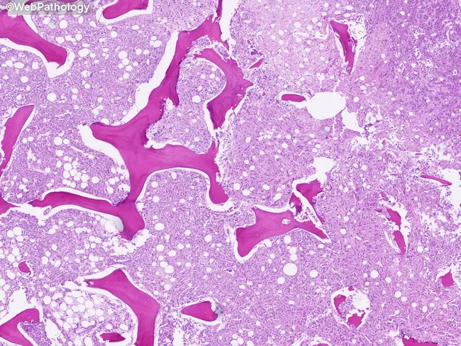
Light-microscopy histology of liver tissue demonstrates macrovesicular steatosis. Imaging modality: Histology using Hematoxylin and Eosin (H&E) stain on paraffin-embedded sections. Anatomical localization: hepatic parenchyma of the liver, with hepatocytes arranged in plates separated by sinusoids and preserved lobular architecture visible at low magnification. Visual features: numerous intracellular lipid vacuoles produce large clear droplets that displace hepatocyte nuclei to the periphery; cytoplasm otherwise eosinophilic; occasional subtle ballooning is not evident; portal tracts and central veins are discernible but not markedly inflamed. Pathological interpretation: macrovesicular steatosis consistent with fatty liver changes; most compatible with nonalcoholic fatty liver disease (NAFLD) or alcoholic fatty liver disease (AFLD) depending on patient history. In this image, steatosis is the predominant finding with minimal inflammatory infiltrate and no definitive fibrosis at this level of magnification. Clinical significance: fatty change indicates hepatocellular stress from metabolic syndrome or hepatotoxic exposure; may precede steatohepatitis, fibrosis, cirrhosis, and increased risk of hepatocellular carcinoma with ongoing injury. Potential clinical use cases: assessment of steatosis burden in NAFLD/NASH studies, biopsy-guided staging, therapeutic monitoring, and education for trainees. The image serves as an educational reference for recognizing hepatocellular lipid accumulation and distinguishing it from other hepatic pathologies such as steatohepatitis and cholestatic injury.
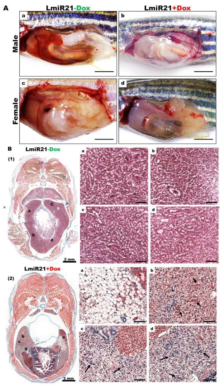
This composite image presents a preclinical study of nonalcoholic steatohepatitis (NASH) using a zebrafish model (LmiR21). Section A displays gross clinical photographs comparing livers from control (LmiR21-Dox) and experimental (LmiR21+Dox) groups at 6 months post-fertilization (mpf). In both males and females, the miR-21 induced livers (+Dox) appear enlarged, pale red, and greasy, consistent with hepatomegaly and steatosis. Section B provides histopathological analysis using Masson’s trichrome staining. The control liver (B1) shows normal hepatic architecture and cellularity. In contrast, the miR-21 overexpressing liver (B2) exhibits hallmark NASH phenotypes, including extensive hepatocyte ballooning (a), increased Mallory-Denk bodies (b, arrows), and significant lobular inflammation (c, d, arrows). These histopathological features demonstrate the progression from simple steatosis to steatohepatitis and early fibrosis, evidenced by blue-stained collagen deposition. This image serves as a translational model for understanding the role of microRNA-21 in hepatic lipid metabolism, inflammation, and fibrogenesis.
![<table><thead><tr><th>Abbreviation</th><th>Full Term</th><th>Description</th></tr></thead><tbody><tr><td>NAFLD<sup>a</sup></td><td>Nonalcoholic fatty liver disease</td><td>Term used for the broad spectrum of the disease, ranging from hepatic steatosis only to steatohepatitis (NASH) to cirrhosis, in the absence of ongoing or recent consumption of significant amounts of alcohol or the presence of other secondary causes of fatty liver disease.</td></tr><tr><td>NASH<sup>a</sup></td><td>Nonalcoholic steatohepatitis</td><td>Presence of ≥5% hepatic steatosis with inflammation and hepatocyte injury (also known as hepatocyte ballooning), with or without evidence of liver fibrosis.</td></tr><tr><td>NASH cirrhosis<sup>a</sup></td><td></td><td>Cirrhosis with histologic evidence of steatosis or steatohepatitis.</td></tr><tr><td>NAS<sup>a</sup></td><td>NAFLD activity score</td><td>An unweighted composite of steatosis, lobular inflammation, and ballooning scores.</td></tr><tr><td>Significant alcohol consumption<sup>a,b</sup></td><td>...</td><td>Defined as ingestion of >21 standard drinks per week in men and >14 standard drinks per week in women over a 2-year period preceding baseline liver histology.</td></tr><tr><td>FIB-4</td><td>Fibrosis-4 index</td><td>An index to estimate the risk of hepatic cirrhosis calculated from the computation of age, plasma aminotransferases (AST and ALT), and platelet count. This noninvasive estimate of liver scarring is used to assess the need for biopsy. The score is calculated using a person's age, AST level, platelet count (PLT), and ALT level. FIB-4 score = age (years) × AST (U/L)/[PLT (10<sup>9</sup>/L) × ALT ½ (U/L).</td></tr><tr><td>ELF</td><td>Enhanced liver fibrosis test</td><td>This blood test measures the levels of tissue inhibitor of metalloproteinases 1, amino-terminal propeptide of type III procollagen, and hyaluronic acid and is used to estimate the rate of liver extracellular matrix metabolism reflecting the severity of liver fibrosis.</td></tr><tr><td>NFS</td><td>NAFLD fibrosis score</td><td>−1.675 + 0.037 × age (years) + 0.094 × BMI (kg/m<sup>2</sup>) + 1.13 × (impaired fasting glucose or DM) + 0.99 × (AST/ALT) − 0.013 × platelet (× 10<sup>9</sup>/L) = 0.66 × albumin (g/dL) (where impaired fasting glucose/DM had a value of 1 if the participants had impaired fasting glucose and 0 if they did not)</td></tr><tr><td>APRI</td><td>AST-to-platelet ratio index</td><td>[AST level (IU/L)/AST (upper limit of normal AST range (IU/L) × 100] divided by platelet count (10<sup>9</sup>/L)</td></tr><tr><td><sup>1</sup>H-MRS</td><td>Proton magnetic resonance spectroscopy</td><td>A technique for quantifying hepatic steatosis</td></tr><tr><td>MRI-PDFF</td><td>Magnetic resonance imaging-proton density fat fraction</td><td>A technique for quantifying hepatic steatosis</td></tr><tr><td>VCTE</td><td>Vibration-controlled transient elastography</td><td>A technique for liver stiffness measurement that is correlated with the severity of liver fibrosis on histology.</td></tr><tr><td>MRE</td><td>Magnetic resonance elastography</td><td>Technology that combines MRI with low-frequency vibrations to assess liver stiffness.</td></tr></tbody></table>](https://cdn.orris.care/cdss_images/GLGCA_1754385_1766497042024_647bfd0f-d397-4e21-b10b-d3a3bfe1bb3f_f054b100-b2da-4fc2-b330-84c18b45b76a.png)
<table><thead><tr><th>Abbreviation</th><th>Full Term</th><th>Description</th></tr></thead><tbody><tr><td>NAFLD<sup>a</sup></td><td>Nonalcoholic fatty liver disease</td><td>Term used for the broad spectrum of the disease, ranging from hepatic steatosis only to steatohepatitis (NASH) to cirrhosis, in the absence of ongoing or recent consumption of significant amounts of alcohol or the presence of other secondary causes of fatty liver disease.</td></tr><tr><td>NASH<sup>a</sup></td><td>Nonalcoholic steatohepatitis</td><td>Presence of ≥5% hepatic steatosis with inflammation and hepatocyte injury (also known as hepatocyte ballooning), with or without evidence of liver fibrosis.</td></tr><tr><td>NASH cirrhosis<sup>a</sup></td><td></td><td>Cirrhosis with histologic evidence of steatosis or steatohepatitis.</td></tr><tr><td>NAS<sup>a</sup></td><td>NAFLD activity score</td><td>An unweighted composite of steatosis, lobular inflammation, and ballooning scores.</td></tr><tr><td>Significant alcohol consumption<sup>a,b</sup></td><td>...</td><td>Defined as ingestion of >21 standard drinks per week in men and >14 standard drinks per week in women over a 2-year period preceding baseline liver histology.</td></tr><tr><td>FIB-4</td><td>Fibrosis-4 index</td><td>An index to estimate the risk of hepatic cirrhosis calculated from the computation of age, plasma aminotransferases (AST and ALT), and platelet count. This noninvasive estimate of liver scarring is used to assess the need for biopsy. The score is calculated using a person's age, AST level, platelet count (PLT), and ALT level. FIB-4 score = age (years) × AST (U/L)/[PLT (10<sup>9</sup>/L) × ALT ½ (U/L).</td></tr><tr><td>ELF</td><td>Enhanced liver fibrosis test</td><td>This blood test measures the levels of tissue inhibitor of metalloproteinases 1, amino-terminal propeptide of type III procollagen, and hyaluronic acid and is used to estimate the rate of liver extracellular matrix metabolism reflecting the severity of liver fibrosis.</td></tr><tr><td>NFS</td><td>NAFLD fibrosis score</td><td>−1.675 + 0.037 × age (years) + 0.094 × BMI (kg/m<sup>2</sup>) + 1.13 × (impaired fasting glucose or DM) + 0.99 × (AST/ALT) − 0.013 × platelet (× 10<sup>9</sup>/L) = 0.66 × albumin (g/dL) (where impaired fasting glucose/DM had a value of 1 if the participants had impaired fasting glucose and 0 if they did not)</td></tr><tr><td>APRI</td><td>AST-to-platelet ratio index</td><td>[AST level (IU/L)/AST (upper limit of normal AST range (IU/L) × 100] divided by platelet count (10<sup>9</sup>/L)</td></tr><tr><td><sup>1</sup>H-MRS</td><td>Proton magnetic resonance spectroscopy</td><td>A technique for quantifying hepatic steatosis</td></tr><tr><td>MRI-PDFF</td><td>Magnetic resonance imaging-proton density fat fraction</td><td>A technique for quantifying hepatic steatosis</td></tr><tr><td>VCTE</td><td>Vibration-controlled transient elastography</td><td>A technique for liver stiffness measurement that is correlated with the severity of liver fibrosis on histology.</td></tr><tr><td>MRE</td><td>Magnetic resonance elastography</td><td>Technology that combines MRI with low-frequency vibrations to assess liver stiffness.</td></tr></tbody></table>
Morphological Spectrum of NAFLD/NASH - MD Pathology Exam
Overview
The Disease Spectrum (Sequential Steps)
Simple Steatosis → NASH → Bridging Fibrosis → Cirrhosis → HCC
1. Simple Steatosis (NAFL)
- Macrovesicular steatosis (predominant): large fat droplets displacing the nucleus to the periphery - predominantly centrilobular (Zone 3)
- Microvesicular steatosis: smaller droplets, nucleus remains central - indicates more acute/severe metabolic stress
- No ballooning degeneration
- No significant fibrosis
- Generally reversible; 25-50% may develop progressive fibrosis
| Grade | % Hepatic Involvement |
|---|---|
| 0 | <5% |
| 1 | 5-33% |
| 2 | 33-67% |
| 3 | >67% |
2. NASH (Nonalcoholic Steatohepatitis)
Key Morphological Features
A. Hepatocyte Ballooning (pathognomonic feature)
- Enlarged hepatocytes with irregular clumped cytoplasm and optically clear non-vesiculated areas
- Cytoskeletal damage - loss of normal keratin intermediate filaments
- May contain Mallory-Denk Bodies (MDB): eosinophilic, ropey cytoplasmic inclusions of hyperphosphorylated, misfolded cytokeratin filaments
- Predominantly in Zone 3 (perivenular)
B. Lobular Inflammation
- Mixed infiltrate: lymphocytes, macrophages, and neutrophils
- Microgranulomas / lipogranulomas may be present
- Neutrophils around ballooned hepatocytes ("satellitosis") - similar to alcoholic hepatitis
C. Steatosis
- Both macrovesicular and microvesicular
- Predominantly Zone 3 (centrilobular)
D. Fibrosis (Perisinusoidal/Chicken-wire)
- Begins in Zone 3 as delicate strands of collagen in perisinusoidal distribution ("chicken-wire fibrosis")
- This is the hallmark pattern distinguishing NASH from viral hepatitis (which is portal-based)
Additional Features Seen in NASH
| Feature | Significance |
|---|---|
| Megamitochondria | Mitochondrial dysfunction |
| Glycogenated nuclei | Insulin resistance/diabetes |
| Lipogranulomas | Fat-laden macrophage clusters |
| Microvesicular steatosis patches | Nonzonal, variable significance |
| Hepatocellular glycogenosis | In pediatric NAFLD |
3. Fibrosis Stages (NASH-CRN Classification)
| Stage | Description |
|---|---|
| F0 | No fibrosis |
| F1a | Mild perisinusoidal fibrosis, Zone 3 |
| F1b | Moderate perisinusoidal fibrosis, Zone 3 |
| F1c | Portal/periportal fibrosis only |
| F2 | Perisinusoidal AND periportal fibrosis |
| F3 | Bridging fibrosis |
| F4 | Cirrhosis |
Key exam point: As fibrosis progresses to Stage 3-4, steatosis, ballooning, and inflammation paradoxically decrease ("burnt-out NASH"). Cirrhosis from NASH may appear "cryptogenic" if steatohepatitis features regress.
4. NASH Cirrhosis
- Regenerative nodules within a fibrotic network
- Steatosis and ballooning may be absent at this stage (burnt-out NASH)
- Histologically indistinguishable from other causes of cirrhosis unless residual steatohepatitis features remain
- 15-25% of NASH patients progress to cirrhosis
NAFLD Activity Score (NAS) - NASH-CRN System
| Component | Score |
|---|---|
| Steatosis | 0-3 |
| Lobular inflammation | 0-2 |
| Hepatocyte ballooning | 0-2 |
| Total NAS | 0-8 |
- NAS ≥5 = correlates with NASH (sensitivity 75%, specificity 83%)
- NAS ≤3 = usually not NASH
- Fibrosis staged separately (F0-F4), not included in NAS
NAS alone cannot diagnose NASH - pattern recognition is essential. 29% of biopsies with NAS ≤4 can still show definite steatohepatitis.
SAF Scoring System (Alternative)
NASH vs. Alcoholic Steatohepatitis (ASH) - Key Distinction
| Feature | NASH | ASH |
|---|---|---|
| Mallory-Denk bodies | Present | Present (more abundant) |
| Neutrophilic infiltrate | Present | Present (more prominent) |
| Perisinusoidal fibrosis | Zone 3 | Zone 3 |
| Megamitochondria | Yes | Less common |
| Clinical history | No significant alcohol | Significant alcohol use |
| Histology | Identical | Identical |
Pediatric NAFLD - Two Distinct Patterns (Schwimmer Classification)
| Type | Features |
|---|---|
| Type 1 | Resembles adult NASH - ballooning + perisinusoidal fibrosis |
| Type 2 | Steatosis + portal inflammation + portal fibrosis, WITHOUT ballooning or perivenular fibrosis |
Histology Images
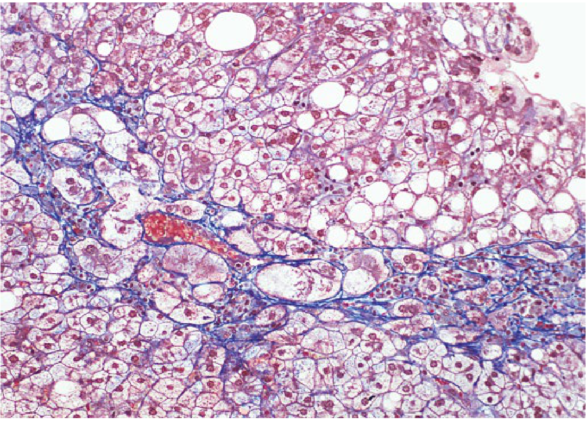
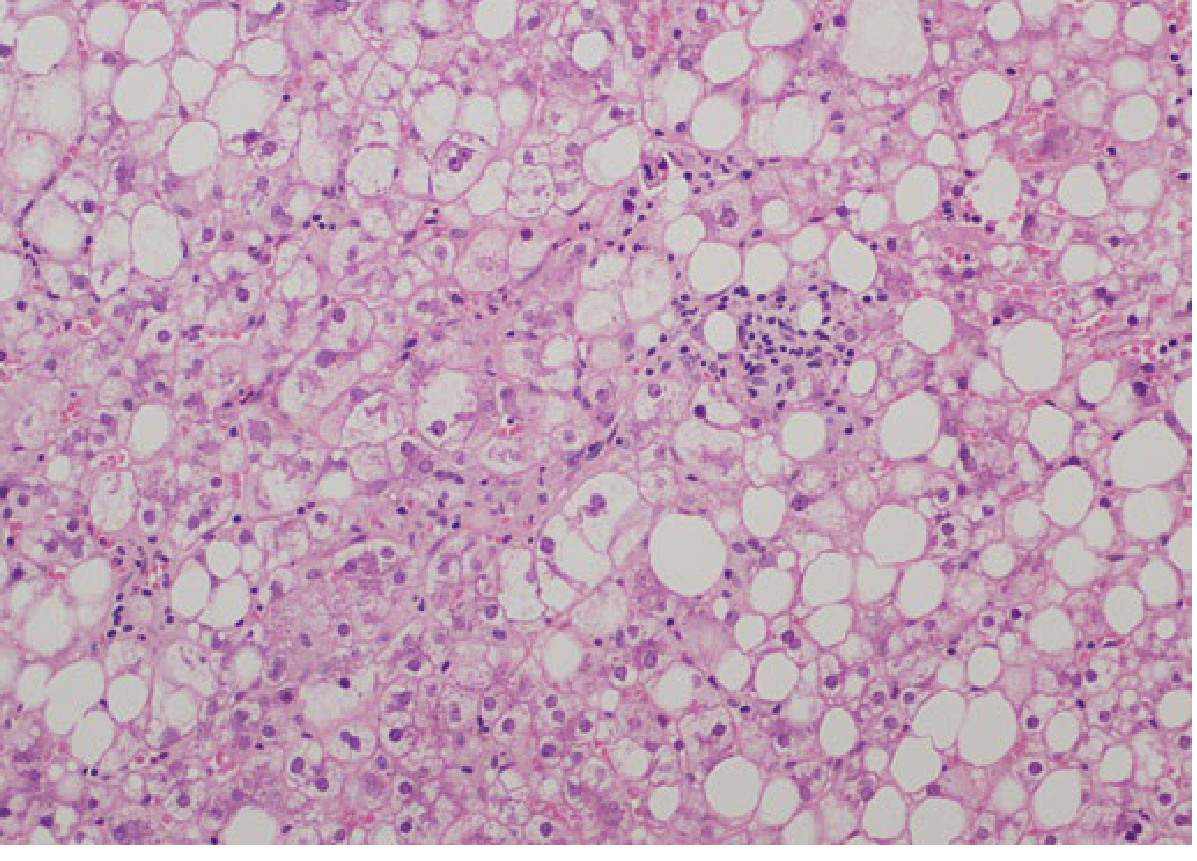
High-Yield Exam Summary Points
- Minimum for NAFLD diagnosis: >5% hepatic steatosis
- NASH triad: Steatosis + ballooning + lobular inflammation ± fibrosis
- Earliest fibrosis in NASH: Perisinusoidal Zone 3 ("chicken-wire") - distinguishes NASH from viral hepatitis (portal fibrosis)
- Mallory-Denk bodies: Misfolded CK-8/18, more abundant in ASH than NASH
- Burnt-out NASH: Stage F3-F4 paradoxically shows LESS steatosis/ballooning/inflammation
- NAS ≥5 = likely NASH; NAS ≤3 = likely NOT NASH (but diagnose by pattern, not just score)
- Pediatric Type 2 NASH: Portal-based inflammation and fibrosis (opposite to adults)
- Genetic risk: PNPLA3 I148M variant - strongest genetic predictor of steatosis and disease severity
Take reference from Robbins for Pathogenesis and clinical feature
NAFLD/NASH - Pathogenesis & Clinical Features
Robbins & Kumar Basic Pathology
Nomenclature Note
- Steatosis (simple fatty liver)
- Steatohepatitis (NASH)
- Cirrhosis
Risk Factors / Associations
- Insulin resistance and metabolic syndrome
- Type 2 diabetes (or family history)
- Obesity - primarily central/visceral
- Dyslipidemia - hypertriglyceridemia, low HDL-C, high LDL-C
- Hypertension
Pathogenesis (Fig. 14.19, Robbins Basic Pathology)
Step 1 - Steatosis ("First Hit")
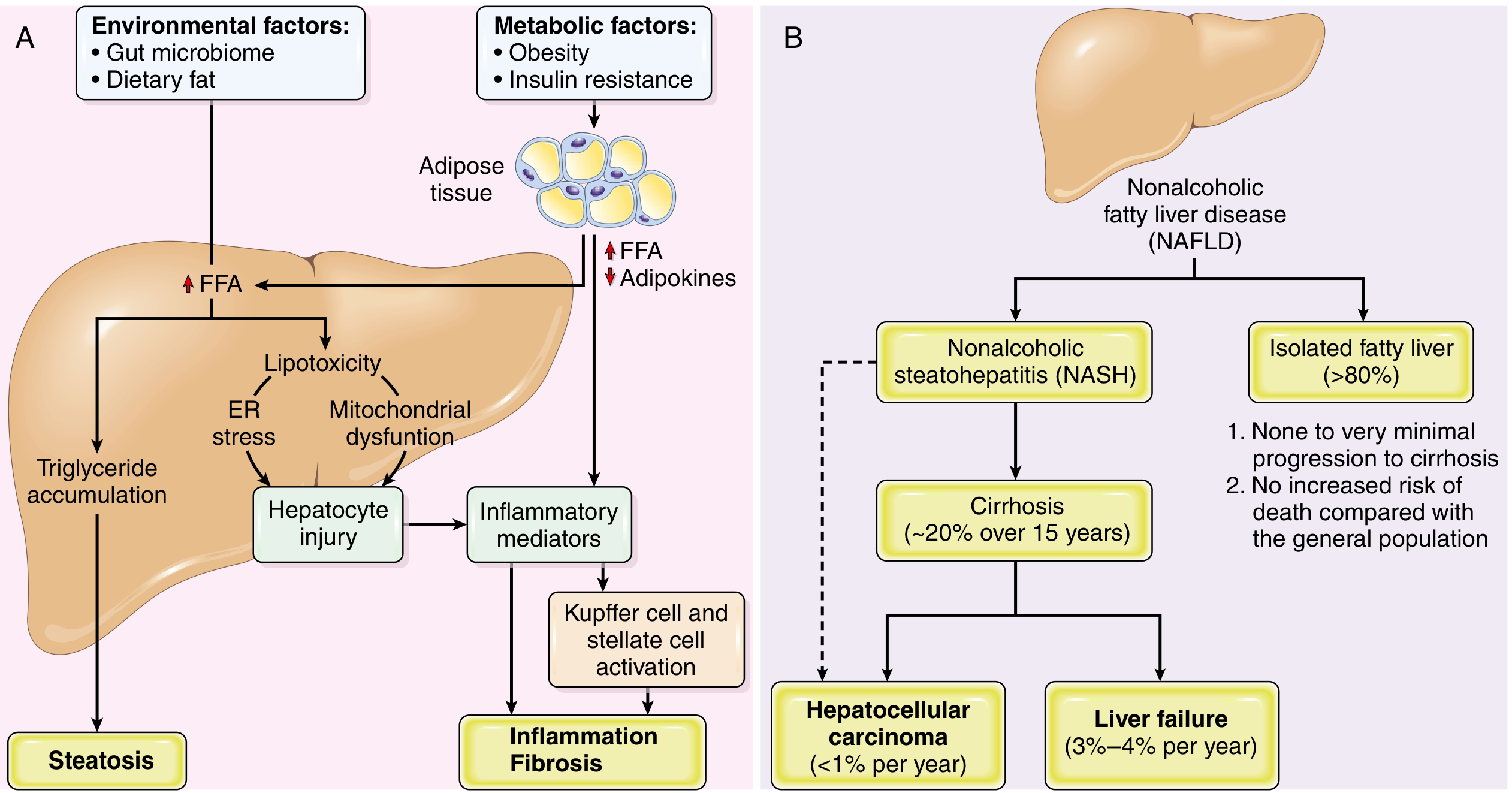
- Insulin resistance leads to increased release of free fatty acids (FFAs) from adipocytes via overactivity of lipoprotein lipase
- Simultaneously, reduced adiponectin production from adipocytes - adiponectin normally promotes hepatic fatty acid oxidation; its deficiency decreases FFA oxidation
- FFAs flood the liver → triglyceride accumulation → steatosis (predominantly Zone 3/centrilobular)
- Environmental factors compound this: dietary fat and altered gut microbiome contribute to FFA delivery to the liver
Step 2 - Hepatocyte Injury, Inflammation & Fibrosis ("Second Hit / Multiple Hits")
- Excess intrahepatic FFAs cause lipotoxicity via two mechanisms:
- ER (endoplasmic reticulum) stress - misfolded protein response, apoptosis
- Mitochondrial dysfunction - impaired beta-oxidation, excess ROS generation
- Hepatocyte injury triggers release of inflammatory mediators
- Inflammatory mediators activate Kupffer cells (hepatic macrophages) and hepatic stellate cells
- Activated stellate cells → collagen deposition → fibrosis
Summary Flow
Obesity/Insulin Resistance
    ↓
↑ FFA from adipose + ↓ Adiponectin
    ↓
Hepatic FFA/TG accumulation
    ↓
STEATOSIS
    ↓
Lipotoxicity → ER Stress + Mitochondrial Dysfunction
    ↓
Hepatocyte Injury
    ↓
Inflammatory Mediators
    ↓
Kupffer Cell + Stellate Cell Activation
    ↓
INFLAMMATION + FIBROSIS (NASH)
    ↓
CIRRHOSIS → HCC / Liver Failure
Histology - Robbins (eFig. 14.2)
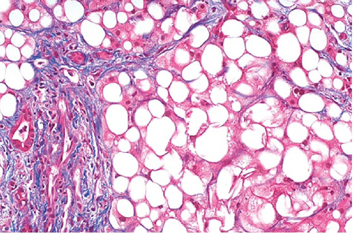
Clinical Features (Robbins)
| Feature | Detail |
|---|---|
| Most common presentation | Incidental elevation of serum transaminases (AST, ALT) |
| AST:ALT ratio | Typically <1 in NAFLD (contrast: >2 in alcohol-related liver disease - key differentiating point) |
| Simple steatosis | Usually asymptomatic |
| NASH/fibrosis | May also be asymptomatic, OR present with fatigue, malaise, right upper quadrant discomfort, symptoms of chronic liver disease |
| Diagnosis | Liver biopsy required to confirm NASH and distinguish it from uncomplicated NAFLD |
| Cardiovascular risk | Incidence of coronary artery disease is increased in NAFLD (shared metabolic risk factors) |
| Cryptogenic cirrhosis | NAFLD is a significant contributor - burnt-out NASH leaves no diagnostic footprint |
| HCC | A worrisome complication; rising proportion as HCV is treated - NASH likely to overtake HCV as leading risk factor for HCC in the US |
Natural History (Fig. 14.19B, Robbins)
| Pathway | Risk |
|---|---|
| Isolated fatty liver (>80% of NAFLD) | None to minimal progression to cirrhosis; NO increased risk of death vs. general population |
| NASH | ~20% progress to cirrhosis over 15 years; increased overall mortality |
| NASH cirrhosis | Liver failure risk: 3-4% per year; HCC risk: <1% per year |
Pediatric NAFLD (Robbins)
- Inflammation and scarring more prominent in portal tracts and periportal regions (vs. Zone 3 in adults)
- Mononuclear infiltrates predominate (vs. neutrophils in adult NASH)
- Becoming an increasing problem as childhood obesity and metabolic syndrome reach epidemic proportions
Treatment (Robbins)
- Weight reduction - most effective
- Reversal of insulin resistance
- Lifestyle modification (diet + exercise) - most effective form
- Bariatric surgery - in selected cases
High-Yield Exam Points from Robbins
- Key initiating events: Obesity + insulin resistance → ↑ FFA + ↓ adiponectin
- Lipotoxicity via ER stress + mitochondrial dysfunction drives the transition from simple steatosis to NASH
- Kupffer cells + stellate cells - activated by inflammatory mediators - mediate inflammation and fibrosis respectively
- AST:ALT <1 in NAFLD (vs. >2:1 in alcohol-related liver disease)
- Liver biopsy - required to diagnose NASH
- Isolated steatosis - minimal progression risk, no increased mortality
- NASH - ~20% cirrhosis risk over 15 years
- NASH + HCC: increasingly the leading cause of HCC, likely to surpass HCV
- Pediatric NASH: portal-based inflammation and fibrosis, mononuclear (not neutrophilic) infiltrate
- MAFLD - new proposed terminology emphasizing metabolic basis
Combine both the contents Robbins and morphological spectrum
NAFLD / NASH - Complete MD Pathology Answer
Robbins & Kumar Basic Pathology + Yamada's Textbook of Gastroenterology + Goldman-Cecil Medicine
1. Definition & Nomenclature
- Encompasses a spectrum: simple steatosis → NASH → fibrosis → cirrhosis → HCC
- NASH = overt clinical liver injury with histological features of steatohepatitis
- MAFLD (Metabolic-Associated Fatty Liver Disease) - newer proposed term emphasizing the metabolic basis
- Histologically identical to alcohol-related liver disease; distinction is by clinical history
2. Risk Factors / Associations
- Obesity (primarily central/visceral)
- Insulin resistance and metabolic syndrome
- Type 2 diabetes mellitus (or family history)
- Dyslipidemia (↑ TG, ↓ HDL, ↑ LDL)
- Hypertension
- Also associated with: PCOS, hypothyroidism, sleep apnea, hypopituitarism
3. Pathogenesis (Robbins, Fig. 14.19)
Step 1 - Steatosis ("First Hit")
- Insulin resistance → ↑ FFA release from adipocytes (overactive lipoprotein lipase)
- ↓ Adiponectin from adipose tissue → decreased hepatic FFA oxidation
- Excess FFAs → hepatic triglyceride accumulation → steatosis (predominantly Zone 3/centrilobular)
- Environmental cofactors: dietary fat and gut microbiome dysbiosis augment FFA delivery to liver
- De novo lipogenesis also increased; impaired VLDL export contributes
Step 2 - Hepatocyte Injury → NASH ("Multiple Hits")
- Excess intrahepatic FFAs → lipotoxicity via:
- ER stress → unfolded protein response → hepatocyte apoptosis
- Mitochondrial dysfunction → impaired β-oxidation, excess reactive oxygen species (ROS)
- Hepatocyte injury → release of inflammatory mediators (TNF-α, IL-6, IL-1β)
- Inflammatory mediators → Kupffer cell activation (hepatic macrophages) → amplify inflammation
- Inflammatory signals → hepatic stellate cell activation → collagen deposition → fibrosis
Pathogenesis Summary Flow
Obesity + Insulin Resistance + Gut Microbiome/Dietary Fat
              ↓
  ↑ FFA from adipose  +  ↓ Adiponectin
              ↓
   Hepatic FFA/TG Accumulation
              ↓
         STEATOSIS
              ↓
  Lipotoxicity → ER Stress + Mitochondrial Dysfunction
              ↓
       Hepatocyte Injury (ROS, apoptosis)
              ↓
   Inflammatory Mediators (TNF-α, IL-6, IL-1β)
              ↓
  Kupffer Cell + Stellate Cell Activation
              ↓
    INFLAMMATION + FIBROSIS  =  NASH
              ↓
    CIRRHOSIS → HCC / Liver Failure
4. Morphological Spectrum
Simple Steatosis → NASH → Bridging Fibrosis → Cirrhosis → HCC
Stage 1 - Simple Steatosis (NAFL)
- Large single fat droplet displacing nucleus to periphery
- Predominantly centrilobular (Zone 3)
- Generally reversible with treatment
- Numerous small fat droplets, nucleus remains central
- Indicates more acute/severe metabolic stress
- Nonzonal small patches
Stage 2 - NASH (Nonalcoholic Steatohepatitis)
Steatosis + Hepatocyte Ballooning + Lobular Inflammation ± fibrosis
A. Hepatocyte Ballooning (pathognomonic)
- Enlarged hepatocytes with irregular clumped cytoplasm and optically clear non-vesiculated areas
- Represents cytoskeletal damage - loss of keratin intermediate filaments (CK-8/18)
- Predominantly Zone 3 (perivenular)
- May contain Mallory-Denk Bodies (MDB): eosinophilic, ropey cytoplasmic inclusions of hyperphosphorylated, misfolded cytokeratin filaments
B. Lobular Inflammation
- Mixed infiltrate: lymphocytes + macrophages + neutrophils
- Neutrophils around ballooned hepatocytes ("satellitosis")
- Microgranulomas / lipogranulomas may be present
C. Steatosis
- Both macro- and microvesicular components
- Predominantly Zone 3
D. Perisinusoidal Fibrosis ("Chicken-wire") - Zone 3
- Begins as delicate strands of collagen in perisinusoidal distribution - Zone 3
- This is the hallmark pattern of NASH - distinguishes it from viral hepatitis (portal-based fibrosis)
Additional Features in NASH
| Feature | Significance |
|---|---|
| Megamitochondria | Mitochondrial dysfunction |
| Glycogenated nuclei | Insulin resistance / diabetes |
| Lipogranulomas | Fat-laden macrophage clusters |
| Microvesicular steatosis patches | Nonzonal, variable significance |
| Hepatocellular glycogenosis | Especially in pediatric NAFLD |
Stage 3 - Progressive Fibrosis (NASH-CRN Staging)
| Stage | Description |
|---|---|
| F0 | No fibrosis |
| F1a | Mild perisinusoidal fibrosis, Zone 3 |
| F1b | Moderate perisinusoidal fibrosis, Zone 3 |
| F1c | Portal/periportal fibrosis only |
| F2 | Perisinusoidal AND periportal fibrosis |
| F3 | Bridging fibrosis |
| F4 | Cirrhosis |
Key: Unlike viral hepatitis (portal → bridging), bridging fibrosis in NASH may result from extension of perisinusoidal fibrosis with retention of hepatocytes within the fibrotic network ("trapped hepatocytes").
As fibrosis progresses to F3-F4, steatosis, ballooning, and inflammation paradoxically decrease - this is "burnt-out NASH."
Stage 4 - NASH Cirrhosis
- Regenerative nodules within a fibrotic network
- Steatohepatitis features may be absent → "cryptogenic cirrhosis"
- Robbins: NAFLD is a significant contributor to pathogenesis of cryptogenic cirrhosis
- 15-25% of NASH patients progress to cirrhosis (~20% over 15 years per Robbins)
Stage 5 - Hepatocellular Carcinoma (HCC)
- Can arise even without cirrhosis in NASH (unlike most other causes)
- Risk: <1% per year from NASH cirrhosis
- NASH is increasingly overtaking HCV as the leading cause of HCC in the United States (Robbins)
5. NAFLD Activity Score (NAS) - NASH-CRN System
| Component | Score Range |
|---|---|
| Steatosis | 0-3 |
| Lobular inflammation | 0-2 |
| Hepatocyte ballooning | 0-2 |
| Total NAS | 0-8 |
| Fibrosis (staged separately) | F0-F4 |
- NAS ≥5 = correlates with NASH (sensitivity 75%, specificity 83%)
- NAS ≤3 = usually not NASH
- NAS alone cannot diagnose NASH - 29% of biopsies with NAS ≤4 can still show definite steatohepatitis
Steatosis Grading
| Grade | % Hepatic Involvement |
|---|---|
| 0 | <5% |
| 1 | 5-33% |
| 2 | 33-67% |
| 3 | >67% |
6. Clinical Features (Robbins)
| Feature | Detail |
|---|---|
| Most common presentation | Incidental elevation of serum transaminases |
| AST:ALT ratio | <1 in NAFLD (contrast: >2:1 in alcohol-related liver disease - key differentiator) |
| Simple steatosis | Usually asymptomatic |
| NASH / fibrosis | May be asymptomatic OR: fatigue, malaise, RUQ discomfort, symptoms of chronic liver disease |
| Diagnosis of NASH | Liver biopsy required - no non-invasive test can distinguish NASH from simple steatosis |
| Cardiovascular risk | ↑ incidence of coronary artery disease (shared metabolic risk factors) |
| Cryptogenic cirrhosis | NAFLD is a major contributor |
| HCC | Increasingly the leading cause, overtaking HCV in the US |
7. Natural History (Robbins, Fig. 14.19B)
| Pathway | Risk |
|---|---|
| Isolated fatty liver (>80% of NAFLD) | None to minimal progression to cirrhosis; NO increased mortality vs. general population |
| NASH | ~20% progress to cirrhosis over 15 years; increased overall mortality |
| NASH cirrhosis: liver failure | 3-4% per year |
| NASH cirrhosis: HCC | <1% per year |
8. NASH vs. ASH (Alcoholic Steatohepatitis) - Differential
| Feature | NASH | ASH |
|---|---|---|
| Mallory-Denk bodies | Present | Present (more abundant) |
| Neutrophilic infiltrate | Present | Present (more prominent) |
| Perisinusoidal fibrosis Zone 3 | Yes | Yes |
| Megamitochondria | Yes | Less common |
| AST:ALT ratio | <1 | >2:1 |
| Histology | Identical | Identical |
| Distinction | Clinical history only | Clinical history only |
9. Pediatric NAFLD - Special Pattern (Robbins + Yamada/Schwimmer)
| Adult NASH | Pediatric NASH (Type 2) | |
|---|---|---|
| Fibrosis location | Zone 3 perisinusoidal | Portal/periportal |
| Ballooning | Zone 3 | Absent |
| Inflammation | Lobular + neutrophilic | Portal, mononuclear |
- Type 1 pediatric NASH: Resembles adult NASH (ballooning + perisinusoidal fibrosis)
- Type 2 pediatric NASH: Steatosis + portal inflammation + portal fibrosis, NO ballooning
10. Treatment (Robbins)
- Weight reduction + lifestyle modification (diet + exercise) - most effective
- Reversal of insulin resistance
- Bariatric surgery - in selected cases
High-Yield Exam Summary
| Point | Detail |
|---|---|
| Key initiating event | Obesity + insulin resistance |
| FFA source | Adipose tissue (lipoprotein lipase overactivity) |
| Adiponectin | Decreased → less FFA oxidation |
| Lipotoxicity mechanisms | ER stress + mitochondrial dysfunction |
| Cells mediating fibrosis | Hepatic stellate cells (activated by inflammatory mediators) |
| Earliest fibrosis | Perisinusoidal, Zone 3 (chicken-wire) |
| Distinguishes NASH fibrosis | Zone 3 perisinusoidal vs. portal in viral hepatitis |
| AST:ALT in NAFLD | <1 (vs. >2 in alcohol) |
| NAS ≥5 | Likely NASH |
| Burnt-out NASH | ↓ steatosis + ballooning as fibrosis advances → cryptogenic cirrhosis |
| HCC risk | Can arise without cirrhosis; NASH overtaking HCV as leading cause |
| Pediatric pattern | Portal fibrosis + mononuclear infiltrate (no ballooning) |
| Genetic risk | PNPLA3 I148M - strongest predictor |