
Give me in detail explanation of cardiovascular system examination with which conditions contain which type of signs and symptoms in order wise and differentiation between diseases if any diseases are similar
Reading File
Now let me search the medical library for comprehensive cardiovascular examination content in parallel.
Finding Sources
Finding Sources
Finding Sources
Reading File
Finding Sources
Finding Sources
Reading File
Reading File
Reading File
Reading File
Reading File
Reading File
Reading File
Reading File
Finding Sources
Reading File
Reading File
Reading File
Reading File
I now have comprehensive data from Harrison's and Goldman-Cecil. Let me compile the complete detailed answer.
Loading Image
Loading Image
Loading Image
I now have all the content needed to write a comprehensive, detailed answer. Let me compile everything.
Cardiovascular System Examination - Complete Guide
Sources: Harrison's Principles of Internal Medicine 22E (2025), Goldman-Cecil Medicine
ORDER OF EXAMINATION
The cardiovascular exam follows a structured sequence:
- General Appearance
- Vital Signs (BP, HR, RR)
- Ophthalmologic Exam
- Jugular Venous Pressure (JVP)
- Carotid Pulse
- Precordial Inspection and Palpation
- Cardiac Auscultation
- Abdominal and Peripheral Exam
1. GENERAL APPEARANCE
Before touching the patient, observe for features pointing to specific diagnoses:
| Finding | Associated Condition |
|---|---|
| Tachypnea, labored breathing | Heart failure, pulmonary edema |
| Cheyne-Stokes respirations | Advanced heart failure |
| Exophthalmos, stare | Hyperthyroidism (atrial fibrillation, high-output failure) |
| Ptosis, expressionless face | Myotonic dystrophy (AV block, arrhythmia) |
| Tall, thin build with arm span > height | Marfan syndrome (aortic root dilation, MVP) |
| Blue sclerae | Osteogenesis imperfecta (aortic dilation, MVP) |
| Webbed neck, low hairline | Turner syndrome (coarctation, bicuspid aortic valve) |
| Short stature, flat face | Down syndrome (ASD, VSD, AV canal) |
| Malar rash, joint swelling | SLE, rheumatoid arthritis (pericarditis, endocarditis) |
| Tight skin, sclerodactyly | Scleroderma (pulmonary hypertension) |
| Skin bronzing | Hemochromatosis (cardiomyopathy, heart block) |
2. VITAL SIGNS
Blood Pressure
Technique: Seated, arm at heart level, 5-10 min rest, appropriately sized cuff. Cuff bladder length 80%, width 40% of arm circumference.
| BP Finding | Interpretation |
|---|---|
| BP difference >10 mmHg between arms | Subclavian artery atherosclerosis/inflammation, aortic dissection, supravalvular aortic stenosis, coarctation |
| Arm BP >> leg BP | Normal: leg systolic is ~20 mmHg higher; reversed ratio suggests coarctation of the aorta |
| Very low diastolic (near 0) | Chronic severe aortic regurgitation (AR) or large arteriovenous fistula |
| Wide pulse pressure | Severe AR, hyperthyroidism, fever, anemia, high-output states |
| Narrow pulse pressure | Severe aortic stenosis (AS), cardiac tamponade, severe LV dysfunction |
Pulsus Paradoxus
A fall in systolic BP >10 mmHg with inspiration.
| Cause | Key Distinguishing Feature |
|---|---|
| Pericardial tamponade | Most classic cause; also has elevated JVP, muffled heart sounds |
| Massive pulmonary embolism | Tachycardia, hypoxia, pleuritic chest pain |
| Severe obstructive lung disease | Wheeze, prolonged expiratory phase |
| Tension pneumothorax | Absent breath sounds, tracheal deviation |
| Hemorrhagic shock | Hypotension, bleeding source |
Pulsus Alternans
Beat-to-beat variability of pulse amplitude at regular rhythm. Indicates severe LV systolic dysfunction. Every other Korotkoff sound is audible as cuff deflates.
3. OPHTHALMOLOGIC EXAM
| Finding | Associated Condition |
|---|---|
| Diabetic retinopathy | Ischemic cardiomyopathy |
| Hypertensive retinopathy | Hypertensive heart disease, LVH |
| Roth spots (white-centered hemorrhages) | Infective endocarditis |
| Beading of retinal arteries | Severe hypercholesterolemia |
| Retinal artery occlusion | Embolus from left atrial thrombus, myxoma, aortic atheroembolism |
4. JUGULAR VENOUS PRESSURE (JVP)
Measurement: Vertical distance from sternal angle (angle of Louis) to top of jugular pulsation at 30-45 degrees. Normal <4.5 cm at 30°; add 5 cm to get central venous pressure in cm H₂O. Normal CVP = 5-10 cm H₂O. Pulsations above the clavicle in the sitting position are always abnormal.
Distinguishing JVP from carotid:
- JVP: biphasic (two peaks), not palpable, changes with posture and respiration
- Carotid: monophasic, palpable, does not change with respiration
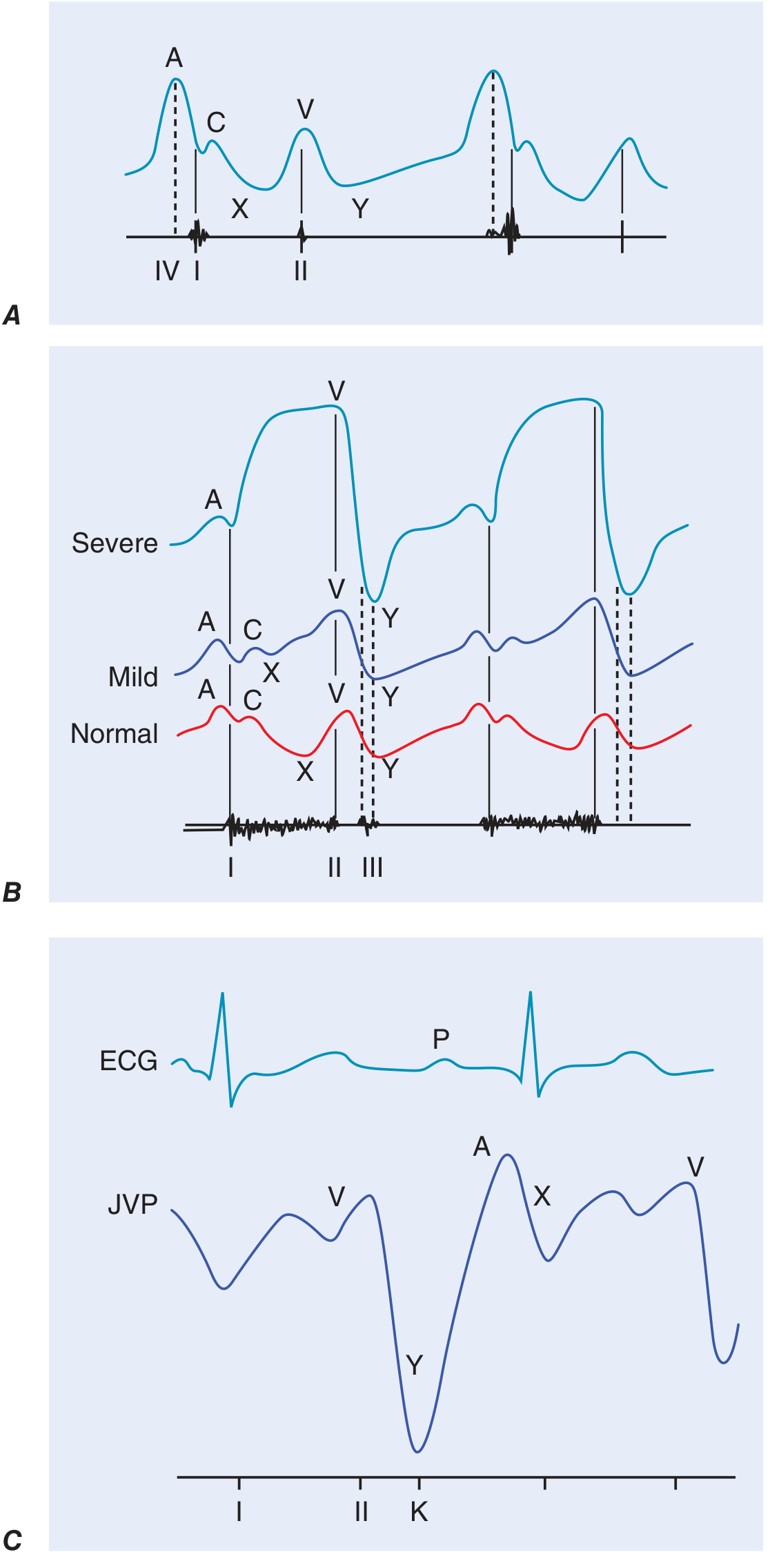
JVP Waveform Components and Their Abnormalities
| Wave/Descent | Normal Meaning | Abnormality | Disease |
|---|---|---|---|
| a wave | RA presystolic contraction (after P wave, before S1) | Prominent a wave | Reduced RV compliance (pulmonic stenosis, pulmonary hypertension, RV hypertrophy) |
| Cannon a wave | AV dissociation (VT, complete heart block) - RA contracts against closed tricuspid valve | ||
| Absent a wave | Atrial fibrillation | ||
| x descent | Fall in RA pressure after tricuspid opening | Absent/blunted | Atrial fibrillation, tamponade |
| v wave | Atrial filling during ventricular systole | Accentuated v wave (cv wave) | Tricuspid regurgitation - waveform becomes "ventriculized" |
| y descent | After v peak, tricuspid valve opens | Blunted/prolonged y descent | Tricuspid stenosis, pericardial tamponade |
| Rapid, prominent y descent | Constrictive pericarditis, severe TR |
Elevated JVP: Differential Diagnosis
| JVP Pattern | Diagnosis |
|---|---|
| Elevated JVP + blunted/absent y descent + pulsus paradoxus + quiet heart | Cardiac tamponade - echocardiography urgently needed |
| Elevated JVP + sharp prominent y descent + Kussmaul sign + quiet precordium | Constrictive pericarditis - CT/MRI/catheterization needed |
| Elevated JVP + sharp brief y descent + Kussmaul sign + evidence of pulmonary hypertension + TR | Restrictive cardiomyopathy |
| Elevated JVP without y descent | Right heart failure of any cause |
| Prominent a wave (no elevated mean JVP) | Tricuspid stenosis, pulmonic stenosis, pulmonary hypertension |
| Prominent v wave + sharp y descent | Tricuspid regurgitation |
Kussmaul's sign (JVP rises or fails to fall with inspiration): Constrictive pericarditis (classic), restrictive cardiomyopathy, massive PE, RV infarction, advanced LV systolic failure.
Abdominojugular (hepatojugular) reflux: Firm pressure over RUQ for >15 seconds; positive = sustained JVP rise >3 cm. Indicates volume-overloaded state; predicts pulmonary artery wedge pressure >15 mmHg in heart failure.
5. CAROTID PULSE (CHARACTER OF PULSE)
Best assessed at the carotid artery. Describes pulse volume and contour.
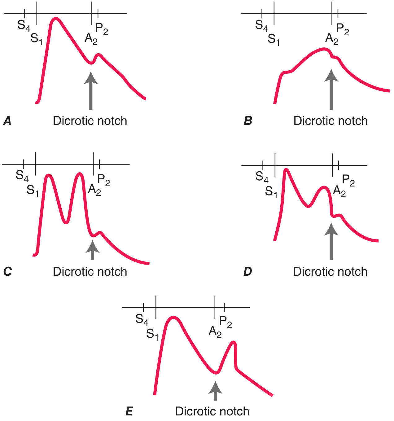
| Pulse Character | Description | Disease(s) |
|---|---|---|
| Pulsus parvus et tardus | Weak, delayed upstroke to a small peak | Severe Aortic Stenosis |
| Anacrotic pulse | Slow notched upstroke, peak near S2 | Severe Aortic Stenosis |
| Corrigan's (water-hammer) pulse | Sharp rapid rise, collapsing fall | Chronic severe Aortic Regurgitation |
| Bisferiens pulse (two systolic peaks) | Percussion wave + tidal wave | Severe AR; HOCM; combined AS+AR with dominant AR |
| Dicrotic pulse (one systolic + one diastolic peak) | Exaggerated dicrotic wave | Sepsis, severe heart failure, hypovolemic shock, tamponade, after AVR |
| Hyperkinetic pulse | Increased amplitude and frequency | AR, AV fistula, hyperthyroidism, fever, anemia |
| Normal contour, reduced amplitude | Normal shape but weak | Any cause of reduced stroke volume |
Differentiating Aortic Stenosis vs Aortic Regurgitation by Pulse
| Feature | Aortic Stenosis | Aortic Regurgitation |
|---|---|---|
| Pulse character | Parvus et tardus (weak, delayed) | Water-hammer (sharp rise, collapsing) |
| Pulse pressure | Narrow | Wide |
| BP diastolic | Normal/elevated | Low (near 0 in severe) |
| Systolic murmur | Ejection (crescendo-decrescendo), midsystolic | None (unless combined lesion) |
| Diastolic murmur | None | Decrescendo, high-pitched, early diastolic |
6. PRECORDIAL INSPECTION AND PALPATION
Inspection
| Finding | Meaning |
|---|---|
| Visible apex beat at 5th ICS mid-clavicular line | Normal in thin adults |
| Apex beat displaced leftward/downward | LV enlargement |
| Visible sternal/parasternal pulsation | RV enlargement |
| Right upper parasternal pulsation | Ascending aortic aneurysm |
| Epigastric pulsation | Cardiac impulse displaced (COPD, emphysema) vs. pulsatile liver |
| Unilateral left chest asymmetry | RV hypertrophy developing before puberty |
Palpation (Technique: supine at 30°, left lateral decubitus to enhance)
| Finding | Meaning | Disease |
|---|---|---|
| Normal apex (<2 cm, brief outward movement) | Normal LV | - |
| Sustained (heaving) apex | Pressure overload | AS, chronic hypertension |
| Displaced, enlarged apex | Volume overload + LV dilation | Severe MR, severe AR, DCM |
| Palpable S4 (presystolic impulse) | Reduced LV compliance | LV hypertrophy, active ischemia, AS |
| Palpable S3 | Rapid early filling (advanced HF) | Dilated cardiomyopathy, severe MR |
| Ectopic dyskinetic impulse (separate from apex) | LV aneurysm | Post-MI aneurysm |
| Triple cadence at apex (S4 + bisferiens) | Very rare | HOCM |
| Sternal/parasternal lift | RV pressure or volume overload | Pulmonary hypertension, RV failure, ASD |
| Loud palpable P2 | Pulmonary hypertension | |
| Systolic thrill | Turbulent flow | Grade ≥4 murmur: severe AS, VSD, MR |
| Diastolic thrill | Turbulent diastolic flow | Severe MS |
7. CARDIAC AUSCULTATION
Heart Sounds
First Heart Sound (S1)
Produced by mitral and tricuspid valve closure.
| S1 Character | Disease |
|---|---|
| Loud S1 | Early rheumatic mitral stenosis (leaflets pliable but narrowed), hyperkinetic states, short PR interval |
| Soft S1 | Late MS (leaflets rigid/calcified), after beta-blockers, long PR interval, LV contractile dysfunction |
| Variable S1 | Complete AV block, atrial fibrillation |
| Wide splitting of S1 | RBBB (delayed tricuspid closure) |
Second Heart Sound (S2)
A2 (aortic closure) + P2 (pulmonic closure). Normally A2 before P2. Splitting increases with inspiration.
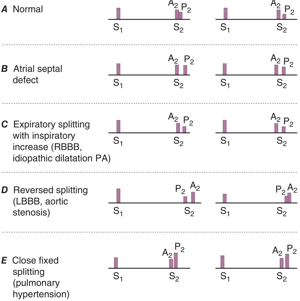
| S2 Pattern | Cause | Disease |
|---|---|---|
| Fixed splitting (A2-P2 gap same in inspiration and expiration) | Equalization of RV/LV filling | Atrial Septal Defect (ASD) |
| Wide splitting (physiologic, inspiratory increase) | Delayed P2 | RBBB, idiopathic PA dilation |
| Paradoxical (reversed) splitting (splits on expiration, closes on inspiration) | Delayed A2 | LBBB, severe AS (mechanical delay), HOCM |
| Close fixed splitting | Loud P2 + fixed gap | Pulmonary hypertension |
| Single S2 | Only one valve audible | Severe AS (A2 absent/inaudible), severe pulmonary hypertension (P2 overwhelms A2) |
| Loud A2 | Increased aortic pressure | Systemic hypertension |
| Soft A2 | Reduced aortic valve excursion | Aortic stenosis |
| Loud P2 | Increased pulmonary artery pressure | Pulmonary hypertension, large ASD |
Third Heart Sound (S3) - Early Diastolic Gallop
Low-pitched sound after S2, in early diastole (rapid ventricular filling phase).
| Context | Significance |
|---|---|
| In patient <40 years | Often normal (physiologic) |
| In adults with dyspnea + dilated LV | LV systolic failure - high sensitivity for elevated wedge pressure |
| After MR | Chronic severe Mitral Regurgitation |
| Right-sided S3 (louder with inspiration) | RV failure, severe TR |
Fourth Heart Sound (S4) - Presystolic Gallop
Low-pitched sound before S1 (late diastole), due to atrial contraction into non-compliant ventricle. Not present with atrial fibrillation.
| Cause | Disease |
|---|---|
| Reduced LV compliance | LV hypertrophy, hypertensive heart disease, AS |
| Active myocardial ischemia | Acute MI, angina |
| Hypertrophic cardiomyopathy | HOCM |
| Right-sided S4 | RV hypertrophy (pulmonary hypertension, pulmonic stenosis) |
Differentiating S3 vs S4
| Feature | S3 | S4 |
|---|---|---|
| Timing | Early diastole (after S2) | Late diastole (before S1) |
| Mechanism | Rapid filling into diseased ventricle | Atrial kick into non-compliant ventricle |
| Present in AF | Yes | NO |
| Implies | LV systolic dysfunction / volume overload | Reduced compliance / pressure overload |
| Pitch | Low (bell) | Low (bell) |
Additional Sounds
| Sound | Timing | Disease |
|---|---|---|
| Ejection click (early systolic) | Just after S1 | Bicuspid aortic valve, mobile aortic valve in congenital AS; pulmonic valve stenosis (click disappears with inspiration); pulmonary hypertension with forceful valve opening |
| Mid-late systolic click | Variable during systole | Mitral Valve Prolapse (MVP) - moves toward S1 with standing/Valsalva; moves toward S2 with squatting |
| Opening snap (OS) | Early diastole (after A2) | Mitral Stenosis - shorter A2-OS interval = more severe MS |
| Pericardial knock | Early diastole | Constrictive pericarditis - high-pitched, occurs at the nadir of Y descent |
| Tumor plop | Early diastole | Atrial myxoma (movement of tumor through mitral valve) |
| Pericardial friction rub | Two or three component (systolic + diastolic) scratching sound | Pericarditis - best heard with patient leaning forward; may disappear with effusion |
Heart Murmurs
Graded 1-6 (thrill present at grade 4+).
SYSTOLIC MURMURS
Holosystolic (Pansystolic) Murmurs
Persist throughout systole (S1 to S2), flat or decrescendo.
| Disease | Location | Radiation | Character | Key Signs |
|---|---|---|---|---|
| Mitral Regurgitation (MR) | Apex | Left axilla (anterior leaflet to back; posterior leaflet to base - can mimic AS) | Holosystolic blowing | Displaced apex, S3, reduced S1 |
| Tricuspid Regurgitation (TR) | Left lower sternal border | None | Holosystolic, increases with inspiration (Carvallo sign) | CV waves in JVP, pulsatile liver, peripheral edema |
| Ventricular Septal Defect (VSD) | Left sternal border 3rd-4th ICS | Right sternal border | Holosystolic harsh | Thrill common; if large: signs of pulmonary hypertension |
Midsystolic (Ejection) Murmurs
Crescendo-decrescendo, begin after S1, end before S2.
| Disease | Location | Radiation | Key Signs | Differentiation |
|---|---|---|---|---|
| Aortic Stenosis (AS) | Right 2nd ICS (aortic area) | Carotids (Gallavardin - to apex, can mimic MR) | Parvus et tardus pulse, sustained apex, soft/absent A2, paradoxical S2 split, S4 | Late-peaking murmur = more severe |
| Pulmonary Stenosis | Left 2nd ICS (pulmonic area) | Left shoulder | Loud P2 if mild (soft if severe), ejection click (decreases with inspiration) | Right-sided signs; no carotid radiation |
| Hypertrophic Obstructive Cardiomyopathy (HOCM) | Left lower sternal border | Does NOT radiate to carotids well | Bisferiens pulse, S4, dynamic murmur | Increases with Valsalva/standing; decreases with squatting/hand-grip |
| ASD (relative pulmonic stenosis) | Left 2nd ICS | - | Fixed split S2 | Soft murmur (flow); fixed S2 splitting |
| Benign flow murmur | Left sternal border/pulmonary area | None | No abnormal signs | Grade 1-2, vibratory (Still's murmur in children) |
Differentiating AS vs MR vs HOCM (all are systolic murmurs)
| Feature | AS | MR | HOCM |
|---|---|---|---|
| Timing | Midsystolic (ejection) | Holosystolic | Mid-late systolic |
| Location | Right 2nd ICS | Apex | Left lower sternal border |
| Radiation | Carotids | Axilla | Not carotids |
| Pulse | Parvus et tardus | Normal or hyperdynamic | Bisferiens |
| S2 | Soft A2, paradoxical split | Wide physiologic split (early A2) | Normal |
| Valsalva | Decreases | Decreases | Increases |
| Squatting | Increases | Increases | Decreases |
| Standing | Decreases | Decreases | Increases |
| Handgrip | Increases | Increases | Decreases |
Late Systolic Murmur
| Disease | Feature |
|---|---|
| Mitral Valve Prolapse | Preceded by mid-systolic click; click-murmur complex moves toward S1 with standing/Valsalva; moves toward S2 with squatting |
DIASTOLIC MURMURS (always pathological)
Early Diastolic (Decrescendo)
| Disease | Location | Radiation | Quality | Key Signs |
|---|---|---|---|---|
| Aortic Regurgitation (AR) | Left sternal border (3rd ICS), also right 2nd ICS if aortic root dilated | Apex | High-pitched blowing decrescendo; best heard with patient sitting forward | Water-hammer pulse, wide pulse pressure, Austin Flint murmur (mid-diastolic low-pitched rumble at apex in severe AR), displaced apex, S3 |
| Pulmonary Regurgitation (Graham Steell murmur) | Left 2nd-3rd ICS | - | High-pitched decrescendo | Loud P2, signs of pulmonary hypertension |
Mid-Diastolic (Rumbling)
| Disease | Location | Radiation | Quality | Key Clues |
|---|---|---|---|---|
| Mitral Stenosis (MS) | Apex | None | Low-pitched rumble (use bell); follows opening snap | Loud S1, OS, shorter A2-OS interval = more severe; pre-systolic accentuation in sinus rhythm; AF eliminates presystolic component |
| Tricuspid Stenosis | Left lower sternal border | None | Rumble; increases with inspiration | Prominent a wave in JVP, hepatomegaly |
| Austin Flint murmur | Apex | None | Low-pitched rumble | In context of severe AR; no OS; no loud S1 |
Differentiating MS vs Austin Flint Murmur
| Feature | Mitral Stenosis | Austin Flint (AR) |
|---|---|---|
| Opening snap | Present | Absent |
| S1 | Loud (early), soft (late) | Normal or soft |
| AR signs | Absent | Present (water-hammer pulse, wide PP) |
| Response to amyl nitrite | Louder (MS) | Softer (Flint - less AR) |
CONTINUOUS MURMURS (systole + diastole)
| Cause | Feature |
|---|---|
| Patent Ductus Arteriosus (PDA) | Left infraclavicular area; "machinery" murmur; wide pulse pressure |
| Ruptured sinus of Valsalva aneurysm | Sudden onset, may have thrill |
| Arteriovenous fistula | Over fistula site |
| Mammary souffle | Over breast in pregnancy |
| Combined AS + AR | Can mimic continuous; two separate murmurs |
8. ABDOMINAL AND PERIPHERAL EXAMINATION
| Finding | Disease |
|---|---|
| Hepatomegaly | Right heart failure (congestive hepatopathy) |
| Pulsatile liver | Tricuspid regurgitation |
| Hepatojugular reflux | Advanced RV failure / obstruction to RV filling |
| Splenomegaly | Infective endocarditis, portal hypertension from chronic HF |
| Pulsatile abdominal mass | Abdominal aortic aneurysm |
| Ascites | Advanced right heart failure, constrictive pericarditis |
| Peripheral edema (pitting) | Right heart failure |
| Ankle-brachial index <0.9 | Peripheral arterial disease |
| Femoral/popliteal aneurysm | Associated with AAA |
| Absent foot pulses | PAD, aortic coarctation (in legs) |
DISEASE-BY-DISEASE SUMMARY: SIGNS IN ORDER
Aortic Stenosis
- General: symptoms on exertion (angina, syncope, dyspnea = classic triad)
- Pulse: parvus et tardus; narrow pulse pressure
- JVP: may show prominent a wave (if pulmonary hypertension develops)
- Apex: sustained (heaving), not displaced (unless LV dilation in late disease)
- Palpation: systolic thrill at 2nd right ICS
- S1: normal; S2: single or paradoxically split (P2 soft/absent A2)
- S4 gallop present
- Murmur: midsystolic ejection crescendo-decrescendo at right 2nd ICS, radiating to carotids
Aortic Regurgitation (Chronic)
- Pulse: water-hammer (Corrigan's); wide pulse pressure; bisferiens if severe
- BP: high systolic, very low diastolic
- Apex: displaced laterally and inferiorly (volume overloaded LV)
- S1: normal; early diastolic decrescendo murmur at left sternal border
- Austin Flint murmur at apex (in severe AR)
- S3 in decompensated AR
- Peripheral signs: pistol-shot femoral pulse (Traube's), Duroziez sign, Quincke pulses, de Musset's sign (head nodding)
Mitral Stenosis
- General: dyspnea, hemoptysis, AF, systemic emboli
- Pulse: irregular if AF
- JVP: elevated if pulmonary hypertension present
- Apex: not displaced; "tapping" quality (palpable S1)
- S1: loud (pliable leaflets); S2: loud P2 if pulmonary hypertension
- Opening snap (shortly after S2)
- Mid-diastolic rumble at apex (bell of stethoscope, left lateral decubitus)
- Presystolic accentuation (in sinus rhythm)
Mitral Regurgitation (Chronic)
- Pulse: brisk, hyperdynamic
- Apex: displaced laterally (volume overload); palpable S3
- S1: soft; S2: wide splitting (early A2 due to rapid LV emptying); S3 present
- Holosystolic blowing murmur at apex, radiating to axilla
- If posterior leaflet: murmur radiates anteriorly to base (mimics AS)
Hypertrophic Obstructive Cardiomyopathy (HOCM)
- General: dyspnea, syncope, chest pain, palpitations in young patient; family history of sudden death
- Pulse: bisferiens; spike-and-dome character
- Apex: triple cadence (S4 + bisferiens); sustained but not displaced
- S4 prominent
- Midsystolic murmur at left lower sternal border
- Dynamic character is key: increases with Valsalva and standing; decreases with squatting and handgrip
- Coexistent MR murmur often present
Constrictive Pericarditis
- General: fatigue, peripheral edema, ascites - often misdiagnosed as liver disease
- JVP: elevated with prominent/rapid y descent; Kussmaul sign (JVP rises with inspiration)
- Pulse: may have pulsus paradoxus (mild)
- Precordium: quiet precordium (no heave)
- S3 equivalent = pericardial knock (high-pitched early diastolic sound; earlier than usual S3, occurs at nadir of rapid Y descent)
- Hepatomegaly, ascites, peripheral edema
Cardiac Tamponade
- General: Beck's triad - Hypotension + Elevated JVP + Muffled heart sounds
- JVP: elevated; blunted/absent Y descent (impaired diastolic filling)
- Pulse: pulsus paradoxus >10 mmHg (may be palpable >15 mmHg)
- Precordium: quiet, no heave, heart sounds muffled
- No Kussmaul sign
Differentiating Tamponade vs Constrictive Pericarditis
| Feature | Tamponade | Constrictive Pericarditis |
|---|---|---|
| Pulsus paradoxus | Present (prominent) | Mild or absent |
| JVP Y descent | Absent/blunted | Prominent, rapid |
| Kussmaul sign | Absent | Present |
| Heart sounds | Muffled | Normal intensity + pericardial knock |
| Pericardial knock | Absent | Present |
| Echo | Pericardial effusion + RV collapse | Thickened/calcified pericardium |
Pulmonary Hypertension
- Pulse: normal or reduced volume
- JVP: elevated; prominent a wave; if TR develops - prominent v wave
- RV heave at left parasternal area
- Palpable P2 at left 2nd ICS
- S2: loud P2, narrow or single splitting
- Right-sided S4; then right-sided S3 if RV fails
- Ejection click in pulmonic area
- TR murmur (holosystolic, left lower sternal border, increases with inspiration) develops late
- PR murmur (Graham Steell) - high-pitched early diastolic decrescendo at left 2nd-3rd ICS
Infective Endocarditis
- General: fever, weight loss, fatigue, embolic phenomena
- Eyes: Roth spots (fundus), petechiae
- Hands: Osler nodes (painful tender nodules - fingertips), Janeway lesions (non-tender hemorrhagic macules - palms/soles), splinter hemorrhages (nails)
- New or changing murmur (most commonly MR or AR)
- Splenomegaly
- Signs of heart failure if severe valvular destruction
NYHA FUNCTIONAL CLASSIFICATION (Summary)
| Class | Limitation |
|---|---|
| I | No limitation; ordinary activity does not cause symptoms |
| II | Slight limitation; ordinary activity causes fatigue/palpitation/dyspnea/angina |
| III | Marked limitation; less than ordinary activity causes symptoms |
| IV | Unable to do any activity without discomfort; symptoms at rest |
Harrison's Principles of Internal Medicine 22E (2025), Goldman-Cecil Medicine
Cortication of aorta arm bp >leg bp why ?
You're asking about coarctation of the aorta - why arm BP is higher than leg BP. Let me give you the quick answer first, then verify from the textbooks.
Quick answer: Coarctation causes a narrowing in the aorta (usually just distal to the left subclavian artery origin). Blood flows easily to the arms (above the coarctation) but flow to the legs is restricted - so arm BP is high and leg BP is low or absent.
Searching sources...
Finding Sources
Reading File
Reading File
Finding Sources
Here is the complete explanation:
Coarctation of the Aorta: Why Arm BP > Leg BP
The Anatomy - This is the Key
In 95%+ of cases, the coarctation (narrowing) is juxtaductal - located at the junction of the distal aortic arch and the descending aorta, just distal to (beyond) the origin of the left subclavian artery.
Heart → Ascending Aorta
         ↓
    Brachiocephalic artery → Right arm
    Left common carotid artery → Brain
    Left subclavian artery → Left arm
         ↓
    *** COARCTATION HERE ***  ← narrowing at this point
         ↓
    Descending aorta → Legs, abdomen
Why Arms Have HIGH BP
The three main arteries that branch before the coarctation - the brachiocephalic (right arm), left carotid, and left subclavian (left arm) - receive blood from the aorta proximal to the obstruction. The heart has to pump harder to push blood past the narrowing. This increased workload raises pressure in the entire segment upstream, which includes the arms and head.
- Chronic increased afterload → LV hypertrophy
- Activation of renin-angiotensin-aldosterone system (kidneys sense reduced perfusion downstream) → further raises BP systemically (arms)
Why Legs Have LOW BP
Blood reaching the legs must pass through the narrowed coarctation. The obstruction reduces flow and pressure beyond it. So the legs receive blood at a lower pressure and reduced volume.
- Femoral pulses are weak and delayed (brachial-femoral pulse delay - you feel the radial pulse before the femoral pulse in the same patient)
- Systolic BP gradient between right arm and lower limb ≥20 mmHg = significant coarctation (Braunwald's Heart Disease)
- Legs may be cool, pale, and the patient may have leg claudication
Important Clinical Nuance: WHICH ARM to Use
Always use the RIGHT arm for BP measurement in coarctation. Why?
Because occasionally the left subclavian artery itself arises at or distal to the coarctation. In that case, the left arm also has lower BP - making it falsely reassuring. The right subclavian/brachiocephalic artery always arises proximal to the coarctation, so the right arm always gives the "high" pre-coarctation pressure.
Summary Table
| Location | Relation to Coarctation | BP | Pulse |
|---|---|---|---|
| Right arm | Proximal (always before) | HIGH | Normal |
| Left arm | Usually proximal, rarely at/past it | Usually high | Normal (may be low if L subclavian involved) |
| Legs | Distal (always after) | LOW | Weak, delayed |
Associated Radiological Signs (Chest X-ray)
- Rib notching (erosion of undersurfaces of ribs 3-8) - caused by dilated intercostal collateral arteries that form to bypass the obstruction and supply the lower body
- Figure-of-3 sign - the aortic silhouette shows pre-stenotic dilation + the coarctation notch + post-stenotic dilation, forming a "3" shape
- E sign on barium swallow (reverse figure-3)
Collateral Circulation
In postductal coarctation, the body compensates by developing collateral vessels (internal mammary, intercostal, subscapular arteries) to bypass the obstruction and reach the lower body. These cause:
-
Continuous interscapular murmur (collateral flow)
-
Rib notching on CXR
-
Braunwald's Heart Disease, Cardiovascular Medicine 2-Vol Set
-
The Developing Human: Clinically Oriented Embryology
Difference between constructive pericarditis and restrictive pericarditis
Quick answer: Constrictive pericarditis = thick/scarred pericardium squeezes the heart from outside. Restrictive cardiomyopathy = stiff myocardium (heart muscle itself) resists filling from inside. Both cause similar hemodynamics (diastolic failure, elevated filling pressures) which is why they are notoriously difficult to differentiate - but the distinction matters because constrictive pericarditis is surgically curable.
Searching sources...
Finding Sources
Finding Sources
Reading File
Finding Sources
Reading File
Reading File
Reading File
Finding Sources
Reading File
Finding Sources
Reading File
Finding Sources
Reading File
Reading File
Reading File
Reading File
I now have rich, detailed data from Fuster & Hurst's The Heart (15th Ed), Braunwald's, Goldman-Cecil and Robbins. Here is the complete comparison:
Constrictive Pericarditis vs Restrictive Cardiomyopathy
The Core Concept First
| Constrictive Pericarditis (CP) | Restrictive Cardiomyopathy (RCM) | |
|---|---|---|
| Problem | Pericardium (outer sac) is thickened, fibrosed, calcified - squeezes heart from outside | Myocardium (heart muscle itself) is stiff - resists filling from inside |
| Structure | Myocardium is normal | Myocardium is diseased |
| Pericardium | Diseased (thick, scarred) | Normal |
| Why it matters | Surgically curable (pericardiectomy) | Medical management only; poor prognosis |
Causes
Constrictive Pericarditis
- Post-cardiac surgery (most common today - ~29% of cases; previously rare)
- Idiopathic / post-viral pericarditis (~22%)
- Radiation therapy (breast cancer, Hodgkin disease - increasingly common)
- Tuberculous pericarditis (most common cause in Africa/Asia)
- Connective tissue disease (SLE, RA)
- Uremia
- Purulent (bacterial) pericarditis
- Neoplasm, trauma, drugs (procainamide, methylsergide)
Restrictive Cardiomyopathy
- Amyloidosis (most common secondary cause)
- Sarcoidosis
- Radiation-induced myocardial fibrosis
- Hemochromatosis
- Glycogen storage diseases
- Endomyocardial fibrosis (tropical Africa/Asia - most common cause worldwide)
- Loeffler endomyocarditis (hypereosinophilia)
- Metastatic tumors
- Idiopathic (interstitial fibrosis)
Pathophysiology
Constrictive Pericarditis
- Visceral and parietal pericardium fuse into a rigid, inextensible fibro-calcific rind
- This shell restricts ventricular filling from outside but the myocardium itself is normal
- Key concept: ventricular interdependence - because total cardiac volume is fixed by the rigid shell, when the RV fills more (on inspiration), it physically shifts the septum into the LV, reducing LV filling. This respiratory reciprocity between the two ventricles is the defining feature.
- Filling pressures equalize across all four chambers (LVEDP = RVEDP within 5 mmHg)
- Early diastolic filling is rapid (open tricuspid/mitral) then abruptly halted when cardiac volume hits the rigid pericardial shell → produces the square-root sign / pericardial knock
Restrictive Cardiomyopathy
- The myocardium itself is stiff due to infiltration or fibrosis
- Impaired relaxation throughout diastole (not just a mid-diastolic halt)
- Both ventricles cannot relax; both atria dilate markedly trying to push blood in
- No ventricular interdependence - the pericardium is normal, so each ventricle operates independently
- Filling pressures are elevated but do not equalize as strictly; LVEDP often > RVEDP
Clinical Features - Comparison Table
| Feature | Constrictive Pericarditis | Restrictive Cardiomyopathy |
|---|---|---|
| Onset | Gradual, chronic; sometimes subacute post-cardiac surgery | Chronic, progressive |
| Appearance | Chronically ill, muscle wasting, ascites | Chronically ill |
| Dyspnea | Present; orthopnea and PND are rare (important clue!) | Present; orthopnea and PND common |
| Ascites | Disproportionately prominent (more than leg edema) - often misdiagnosed as cirrhosis | Present but less prominent |
| Peripheral edema | Present but often less than ascites | More prominent |
| Pulmonary rales | Uncommon | Common |
| Kussmaul sign (JVP rises on inspiration) | Present - classic | Can be present |
| Pulsus paradoxus | Mild or absent | Absent |
| JVP | Elevated; prominent rapid y descent (W-shaped JVP) | Elevated; prominent y descent but often also prominent a wave |
| Pericardial knock | Present (high-pitched, early diastolic, specific for CP) | Absent |
| S3 gallop | Can be present (low-pitched variant of pericardial knock) | Present (often prominent) |
| S4 gallop | Absent | Present |
| Murmurs | Usually absent | May have MR, TR (secondary) |
| Loud P2 | Unusual - its presence suggests concomitant myocardial disease | Present if pulmonary hypertension develops |
| Heart size | Normal or mildly enlarged (biatrial dilation) | Biatrial dilation prominent; ventricles normal size |
| Precordium | Quiet | May have RV heave if pulmonary hypertension |
Differentiating Features (Key Points)
1. Orthopnea/PND
- Absent in CP - because the pericardium equally restricts all positions; left atrial pressure is not dramatically higher than right
- Present in RCM - especially when LV disease is dominant
2. Ascites out of proportion
- In CP, ascites is often the most striking finding, more than leg edema - frequently misdiagnosed as liver cirrhosis
- In RCM, peripheral edema and ascites are more proportionate
3. Pericardial Knock vs S3
| Pericardial Knock (CP) | S3 (RCM/HF) | |
|---|---|---|
| Pitch | High-pitched | Low-pitched |
| Timing | Early diastole (slightly earlier than S3) | Early diastole |
| Cause | Abrupt halt of rapid early filling when rigid pericardium is hit | Deceleration of blood into compliant but failing ventricle |
| Significance | Specific for CP | Seen in heart failure / volume overload |
4. BNP Levels
- CP: Low BNP (myocardium is not under stretch/stress; it's the pericardium at fault)
- RCM: High BNP (myocardium itself is diseased and stressed)
- This is an important and practical differentiator
5. History
- Prior cardiac surgery, radiation, TB infection → think CP
- Amyloidosis (carpal tunnel, peripheral neuropathy, proteinuria), sarcoidosis → think RCM
ECG
| Feature | CP | RCM |
|---|---|---|
| General | Non-specific ST-T changes | May show low voltage (amyloid) |
| Atrial fibrillation | Common | Common |
| P mitrales (broad notched P in II) | Described (sign of biatrial stretch) | Present |
| Low voltage + thick walls | Absent | Classic for amyloidosis (pseudo-infarction pattern) |
| AV block | Rare | In sarcoidosis, hemochromatosis |
Chest X-Ray
| Feature | CP | RCM |
|---|---|---|
| Pericardial calcification | Present in 25-50% (specific when present) | Absent |
| Cardiac size | Normal or mildly enlarged | Biatrial enlargement |
| Pleural effusions | Frequently present | Less common |
Echocardiography
| Feature | CP | RCM |
|---|---|---|
| Pericardium | Thickened (>4mm suggestive; >5-6mm highly specific) | Normal |
| Ventricular size | Normal | Normal or mildly enlarged |
| Atria | Mild dilation | Severe biatrial enlargement |
| Systolic function (EF) | Normal | Usually normal early; reduced late |
| Myocardium | Normal | May show speckled granular texture (amyloid); thickened walls |
| Septal bounce/shudder | Present (early diastolic oscillation - ventricular interdependence) | Absent |
| Septal respiratory shift | Present (inspiratory shift toward LV) | Absent |
| IVC | Dilated, non-collapsible (<50% collapse) | Dilated, non-collapsible |
Doppler - The Most Specific Echo Differentiator
| Doppler Feature | CP | RCM |
|---|---|---|
| Mitral E/A ratio | >1 (restrictive pattern) | >1 (restrictive pattern) - same! |
| Mitral E velocity respiratory variation | >25% decrease on inspiration | Minimal variation |
| Medial mitral annulus e' (tissue Doppler) | ≥9 cm/s (AUGMENTED) - lateral e' < medial e' = "annulus reversus" | Reduced <8 cm/s (impaired myocardial relaxation) |
| Hepatic vein Doppler | Expiratory diastolic flow reversals | Inspiratory diastolic flow reversals |
Mayo Clinic Echocardiographic Criteria for CP (if septal shift + one of below → sensitivity/specificity ~90%):
- Diastolic respirophasic shift of ventricular septum
- Medial mitral annulus e' ≥9 cm/s
- Diastolic expiratory hepatic vein flow reversal ratio >0.8
Cardiac Catheterization (Gold Standard)
| Feature | CP | RCM |
|---|---|---|
| LVEDP vs RVEDP | Equalization within 5 mmHg | LVEDP often > RVEDP by >5 mmHg |
| Square root sign (dip-and-plateau) | Prominent in both ventricles | Present but less pronounced |
| RV systolic pressure | <55 mmHg | >55 mmHg (pulmonary hypertension develops) |
| RVEDP/RVSP ratio | >1/3 (RVEDP > 1/3 of RVSP) | <1/3 |
| Respiratory ventricular discordance | Present (RV/LV pressure change opposite with respiration) | Absent |
The respiratory discordance at cath is the most specific catheterization finding for CP - in CP, on inspiration, RV pressure rises while LV pressure falls; in RCM both move together.
CT / MRI
| Feature | CP | RCM |
|---|---|---|
| Pericardial thickness | >4 mm suggestive; >5-6 mm highly specific | Normal (<2 mm) |
| Pericardial calcification | Present in 40-50% on CT | Absent |
| Myocardial signal on CMR | Normal | Abnormal - late gadolinium enhancement (amyloid = diffuse subendocardial; sarcoid = mid-wall patchy) |
| MRI | Can show pericardial thickening and inflammation | Characterizes myocardial infiltration |
Important: ~20% of CP patients have normal pericardial thickness on CT/MRI yet still have constrictive physiology.
Treatment
| CP | RCM | |
|---|---|---|
| Definitive treatment | Pericardiectomy (surgical stripping of pericardium) - potentially curative | No cure; treat underlying cause |
| Medical | Anti-inflammatories for subacute CP; diuretics | Diuretics (careful - can cause hypotension); treat amyloid, sarcoid, etc. |
| Prognosis | Good if operated early before myocardial atrophy | Poor; most patients progress |
Quick Memory Summary
CP = Outside problem (pericardium). Normal myocardium. Surgically curable. Ventricular interdependence. Expiratory hepatic reversals. High e'. Low BNP. Pericardial knock. Septal bounce. Calcification on CXR.
RCM = Inside problem (myocardium). Stiff muscle. Not curable. No interdependence. Inspiratory hepatic reversals. Low e'. High BNP. No pericardial knock. No septal bounce. Thick walls/granular texture (amyloid).
- Fuster and Hurst's The Heart, 15th Ed
- Braunwald's Heart Disease, Cardiovascular Medicine
- Robbins & Kumar Pathologic Basis of Disease
- Grainger & Allison's Diagnostic Radiology
Difference between constructive pericarditis and restrictive pericarditis and hocm
Quick answer: All three cause diastolic dysfunction and similar symptoms (dyspnea, fatigue, raised JVP), which makes them notoriously hard to distinguish clinically. The key differentiator: CP = pericardium is the culprit (curable surgically); RCM = stiff myocardium (infiltrative disease); HOCM = hypertrophied myocardium with dynamic outflow obstruction (genetic, very different treatment).
Searching sources...
Finding Sources
Finding Sources
Finding Sources
Finding Sources
Reading File
Reading File
Reading File
Reading File
Reading File
Finding Sources
Finding Sources
Finding Sources
Reading File
Reading File
I have comprehensive data from Harrison's, Robbins, Braunwald's, and Fuster & Hurst. I can now compile the complete three-way comparison.
Constrictive Pericarditis (CP) vs Restrictive Cardiomyopathy (RCM) vs HOCM - Complete Comparison
THE FUNDAMENTAL CONCEPT: WHAT IS THE PROBLEM?
| Constrictive Pericarditis (CP) | Restrictive Cardiomyopathy (RCM) | HOCM | |
|---|---|---|---|
| Culprit structure | Pericardium (outer sac) | Myocardium (heart muscle) | Myocardium (heart muscle) |
| Problem | Rigid scarred pericardium squeezes heart from outside | Stiff infiltrated/fibrosed myocardium resists filling from inside | Massive hypertrophy of septum causes dynamic outflow obstruction + diastolic dysfunction |
| Systolic function | Normal | Usually normal (early) | Hyperdynamic (supranormal EF) |
| Diastolic function | Impaired (externally restricted) | Severely impaired (intrinsic muscle disease) | Impaired (stiff hypertrophied muscle) |
| Outflow obstruction | None | None | Yes - dynamic LVOT obstruction |
| Pericardium | Diseased (thick, scarred, calcified) | Normal | Normal |
| Myocardium | Normal | Diseased (amyloid, fibrosis, etc.) | Hypertrophied (sarcomeric gene mutation) |
| Curability | Surgically curable (pericardiectomy) | Not curable (treat cause) | Medical/surgical/alcohol ablation; ICD for sudden death |
CAUSES / ETIOLOGY
Constrictive Pericarditis
- Post-cardiac surgery (#1 today ~29%)
- Idiopathic/post-viral (~22%)
- Radiation therapy (breast cancer, Hodgkin lymphoma)
- Tuberculosis (#1 in Africa/Asia)
- Connective tissue disease (SLE, RA)
- Uremia, purulent pericarditis, neoplasm
Restrictive Cardiomyopathy
- Amyloidosis (most common secondary cause - AL or TTR)
- Sarcoidosis
- Hemochromatosis
- Radiation-induced myocardial fibrosis
- Endomyocardial fibrosis (tropical regions - most common worldwide)
- Loeffler endomyocarditis (hypereosinophilia)
- Glycogen storage diseases, mucopolysaccharidoses
- Metastatic tumors, idiopathic fibrosis
HOCM
- Autosomal dominant genetic disorder (1 in 500 people - most common genetic cardiovascular disease)
- Mutations in sarcomeric proteins: beta-myosin heavy chain (most common), myosin-binding protein C, troponin T
-
400 causative mutations identified; all are gain-of-function mutations
- Most common cause of sudden cardiac death in athletes under 35 years
PATHOPHYSIOLOGY SUMMARY
CP
- Visceral + parietal pericardium fuse into a rigid fibro-calcific shell
- Myocardium is completely normal
- Total cardiac volume is fixed by the rigid shell → ventricular interdependence: when RV fills more (inspiration), it physically shifts the septum into LV → LV filling falls (and vice versa)
- All chamber pressures equalize (LVEDP ≈ RVEDP within 5 mmHg)
- Early diastolic filling is rapid then abruptly halted mid-diastole (hits the rigid pericardial wall) → square-root sign (dip and plateau)
RCM
- Myocardium itself is stiff due to infiltration (amyloid deposits) or fibrosis
- Impaired relaxation throughout all of diastole - not just mid-diastole
- Both ventricles stiff → massive biatrial dilation (atria work harder to push blood in)
- No ventricular interdependence (pericardium is normal)
- LVEDP often > RVEDP (asymmetric - LV usually more affected)
- BNP is high (myocardium is stressed)
HOCM
- Massive asymmetric septal hypertrophy (90% of cases) → septum bulges into LVOT ("banana-shaped" LV cavity)
- During systole, high-velocity blood flow across narrow LVOT → Venturi effect → systolic anterior motion (SAM) of anterior mitral leaflet → MR + further worsening of obstruction
- Result: dynamic LVOT gradient that increases when cavity is smaller (less blood in LV) or when heart contracts harder
- Myocardium is hypercontractile but does not relax properly → diastolic dysfunction + high filling pressures
- Massive hypertrophy + compromised intramural coronaries → ischemia even without CAD
- Risk of VF → sudden death (myocyte disarray, interstitial fibrosis are arrhythmogenic)
CLINICAL FEATURES - THREE-WAY COMPARISON TABLE
| Feature | CP | RCM | HOCM |
|---|---|---|---|
| Age of presentation | Any age (depends on cause) | Any age | Young (postpubertal); can present at any age |
| Family history | None | Rarely (familial amyloidosis) | Yes - autosomal dominant (50% inheritance) |
| Prior history | Cardiac surgery, radiation, TB, pericarditis | Systemic disease (amyloid, sarcoid, hemochromatosis) | Often none; family history of sudden death |
| Main symptoms | Fatigue, ascites, edema - systemic venous congestion | Dyspnea, edema, fatigue | Dyspnea, angina, syncope (classic triad) - especially on exertion |
| Syncope | Uncommon | Uncommon | Classic - effort syncope (↓ CO + LVOT obstruction) |
| Sudden death risk | Low | Low | HIGH - most common cause of sudden death in young athletes |
| Dyspnea | Present; orthopnea and PND rare | Present; orthopnea and PND common | Present; orthopnea present |
| Ascites | Disproportionately prominent (often > leg edema; mimics cirrhosis) | Present but proportionate | Uncommon |
| Chest pain (angina) | Absent | Absent | Common (ischemia due to massive hypertrophy without adequate coronary supply) |
| Palpitations/AF | Common | Common | Common |
PHYSICAL EXAMINATION - THREE-WAY COMPARISON
Pulse
| Pulse Feature | CP | RCM | HOCM |
|---|---|---|---|
| Volume | Reduced | Reduced | Bisferiens (spike-and-dome, two systolic peaks) |
| Pulsus paradoxus | Mild or absent | Absent | Absent |
| Character | Normal or low volume | Low volume | Bisferiens / brisk upstroke |
JVP
| JVP Feature | CP | RCM | HOCM |
|---|---|---|---|
| Level | Elevated (almost always) | Elevated | Normal or mildly elevated if RV fails |
| a wave | Present | Prominent a wave (non-compliant RV resists RA contraction) | Prominent a wave (stiff LV; S4) |
| y descent | Prominent, rapid (W-shaped JVP) | Prominent | Normal |
| Kussmaul sign | Classic - JVP rises with inspiration | Can be present | Absent |
Precordium
| Palpation Feature | CP | RCM | HOCM |
|---|---|---|---|
| Apex beat | Normal position; quiet precordium | Normal position, may be diffuse | Sustained; triple cadence (S4 + bisferiens = triple apex beat) |
| Apex character | Not displaced | Not displaced (biatrial dilation but ventricles small) | Not displaced but sustained/heaving (pressure overload, not volume) |
| Sternal/parasternal lift | Absent | May have RV heave if pulm. HTN | Uncommon |
| Thrills | Absent | Absent | Systolic thrill at left lower sternal border (if severe obstruction) |
Heart Sounds
| Auscultation | CP | RCM | HOCM |
|---|---|---|---|
| S1 | Normal | Normal | Normal |
| S2 | Normal | Loud P2 if pulm. HTN develops | Normal or paradoxically split (delayed aortic closure due to obstruction) |
| S3 | Low-pitched variant of pericardial knock | Present (volume overload/myocardial failure) | Absent |
| S4 | Absent | May be present | Prominent S4 (stiff, non-compliant LV hypertrophy) |
| Pericardial knock | Present (high-pitched early diastolic, specific for CP) | Absent | Absent |
| Murmur | Usually none | Usually none (unless secondary MR/TR) | Midsystolic ejection murmur at left lower sternal border |
| Murmur radiation | - | - | Does NOT radiate well to carotids (unlike AS) |
| MR murmur | Absent | Secondary MR if advanced | Common - due to SAM of mitral valve |
| Opening snap | Absent | Absent | Absent |
THE CARDINAL DIFFERENTIATING FEATURE: MURMUR BEHAVIOR IN HOCM
HOCM has a DYNAMIC murmur - it changes with maneuvers that alter LV cavity size and contractility. This is the single most important clinical differentiator from CP and RCM, which have no significant murmur.
| Maneuver | Effect on LV cavity | HOCM murmur | AS murmur | MR murmur |
|---|---|---|---|---|
| Valsalva (strain phase) | Smaller (↓ venous return) | Louder | Softer | Softer |
| Standing | Smaller (↓ venous return) | Louder | Softer | Softer |
| Squatting | Larger (↑ venous return + afterload) | Softer | Louder | Louder |
| Hand-grip (isometric) | Larger (↑ afterload → ↑ volume) | Softer | Louder | Louder |
| Amyl nitrite | Smaller (vasodilation) | Louder | Softer | Softer |
| Leg raise | Larger (↑ venous return) | Softer | Louder | Louder |
Memory rule: In HOCM, anything that makes the LV smaller (less volume, more contraction) = louder murmur. Anything making the LV bigger (more volume, less contraction) = softer murmur. CP and RCM have no such dynamic murmur.
ECG COMPARISON
| ECG Feature | CP | RCM | HOCM |
|---|---|---|---|
| Voltage | Normal | Low voltage in amyloidosis (despite thick walls - "voltage/mass dissociation") | High voltage (LVH pattern) |
| Pattern | Non-specific ST-T changes; AF common | Pseudo-infarction (amyloid); AV block (sarcoid/hemochromatosis) | LVH + deep Q waves (septal hypertrophy) in inferior/lateral leads (I, aVL, V4-V6) - "pseudo-infarction" |
| P mitrales | Described (biatrial stretch) | Present | Present (LA enlargement) |
| AF | Common | Common | Common |
| Conduction disease | Rare | AV block, LBBB in sarcoidosis, hemochromatosis | Uncommon |
Key ECG point: In amyloidosis (RCM), there is a paradox: thick walls on echo but low voltage on ECG (amyloid protein does not conduct electricity). In HOCM: thick walls on echo AND high voltage on ECG.
IMAGING COMPARISON
Chest X-Ray
| CXR Feature | CP | RCM | HOCM |
|---|---|---|---|
| Pericardial calcification | Present in 25-50% (very specific) | Absent | Absent |
| Cardiac size | Normal/mild enlargement (biatrial) | Enlarged (severe biatrial) | Normal to mildly enlarged |
| Pulmonary vascular markings | Mild congestion | Pulmonary venous congestion | Pulmonary venous congestion |
Echocardiography
| Echo Feature | CP | RCM | HOCM |
|---|---|---|---|
| Pericardium | Thickened (>4-6 mm) | Normal | Normal |
| LV wall thickness | Normal | May be thick (amyloid, sarcoid) | Markedly thick (especially septum; >15 mm; up to 30 mm) |
| Septal/free wall ratio | Normal (1:1) | Normal (unless amyloid = symmetrical thickening) | >1.3:1 (asymmetric) in 90% of cases |
| LV cavity | Normal size | Normal or slightly reduced | Small, hypercontractile; "banana-shaped" |
| LA/RA size | Mild biatrial dilation | Severe biatrial dilation | LA enlarged; RA normal |
| Systolic function (EF) | Normal | Normal early; reduced late | Supranormal (EF >75%) |
| Septal bounce | Present (respiratory oscillation) | Absent | Absent |
| Septal respiratory shift | Present (inspiratory shift to LV) | Absent | Absent |
| LVOT gradient | None | None | Present (>30 mmHg at rest or >50 mmHg with provocation = significant) |
| SAM (systolic anterior motion of mitral valve) | Absent | Absent | Present (pathognomonic) |
| Myocardial texture | Normal | Granular, speckled (amyloid) | Disorganized (myocyte disarray) |
| IVC | Dilated, non-collapsible | Dilated, non-collapsible | Normal |
Tissue Doppler (e' velocity - most important differentiator between CP and RCM)
| TDI Feature | CP | RCM | HOCM |
|---|---|---|---|
| Medial mitral e' | ≥9 cm/s (AUGMENTED) - myocardium is normal | Reduced <8 cm/s (stiff myocardium) | Reduced (impaired relaxation) |
| Lateral vs medial e' | Lateral e' < medial e' = "annulus reversus" | Lateral > medial (normal pattern) | Lateral reduced |
| Hepatic vein Doppler | Expiratory diastolic flow reversals | Inspiratory diastolic flow reversals | Normal |
| Mitral E velocity variation with respiration | >25% decrease on inspiration | Minimal variation | Minimal variation |
CARDIAC CATHETERIZATION COMPARISON
| Cath Feature | CP | RCM | HOCM |
|---|---|---|---|
| LVEDP vs RVEDP | Equal (within 5 mmHg) - equalization | LVEDP > RVEDP (>5 mmHg difference) | LVEDP elevated; RVEDP normal unless RV involved |
| LVOT gradient | None | None | Present (dynamic; increases with provocations) |
| Square root sign | Prominent in both ventricles | Present but less dramatic | Absent (different physiology) |
| RV systolic pressure | <55 mmHg (can't generate high pressure in constrictive shell) | >55 mmHg (pulmonary HTN develops) | Normal |
| RVEDP/RVSP ratio | >1/3 | <1/3 | Normal |
| Respiratory discordance | Present (RV pressure rises, LV falls on inspiration) | Absent (both move together) | Absent |
MRI / CT COMPARISON
| Imaging | CP | RCM | HOCM |
|---|---|---|---|
| Pericardial thickness | >4-6 mm (diagnostic) | Normal | Normal |
| Pericardial calcification | Present on CT | Absent | Absent |
| Gadolinium enhancement (CMR) | Pericardial enhancement in active inflammation | Subendocardial diffuse (amyloid); Midwall patchy (sarcoid) | Focal septal midwall LGE (fibrosis; marks sudden death risk) |
| Septal thickness | Normal | Normal or symmetrically thick | Grossly thick (>15 mm; asymmetric) |
BNP (Brain Natriuretic Peptide)
| CP | RCM | HOCM | |
|---|---|---|---|
| BNP level | LOW (myocardium is not diseased or stressed) | HIGH (diseased, stretched myocardium) | Elevated (hypertrophied stiff myocardium) |
This is one of the most practical bedside differentiators: low BNP in a patient with signs of heart failure → think CP.
TREATMENT COMPARISON
| Treatment | CP | RCM | HOCM |
|---|---|---|---|
| Definitive | Pericardiectomy (surgical pericardial stripping) - can be curative | No cure; treat underlying disease | Surgical myectomy (Morrow procedure) for severe obstruction |
| Medical | Anti-inflammatory (colchicine/NSAIDs for subacute); diuretics cautiously | Diuretics carefully; treat amyloid (tafamidis for TTR-amyloid); iron chelation for hemochromatosis | Beta-blockers or verapamil (↓ heart rate → ↑ filling time; ↓ contractility → ↓ obstruction); disopyramide (reduces LVOT gradient) |
| Avoid | - | - | Digoxin CONTRAINDICATED (positive inotropy worsens obstruction); avoid nitrates, diuretics (reduce LV volume → worsen obstruction) |
| Arrhythmia | Rate control for AF | Rate control for AF | ICD for sudden death prevention; AF anticoagulation |
| Invasive alternatives | - | Heart transplant (end-stage) | Alcohol septal ablation (catheter-based septal infarction) |
| Prognosis | Good if operated early (before irreversible myocardial atrophy sets in) | Poor; progressive | Variable; 1% annual sudden death rate; improved with therapy |
THE CLASSIC "CANNOT MISS" DIFFERENTIATORS - SUMMARY
| Clue | Think |
|---|---|
| Pericardial knock + low BNP + Kussmaul sign + quiet precordium + ascites > edema | CP |
| Cardiac surgery / radiation / TB in history + above findings | CP |
| Severe biatrial enlargement + low voltage on ECG + thick walls + granular echo texture + high BNP | RCM (amyloidosis) |
| Carpal tunnel syndrome + neuropathy + renal disease + cardiac symptoms | RCM (amyloid) |
| Murmur that gets LOUDER on standing/Valsalva + young patient + syncope on exertion + family history sudden death | HOCM |
| Massive asymmetric septal hypertrophy + SAM + supranormal EF + bisferiens pulse + S4 | HOCM |
| Low BNP + pericardial calcification on CXR/CT + normal myocardium | CP |
| High BNP + no pericardial thickening + speckled myocardium on echo | RCM |
| Dynamic murmur + bisferiens pulse + young patient | HOCM |
Sources: Fuster & Hurst's The Heart 15th Ed; Harrison's Principles of Internal Medicine 22E (2025); Braunwald's Heart Disease; Robbins & Kumar Pathologic Basis of Disease
Pre systolic accentuation meaning
Quick answer: Pre-systolic accentuation is the crescendo increase in the mid-diastolic rumble of mitral stenosis just before S1, caused by atrial contraction pushing blood through the narrowed mitral valve in late diastole. It is only present in sinus rhythm - it disappears in atrial fibrillation.
Searching sources...
Finding Sources
Reading File
Reading File
Reading File
Here is the complete explanation:
Pre-Systolic Accentuation - Meaning
Simple Definition
Pre-systolic accentuation is a crescendo (getting louder) increase in the mid-diastolic rumbling murmur of mitral stenosis that occurs at the very end of diastole, just before S1 (before systole begins).
The word breaks down simply:
- Pre = before
- Systolic = systole (ventricular contraction)
- Accentuation = getting louder / intensified
Why Does It Happen? (Mechanism)
To understand this, recall what happens at the end of diastole:
DIASTOLE (relaxation phase)
|
|--- Early diastole: Mitral valve opens → blood rushes from LA to LV (rapid filling)
|       ↓  (mid-diastolic rumble starts here after opening snap)
|--- Mid diastole: Flow slows → murmur gets softer (decrescendo)
|
|--- Late diastole: LEFT ATRIUM CONTRACTS ← this is the KEY moment
|       ↓
|   Atrial contraction forces more blood through the narrowed mitral valve orifice
|   → Turbulent flow suddenly INCREASES again
|   → Murmur becomes louder just before S1
|
PRE-SYSTOLIC ACCENTUATION here ↑
|
S1 (mitral valve closes → systole begins)
The cause is atrial contraction forcing blood through the narrowed mitral valve in late diastole. This creates a surge of turbulent flow at a time when flow had already slowed, producing a crescendo in the murmur just before S1.
The Cardinal Rule: Only Present in Sinus Rhythm
| Condition | Pre-Systolic Accentuation |
|---|---|
| Sinus rhythm | Present - because coordinated atrial contraction occurs |
| Atrial fibrillation | Absent - no coordinated atrial contraction, so no late diastolic flow surge |
This is one of the most important and frequently tested facts in cardiology. When a patient with mitral stenosis develops AF, the pre-systolic accentuation disappears - it is the first auscultatory feature to be lost.
Where Does It Fit in the Full MS Murmur?
The complete auscultatory sequence of mitral stenosis at the apex (bell, left lateral decubitus):
S1 ——— systole ——— S2 — [Opening Snap] — mid-diastolic rumble (decrescendo)
                                                              ↘
                                                   [crescendo] → S1
                                                   ↑
                                          PRE-SYSTOLIC ACCENTUATION
- Loud S1 (pliable leaflets closing)
- Opening snap (shortly after A2; short A2-OS interval = more severe MS)
- Mid-diastolic low-pitched rumble (best with bell; left lateral decubitus)
- Pre-systolic crescendo (atrial contraction) → then S1
Clinical Significance - What Does It Tell You?
| Finding | Implication |
|---|---|
| Pre-systolic accentuation present | Patient is in sinus rhythm |
| Pre-systolic accentuation absent | Patient may have atrial fibrillation - check pulse for irregularity |
| In mild MS | The murmur may be heard ONLY during the pre-systolic phase (the rest is inaudible) |
| In moderate-severe MS | Full mid-diastolic rumble + prominent presystolic accentuation |
In mild MS, the mid-diastolic rumble may be too soft to hear in early and mid diastole, but the presystolic component from atrial contraction is forceful enough to be audible. So paradoxically, presystolic accentuation may be the first detectable murmur in mild mitral stenosis.
How to Best Hear the Murmur (Including Presystolic Component)
- Patient in left lateral decubitus position (brings apex to chest wall)
- Use the bell of the stethoscope (low pitch)
- Apply light pressure (heavy pressure converts bell to diaphragm - loses low frequencies)
- Quiet room - this is a very soft murmur
- Exercise the patient briefly (sit-ups, leg raises) - increases heart rate and gradient → murmur louder
- Listen specifically for the crescendo just before S1
Conditions Where Presystolic Accentuation is Absent Even with MS
- Atrial fibrillation (most common - no coordinated atrial contraction)
- Complete AV block with long PR (atrial contraction so far from ventricular systole that the crescendo dissipates)
- Severe MS with calcified, immobile valve (very little flow even with atrial contraction)
Austin Flint Murmur - Does It Have Pre-Systolic Accentuation?
This is a common exam question. The Austin Flint murmur (mid-diastolic rumble in severe aortic regurgitation) does NOT have pre-systolic accentuation - this helps distinguish it from true mitral stenosis.
| Feature | Mitral Stenosis Rumble | Austin Flint Murmur (AR) |
|---|---|---|
| Pre-systolic accentuation | Present (in sinus rhythm) | Absent |
| Opening snap | Present | Absent |
| S1 | Loud | Normal/soft |
| Signs of AR | Absent | Present (water-hammer pulse, wide PP) |
- Fuster & Hurst's The Heart, 15th Ed
- Braunwald's Heart Disease, Cardiovascular Medicine
- Frameworks for Internal Medicine
Pulses paradoxus
Quick answer: Pulsus paradoxus is an exaggerated fall in systolic BP of >10 mmHg during inspiration. Normally BP falls slightly (<10 mmHg) on inspiration - pulsus paradoxus is when this fall is abnormally large. Classic cause is cardiac tamponade, but also seen in severe asthma, COPD, massive PE, and tension pneumothorax.
Searching sources...
Finding Sources
Finding Sources
Reading File
Reading File
Reading File
Loading Image
Here is the complete, detailed explanation:
Pulsus Paradoxus - Complete Guide
Definition
Pulsus paradoxus is an abnormally large fall in systolic blood pressure (>10 mmHg) during inspiration.
- Normal: systolic BP falls slightly (<10 mmHg) on inspiration - this is physiological
- Abnormal (pulsus paradoxus): systolic BP falls >10 mmHg on inspiration
Why is it called "paradoxus"?
Adolf Kussmaul (1873) described it as "paradoxical" because he noticed the pulse disappears during inspiration even though the heartbeat continues - the heart keeps beating (you can hear it) but you cannot feel the radial pulse during inspiration. A pulse present on auscultation but absent on palpation during inspiration seemed "paradoxical."
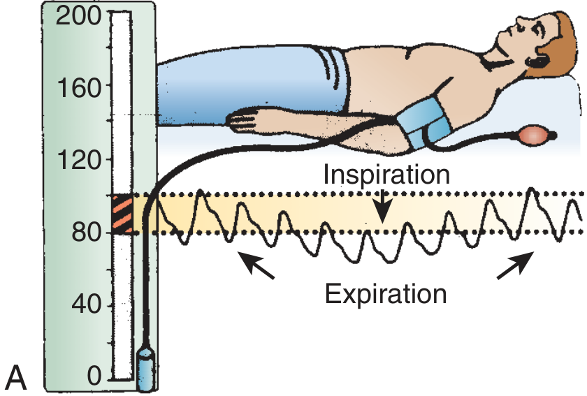
Normal Physiology First (to understand the pathology)
During normal inspiration:
- Intrathoracic pressure falls (diaphragm descends)
- Pulmonary vasculature expands → temporarily pools more blood in lungs
- Right heart: venous return increases (RV fills more)
- Left heart: less blood returns from pulmonary veins → LV fills slightly less
- Net result: small fall in systolic BP (~5-10 mmHg) - this is normal
This normal physiological variation is not called pulsus paradoxus.
Mechanism of Pulsus Paradoxus
There are two main mechanisms acting together, best understood in cardiac tamponade:
Mechanism 1: Ventricular Interdependence (Dominant)
INSPIRATION
  ↓
Intrathoracic pressure falls → RV venous return INCREASES → RV fills MORE
  ↓
Total pericardial volume is FIXED (by rigid effusion / pericardium)
  ↓
RV expands → physically pushes interventricular septum LEFT into LV
  ↓
LV cavity becomes SMALLER → LV stroke volume DECREASES
  ↓
Aortic output FALLS → Systolic BP DROPS on inspiration
This is ventricular interdependence - when one ventricle fills more, the other fills less because total cardiac volume is constrained.
Mechanism 2: Pulmonary Vascular Pooling (Contributing)
INSPIRATION
  ↓
Intrathoracic pressure falls → Pulmonary veins also fall in pressure
  ↓
But: intracardiac pressure (LA) is buffered by pericardial effusion
     so it does NOT fall as much
  ↓
Diastolic filling gradient (pulmonary veins → LA) DECREASES
  ↓
Less blood enters LV → LV stroke volume FALLS further
Both mechanisms together produce the exaggerated fall in systolic BP seen in tamponade.
Causes - Ordered by Importance
Cardiac Causes
| Cause | Mechanism | Severity of PP |
|---|---|---|
| Cardiac tamponade | Classic cause - ventricular interdependence + pericardial constraint | Usually >20 mmHg |
| Constrictive pericarditis | Mild ventricular interdependence | Mild or absent |
| Large pericardial effusion (without tamponade) | Partial constraint | Variable |
| RV infarction | RV dysfunction + ventricular interdependence | Can occur |
Pulmonary/Respiratory Causes
| Cause | Mechanism |
|---|---|
| Severe bronchial asthma | Marked negative intrathoracic pressure on inspiration → hugely exaggerates normal mechanism |
| COPD (severe) | Same as asthma - large intrathoracic pressure swings |
| Tension pneumothorax | Mediastinal shift + reduced venous return |
| Massive pulmonary embolism | Acute RV dilation → interventricular interdependence |
Other Causes
| Cause | Mechanism |
|---|---|
| Hemorrhagic shock | Reduced intravascular volume |
| Severe obesity | Increased intrathoracic pressure swings |
| Superior vena cava obstruction | Impaired venous return variation |
Differentiating the Causes
| Feature | Cardiac Tamponade | Severe Asthma/COPD | Constrictive Pericarditis |
|---|---|---|---|
| Degree of PP | >20 mmHg (often palpable) | Variable, often >10 mmHg | Mild or absent |
| JVP | Elevated, absent Y descent | Normal or elevated | Elevated, prominent Y descent |
| Kussmaul sign | Absent | Absent | Present |
| Heart sounds | Muffled | Normal | Normal + pericardial knock |
| Wheeze | Absent | Present | Absent |
| Breath sounds | Normal | Reduced + wheeze | Normal |
| Echo | Pericardial effusion + RV collapse | Normal heart | Pericardial thickening |
| Response to sitting forward | Better | No change | No change |
How to Measure Pulsus Paradoxus (Step-by-Step)
Equipment: Standard sphygmomanometer (BP cuff)
Position: Patient supine or reclined at 30-45°; breathing normally (not forced)
Steps:
- Inflate cuff well above systolic pressure (e.g., 30 mmHg above expected)
- Slowly deflate at 2-3 mmHg/second
- Note the pressure at which Korotkoff sounds are first heard - they will only appear during expiration at first - record this pressure (A)
- Continue slowly deflating
- Note the pressure at which Korotkoff sounds are heard throughout the entire respiratory cycle (both inspiration and expiration) - record this pressure (B)
- Pulsus paradoxus = A - B (in mmHg)
Interpretation:
| Difference (A-B) | Interpretation |
|---|---|
| <10 mmHg | Normal physiological variation |
| >10 mmHg | Pulsus paradoxus (abnormal) |
| >20-25 mmHg | Severe - highly specific for significant tamponade |
Alternative bedside method:
- Palpate the radial pulse while watching the patient breathe
- If the pulse disappears or markedly weakens on inspiration, pulsus paradoxus is present
- A palpable pulsus paradoxus at the radial or femoral artery indicates the difference is >15-20 mmHg
Pulse oximetry method:
- Watch the pulse oximetry waveform
- If the waveform amplitude visibly decreases during inspiration → suggests pulsus paradoxus
When Pulsus Paradoxus May Be Falsely ABSENT in Tamponade
This is an important clinical trap - tamponade can exist WITHOUT detectable pulsus paradoxus in these situations:
| Situation | Why PP is absent |
|---|---|
| Aortic regurgitation | Aortic run-off maintains diastolic pressure on both sides equally |
| Atrial septal defect (ASD) | RA and LA pressure equalize across defect; ventricular interdependence is blunted |
| LV hypertrophy | Stiff LV resists septal shift |
| Very low cardiac output / very narrow pulse pressure | Even a 10 mmHg fall may represent 100% of the pulse pressure - paradox may be "deceptively small" (5-15 mmHg) |
| Positive pressure ventilation | Respiratory mechanics reversed |
| Tachycardia, AF, tachypnea | Hard to measure accurately |
Pulsus Paradoxus vs Pulsus Alternans - Don't Confuse!
| Feature | Pulsus Paradoxus | Pulsus Alternans |
|---|---|---|
| Definition | BP falls >10 mmHg on inspiration | Beat-to-beat variation in pulse strength (alternating strong/weak) |
| Timing | Varies with respiration | Independent of respiration; regular rhythm |
| Cause | Tamponade, asthma, COPD, PE | Severe LV systolic dysfunction |
| Mechanism | Ventricular interdependence | Incomplete LV recovery in alternate beats |
| Detection | BP cuff (respiratory variation) | BP cuff (every other Korotkoff sound at different pressure) |
Pulsus Paradoxus vs Kussmaul Sign - Related but Different
| Pulsus Paradoxus | Kussmaul Sign | |
|---|---|---|
| What changes | Arterial pressure (BP falls on inspiration) | Venous pressure (JVP rises on inspiration) |
| Normal physiology | Arterial BP falls slightly on inspiration | Venous pressure falls on inspiration |
| Seen in | Cardiac tamponade | Constrictive pericarditis, restrictive cardiomyopathy, RV infarction |
| In tamponade | Present | Absent |
| In constrictive pericarditis | Absent or mild | Present |
This difference is the key bedside differentiator between tamponade and constrictive pericarditis.
Quick Memory Summary
Pulsus paradoxus = systolic BP drops >10 mmHg on inspiration = exaggerated normal physiology
Mechanism = ventricular interdependence (RV fills more → LV fills less) + pulmonary vascular pooling
Classic cause = cardiac tamponade (>20 mmHg)
Also in = severe asthma, COPD, massive PE, tension pneumothorax
Absent in tamponade when = AR, ASD, severe LVH, positive pressure ventilation
Paradoxus = arterial problem; Kussmaul = venous problem - opposite findings in tamponade vs constrictive pericarditis
- Roberts & Hedges' Clinical Procedures in Emergency Medicine
- Braunwald's Heart Disease, Cardiovascular Medicine
- Fuster & Hurst's The Heart, 15th Ed
This is a shared conversation. Sign in to Orris to start your own chat.