
Short notes on torus fracture For a 4th year mbbs student
torus buckle fracture pediatric X-ray radius
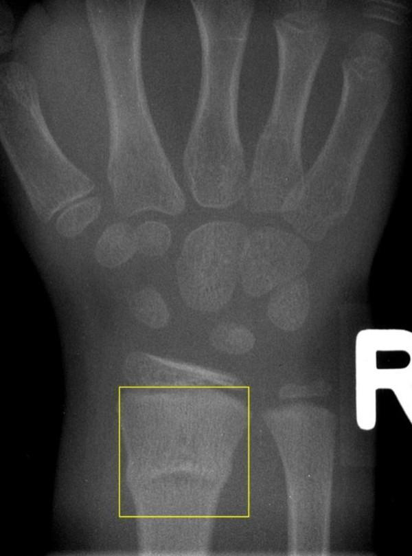
This diagnostic X-ray image (radiography) captures the pediatric wrist and hand, focusing on the distal forearm. The imaging demonstrates a buckle fracture, also known as a torus fracture, of the distal radius metaphysis. Visually, this is characterized by a subtle outward bulging or buckling of the cortical surface without a complete cortical break, typically occurring due to axial compression loads in children with relatively pliable bones. The fracture is localized within the metaphyseal region of the radius, identified here by a yellow bounding box. There is no significant angulation or displacement of the bone, and the distal ulna, carpal bones, and growth plates appear intact and well-aligned. This image is an educational example of a common pediatric orthopedic injury, illustrating the characteristic 'buckle' morphology used for clinical classification and diagnostic identification in emergency and pediatric medicine.
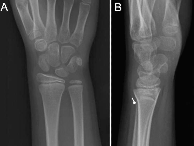
Two-view X-ray series (PA and lateral) of a pediatric right wrist demonstrating an angled distal radius buckle fracture (DRBF). The images show a skeletally immature individual with visible open physes (growth plates). Image A (posteroanterior view) shows subtle cortical irregularities at the distal radial metaphysis. Image B (lateral view) more clearly illustrates the pathology, marked by a white arrow pointing to a single focal cortical deflection or bulge along the dorsal aspect of the distal radius. This visual finding represents plastic deformation of the cortex without complete fracture line extension through the bone. The lack of significant displacement or involvement of the physis distinguishes this buckle fracture from more unstable pediatric injuries like Salter-Harris fractures. This diagnostic image is used to teach the identification of torus-type fractures in pediatric patients following axial loading injuries, such as a fall on an outstretched hand (FOOSH).
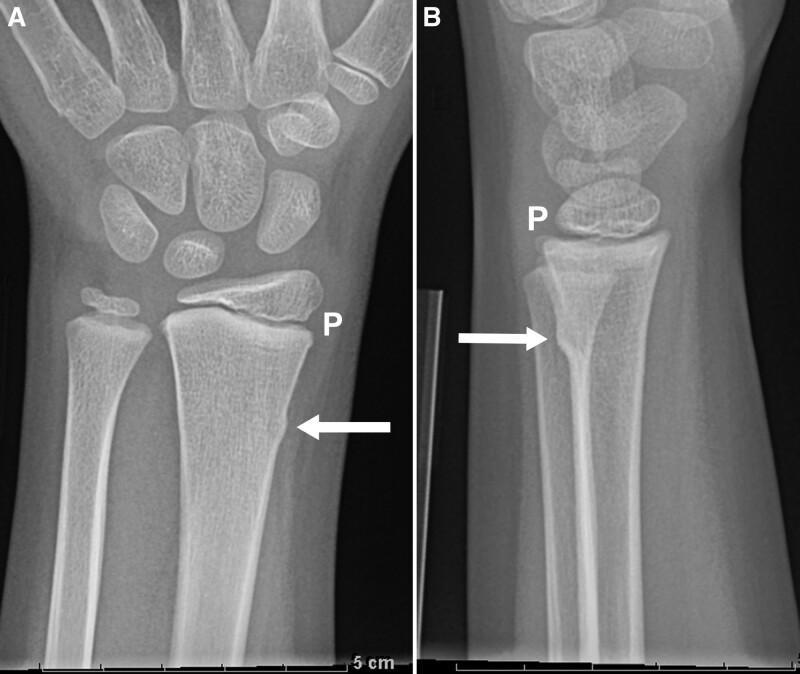
This diagnostic image consists of two X-ray radiographs—posteroanterior (A) and lateral (B) views—of a pediatric wrist belonging to an eight-year-old patient. The images demonstrate a cortical buckle fracture (torus fracture) involving the distal radial metaphysis. In both projections, a white arrow highlights a subtle focal bulging or outward buckling of the bony cortex without frank cortical disruption or angulation. The distal radial physis, labeled 'P', is visible as a clear radiolucent line, indicating the growth plate remains open. A key educational feature is the presence of normal, intact trabecular bone between the fracture site and the physis, distinguishing this as a stable buckle fracture rather than a potentially unstable distal radius fracture or a physeal injury. The carpal bones and distal ulna appear unremarkable. This clinical image is significant in pediatric orthopedics for teaching the morphologic criteria used to differentiate stable cortical deformities from unstable fractures requiring different management protocols.
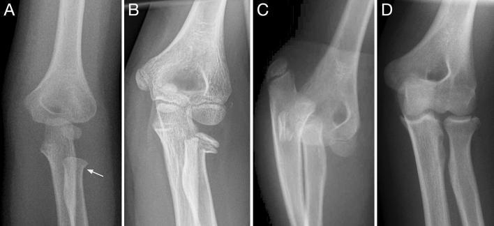
This composite diagnostic image features four anteroposterior and lateral X-ray radiographs (A–D) demonstrating a spectrum of proximal radius fractures in pediatric and adolescent patients. Panel A shows a stable buckle fracture of the radial neck in a 4-year-old, characterized by a focal cortical bulge (indicated by an arrow) with preserved alignment of the radial head and capitellum. Panel B illustrates a Salter-Harris II physeal fracture of the radial neck with moderate lateral displacement of the epiphysis. Panel C depicts a severe, completely displaced metaphyseal radial neck fracture associated with elbow dislocation in a 12-year-old; the radial head is entirely dissociated from both the humeral capitellum and the radial shaft, indicating high risk for avascular necrosis. Panel D displays an intraarticular radial head fracture in a 15-year-old adolescent with closed physes, representing an adult-pattern injury. This series illustrates various injury mechanisms, from stable torus-type fractures to complex articular disruptions and total dislocations, serving as an educational comparison of fracture stability and clinical prognosis in pediatric orthopedics.
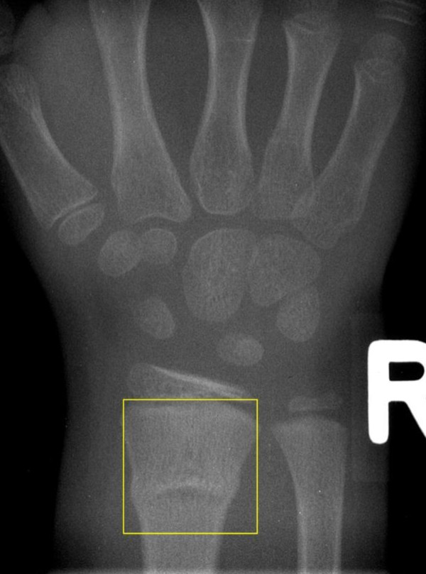
**Imaging Modality:** Posterior-anterior (PA) projection radiograph (X-ray). **Anatomical Region:** Pediatric right wrist and distal forearm. **Observed Pathology:** Buckle (torus) fracture of the distal radius. **Characteristic Visual Features:** The image demonstrates a localized bulging or "buckling" of the cortical bone at the distal radial metaphysis, highlighted by a yellow bounding box. This is characterized by a focal outward protrusion of the cortex without a discrete fracture line extending through the entire bone width. The distal ulna appears intact. The presence of open physes and developing carpal bones confirms the pediatric nature of the skeleton. **Key Diagnostic Features:** - **Type:** Incomplete fracture (torus/buckle). - **Location:** Distal metaphysis of the radius. - **Displacement:** Non-displaced; anatomical alignment of the radial shaft and epiphysis is maintained. - **Pattern:** Cortical compression resulting from axial loading, typical of a pediatric injury. **Clinical Context:** Common pediatric orthopedic injury often resulting from a fall on an outstretched hand (FOOSH), classified under pediatric fracture coding systems as an incomplete metaphyseal compression fracture.
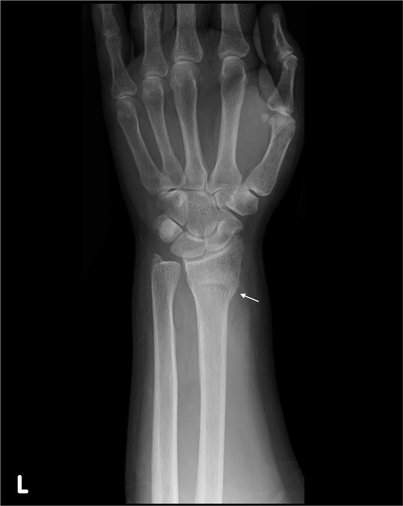
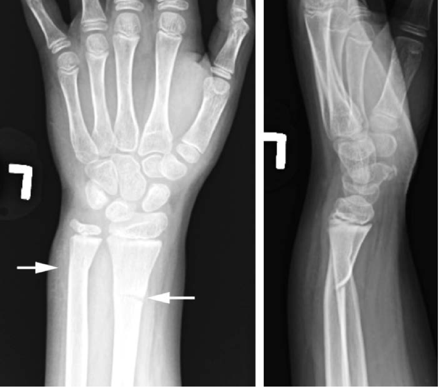
**Imaging Modality:** Posterior-anterior (PA) radiograph (X-ray). **Anatomical Region:** Left wrist and distal forearm, including the distal radius, distal ulna, carpal bones, and proximal metacarpals. **Observed Pathology:** Buckle (torus) fracture of the distal radial metaphysis. **Characteristic Visual Features:** * **Fracture Morphology:** There is a discrete cortical bulging or "buckling" of the lateral cortex of the distal radius, indicated by a white arrow. * **Alignment:** The fracture is incomplete, non-displaced, and demonstrates no significant angulation or shortening of the radial shaft. * **Joint Integrity:** The radiocarpal and distal radioulnar joints (DRUJ) appear maintained with preserved joint spaces. No evidence of carpal bone fracture or dislocation is visualized. * **Secondary Findings:** The distal ulnar epiphysis and shaft remain intact. Surrounding soft tissues appear within normal limits for this projection. **Key Diagnostic Features:** The localized cortical protrusion without a distinct fracture line through the entire bone thickness is characteristic of a torus-type injury, typically seen in pediatric or adolescent populations with incomplete ossification.
Torus (Buckle) Fracture - Short Notes
Definition
Why Only in Children?
Torus (mildest) → Greenstick → Complete fracture (most severe)
Mechanism
- Fall on an outstretched hand (FOOSH) - the most common cause
- Axial compression load is transmitted to the metaphysis (the weakest zone, where cortical bone transitions to cancellous bone)
- The compression cortex buckles inward; no tension-side failure occurs
Common Site
- Distal radius metaphysis - most frequent (accounts for majority of cases)
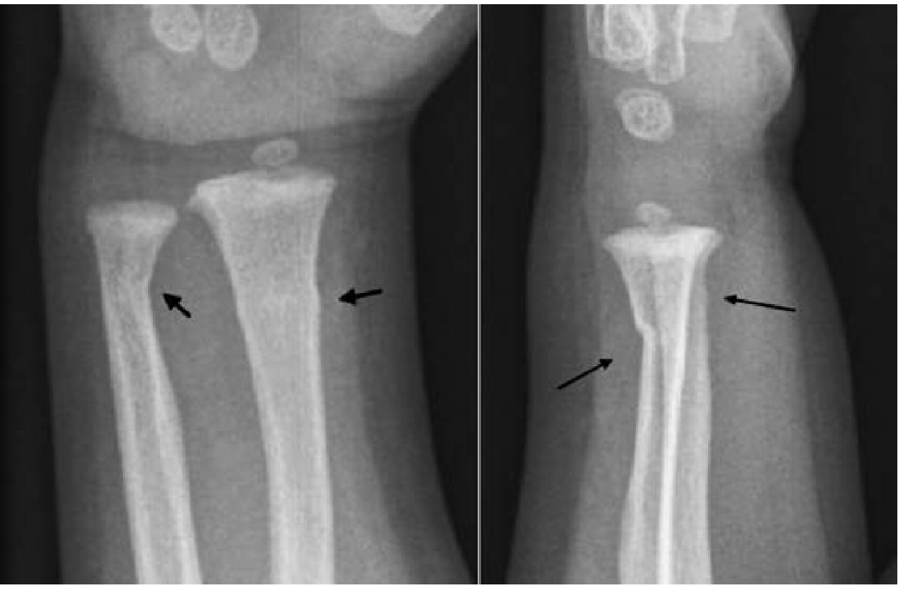
- Distal ulna (often co-exists with distal radius torus fracture - see X-ray below)
- Distal tibia
- Age group: typically 5-10 years
Clinical Features
| Feature | Description |
|---|---|
| Pain | Localized over the fracture site |
| Swelling | Soft tissue swelling over the metaphysis |
| Tenderness | Point tenderness on palpation |
| Deformity | None - no visible bony deformity |
| Neurovascular status | Normal |
Radiological Features
- Inspect the metaphyseal flare carefully on both views
- Look for asymmetry, bulging, or deviation of the cortical margin
- No angulation, no displacement, no rotational abnormality
- The finding can be very subtle - soft tissue swelling may be the only clue
Comparison with Greenstick Fracture
| Feature | Torus | Greenstick |
|---|---|---|
| Cortex involved | Compression cortex only (buckles) | Tension cortex disrupted; compression cortex intact |
| Fracture line | No complete fracture line | Actual cortical crack on one side |
| Severity | Milder | More severe |
| Periosteum | Intact on both sides | Torn on tension side |
| Reduction needed? | No | Sometimes yes |
Management
- No reduction required - torus fractures have no angulation or displacement
- Immobilization with a below-elbow splint or soft cast for 3-4 weeks
- Some guidelines support removable splinting for comfort in stable torus fractures
- Outpatient follow-up with orthopedics is appropriate
- Excellent prognosis - heals completely without growth disturbance (physeal plate is not involved)
- No surgical intervention needed
Key Exam Points
- Torus = buckle = cortical fracture - all synonymous
- Exclusively in children due to bone plasticity
- Most common site: distal radial metaphysis
- Mechanism: FOOSH (axial compression)
- X-ray: subtle cortical bulge at metaphysis, no angulation or displacement
- Management: conservative - simple immobilization, no reduction
- Contrast with greenstick: in torus, only the compression cortex buckles; in greenstick, the tension cortex cracks
Short notes on Greenstick Fracture
greenstick fracture pediatric X-ray forearm
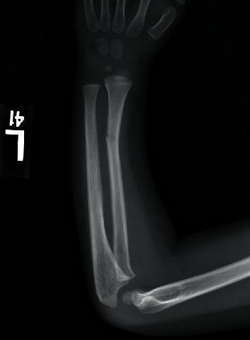
A diagnostic X-ray (radiography) of the pediatric left forearm in a lateral-oblique view. The image depicts the humerus, radius, and ulna, as well as several carpal and metacarpal bones. A greenstick fracture is clearly visible in the distal third of the radial diaphysis. The fracture presents as an incomplete cortical disruption on the dorsal aspect with bowing of the opposite cortex, characteristic of pediatric skeletal flexibility. There is minimal volar angulation of the distal fragment, and no significant displacement is observed. The adjacent ulna remains intact without evidence of plastic deformation or fracture. The presence of visible growth plates (physes) at the distal radius, ulna, and proximal humerus confirms the pediatric nature of the patient. This radiograph is an educational example of common pediatric musculoskeletal trauma resulting from low-energy mechanisms like a fall on an outstretched hand (FOOSH).
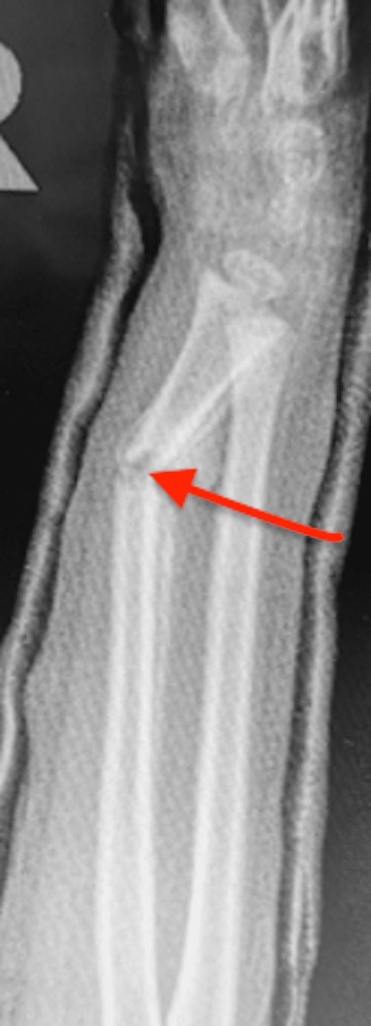
A lateral X-ray of a pediatric forearm demonstrating a displaced fracture of the radius and ulna. The image shows a significant dorsal angulation at the fracture site, specifically identified by a red arrow pointing to an 'angulated slipper fracture'. The fracture appears to be incomplete or greenstick in nature, with an irregular cortex at the apex of the deformity. The forearm is encased in a short arm cast, which is visible as a radio-opaque layer surrounding the soft tissue. The alignment is disrupted, showing a loss of reduction where the distal segments are tilted relative to the proximal shafts. This diagnostic image serves as an educational example of fracture displacement and failure of immobilization following transition from a long arm to a short arm cast in pediatric orthopedic management.
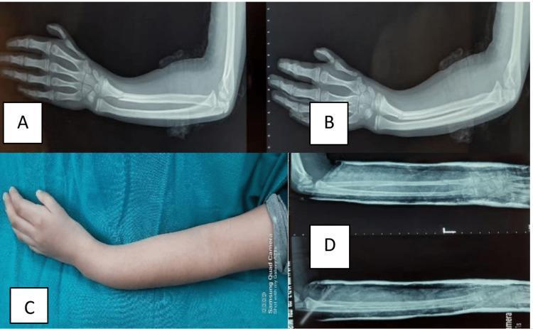
Educational panel illustrating the management of a pediatric forearm fracture, categorized into diagnostic imaging and clinical photography. Panel A displays a pre-reduction lateral X-ray of a pediatric forearm showing a greenstick fracture of the distal radius with characteristic cortical bowing and dorsal angulation; the ulna appears intact. Panel B is a post-reduction lateral X-ray demonstrating corrected alignment of the radial shaft following closed manipulation. Panel C is a clinical photograph of the patient's forearm, showcasing the visible physical deformity often described as a 'dinner fork' or volar-angulated appearance at the distal forearm and wrist, with intact skin and no obvious bruising. Panel D provides post-reduction X-rays within a cast, confirming satisfactory anatomical realignment in multiple planes. This sequence serves as an educational tool for identifying pediatric greenstick fractures, visualizing clinical deformities, and assessing the success of orthopedic closed reduction procedures in the emergency or orthopedic setting.
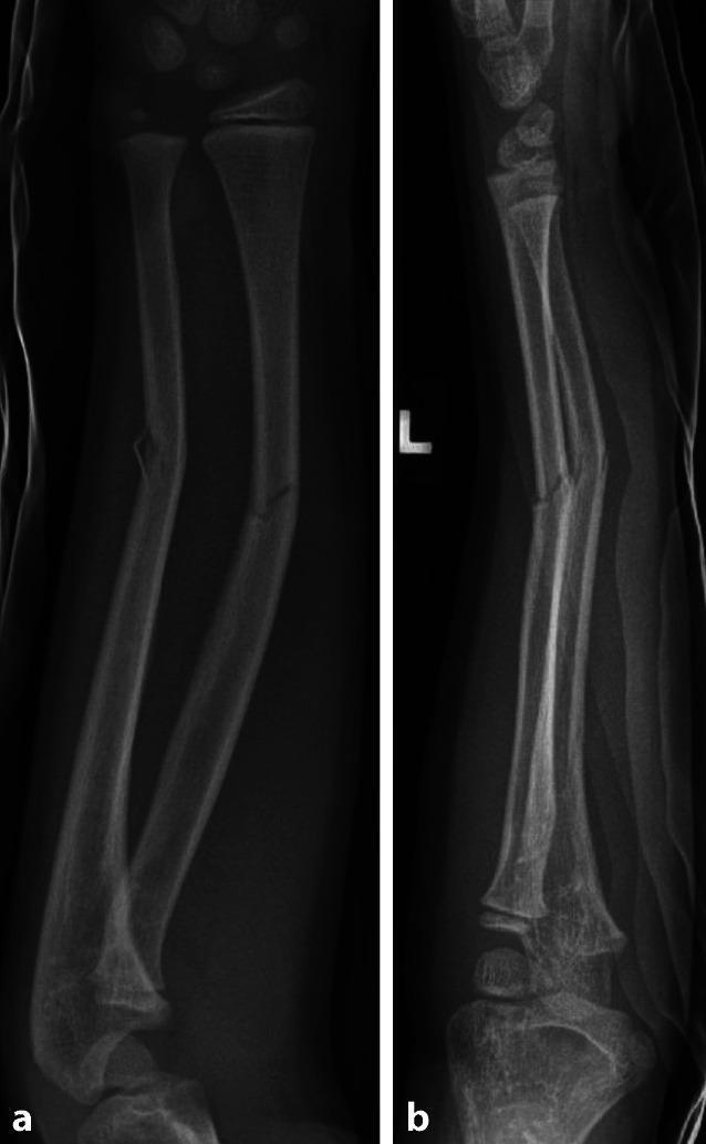
This diagnostic image consists of two X-ray radiographs (a.p. and lateral views) showing the left forearm of a pediatric patient. The images demonstrate an acute greenstick fracture of the mid-diaphyseal shafts of both the radius and ulna. In the a.p. view (a), an incomplete fracture line is visible involving the tension side of the cortex with characteristic plastic deformation and bowing of the opposite (intact) cortex. The lateral view (b) highlights the apex-volar angulation of the fracture segments. Visible open growth plates (physes) at the distal radius, distal ulna, and elbow confirm the pediatric nature of the patient. There is no evidence of complete cortical disruption or significant displacement (apposition is maintained), which is typical of greenstick fractures in children where the thick periosteum remains partially intact. This content is used to teach diagnostic hallmarks of pediatric long bone injuries and the clinical importance of the intact opposite cortex in fracture stability.
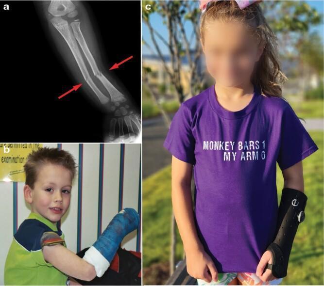
This composite educational resource illustrates pediatric forearm fractures and subsequent immobilization methods. (a) A diagnostic X-ray of a child's forearm shows midshaft (diaphyseal) fractures of both the radius and ulna. Red arrows highlight the specific sites: the radius exhibits a complete angular fracture with disruption of both cortices, while the ulna demonstrates an incomplete greenstick fracture, characterized by cortical buckling and bending without full separation. (b) A clinical photograph shows a young child with a traditional hard fiberglass cast extending from the distal humerus to the metacarpals, providing rigid immobilization of the elbow and wrist. (c) A contemporary clinical photograph depicts a child wearing a modern soft splint, a removable orthotic device that provides support while allowing greater comfort and limited movement compared to a rigid cast. The material emphasizes pediatric resilience and the evolution of orthopedic management for common childhood injuries like those sustained from falls onto an outstretched hand (FOOSH).
![This diagnostic radiographic image consists of two X-ray views (anterior-posterior [a] and lateral [b]) of a pediatric forearm showing evidence of healing fractures in the mid-diaphysis of both the radius and ulna. The images demonstrate a follow-up status after four weeks of immobilization. Visual findings include significant cortical thickening and periosteal reaction at the fracture sites, indicating secondary bone healing with callus formation. In the AP view, the longitudinal alignment is largely maintained, though medial bowing is visible in the ulnar shaft. The lateral view reveals a prominent posterior angulation at the ulnar fracture site and subtle anterior bowing of the radius. The growth plates are open, consistent with a pediatric patient. These findings represent a greenstick fracture progression with incomplete consolidation, highlighting the clinical risk of refracture if mechanical loading is resumed too early. The educational focus is on the assessment of fracture stability, cortical continuity, and secondary ossification during the remodeling phase of pediatric long bone injuries.](https://cdn.orris.care/cdss_images/pmc_clinical_VQA_a664681ed2590a2e23328a46cabad1c173d66a9727212a38d06bf647744e02e1.jpg)
This diagnostic radiographic image consists of two X-ray views (anterior-posterior [a] and lateral [b]) of a pediatric forearm showing evidence of healing fractures in the mid-diaphysis of both the radius and ulna. The images demonstrate a follow-up status after four weeks of immobilization. Visual findings include significant cortical thickening and periosteal reaction at the fracture sites, indicating secondary bone healing with callus formation. In the AP view, the longitudinal alignment is largely maintained, though medial bowing is visible in the ulnar shaft. The lateral view reveals a prominent posterior angulation at the ulnar fracture site and subtle anterior bowing of the radius. The growth plates are open, consistent with a pediatric patient. These findings represent a greenstick fracture progression with incomplete consolidation, highlighting the clinical risk of refracture if mechanical loading is resumed too early. The educational focus is on the assessment of fracture stability, cortical continuity, and secondary ossification during the remodeling phase of pediatric long bone injuries.
Greenstick Fracture - Short Notes
Definition
Why Only in Children?
- Thicker and more elastic periosteum
- Higher water content and lower mineral density
- Active Haversian remodeling
Position in the Spectrum of Pediatric Fractures
Torus (mildest) → Greenstick → Complete fracture (most severe)
| Feature | Torus | Greenstick |
|---|---|---|
| Cortex failed | Compression cortex buckles | Tension cortex cracks |
| Other cortex | Intact | Intact (but may buckle) |
| Periosteum | Intact both sides | Torn on tension side; intact on concave side |
| Severity | Milder | More severe |
Mechanism
- Fall on an outstretched hand (FOOSH) - most common
- A bending force applied to the bone creates a tension side (convex) and a compression side (concave)
- The tension cortex cracks first; the compression cortex bends/buckles but does not completely break
- The intact periosteum on the concave side limits displacement
Common Sites
- Distal radius (most common) - often with distal ulna involvement
- Radius and ulna shaft (both bones forearm)
- Clavicle (in newborns and infants)
- Tibia (distal diaphysis)
- Age: typically 1-10 years, younger children more prone
Clinical Features
| Feature | Description |
|---|---|
| Pain | Localized, often moderate |
| Swelling | Present over fracture site |
| Tenderness | Point tenderness |
| Deformity | May be visible - bowing/angulation of the limb |
| Stability | Relatively stable (intact periosteum limits shift) |
| Neurovascular | Usually intact |
Radiological Features
- Cortical disruption on one side (tension/convex side) - visible fracture line
- Intact or buckled cortex on the opposite side (compression/concave side)
- Angulation is typically present (unlike torus)
- No complete fracture line through both cortices
- Bowing of the bone may be evident
- Look at both AP and lateral views - the fracture line may only be seen on one projection
Management
Conservative (majority of cases)
- Closed reduction - required if angulation is significant (>10-15° depending on site and age)
- Immobilization in a plaster/fibreglass cast for 4-6 weeks (longer than torus)
- In some greenstick fractures, the surgeon may deliberately complete the fracture during reduction to prevent "springing back" - the intact periosteum on the concave side tends to cause the fracture to re-angulate if not fully reduced
Reduction technique
- Apply a 3-point bending force to correct the angulation
- Completing the fracture (breaking the intact cortex) may be needed for unstable angulations to achieve and hold reduction
- Long-arm cast for forearm fractures to control rotation
Follow-up
- Repeat X-ray at 1-2 weeks to check for loss of reduction ("re-angulation")
- Risk of re-fracture is higher than torus fractures
- Excellent remodeling potential in young children
Complications
| Complication | Notes |
|---|---|
| Re-angulation | Intact periosteum springs back; check X-ray at 1-2 weeks |
| Re-fracture | Cortex weakened at fracture site |
| Refracture during reduction | If trying to complete intentionally |
| Mal-union | If angulation not adequately corrected |
| Growth disturbance | Rare; only if physis involved |
Key Exam Points
- Greenstick = tension cortex cracks, compression cortex intact (opposite of torus)
- Named after a green twig analogy - cracks on the outer convex side
- Exclusive to children due to bone plasticity
- Angulation is present (key distinguishing feature from torus)
- May need deliberate completion of fracture during reduction to prevent re-angulation
- Immobilize for 4-6 weeks (longer than torus)
- Monitor closely for re-angulation at 1-2 weeks post-reduction
- More stable than complete fractures due to intact concave periosteum
Toddler’s fracture
toddler's fracture tibia spiral X-ray child
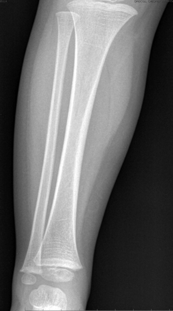
**Imaging Modality:** Projectional radiography (X-ray). **Anatomical Region:** Anteroposterior (AP) view of the pediatric right lower leg, including the shafts of the tibia and fibula, the proximal metaphyses, and the distal epiphyses/physes near the ankle joint. **Observed Pathology:** No acute osseous abnormalities are identified. **Characteristic Visual Features:** * **Bone Morphology:** The cortical margins of the tibial and fibular diaphyses are smooth and continuous, with no evidence of fracture lines, cortical buckling, or periosteal reaction. * **Physeal Presentation:** The visible growth plates (physes) at the proximal and distal ends appear unremarkable for the patient's developmental age. * **Joint Spaces:** The visible portions of the knee and ankle joints demonstrate maintained alignment and normal joint spacing. * **Soft Tissues:** The surrounding soft tissue planes are symmetric without focal swelling or radio-opaque foreign bodies. **Key Diagnostic Features:** This radiograph represents a normal pediatric study of the tibia and fibula. The absence of a visible fracture line or subtle "toddler’s fracture" (undisplaced spiral fracture of the distal tibia) on this specific AP projection is noted.
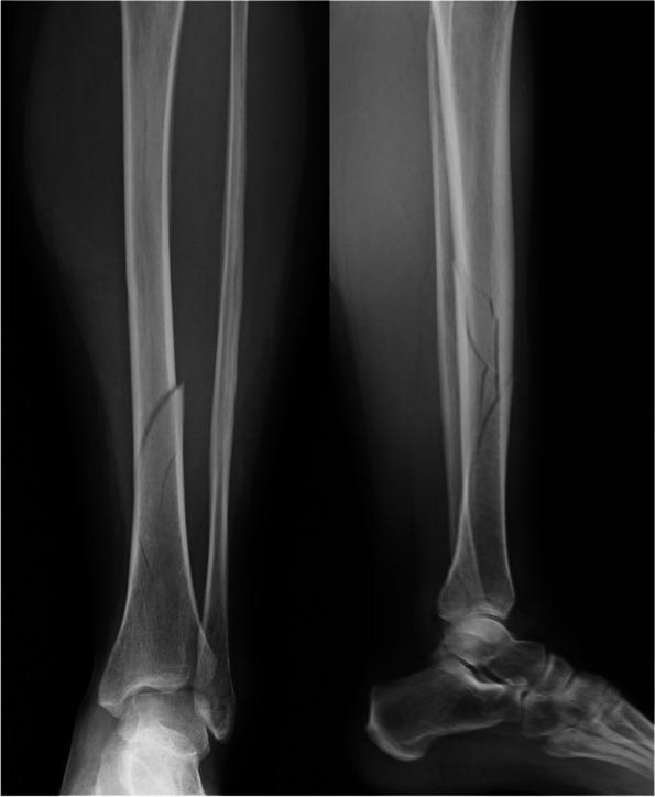
Two-view pre-operative X-ray (Anteroposterior and Lateral) of a left lower leg demonstrating fractures of the tibia and fibula. The tibial fracture is a spiral/oblique diaphyseal fracture (AO 42-A1) located at the junction of the middle and distal thirds. On the AP view, there is lateral displacement and angulation of the distal tibial fragment. The lateral view reveals a posterior displacement of the distal fragment with evidence of cortical comminution. Additionally, a distal fibular fracture (AO 44-B1) is visible near the level of the syndesmosis, appearing non-displaced on these views. Soft tissue swelling is noted around the fracture sites and the ankle joint. This imaging is characteristic of trauma-induced lower limb fractures requiring orthopedic evaluation for stabilization, such as intramedullary nailing or open reduction internal fixation.
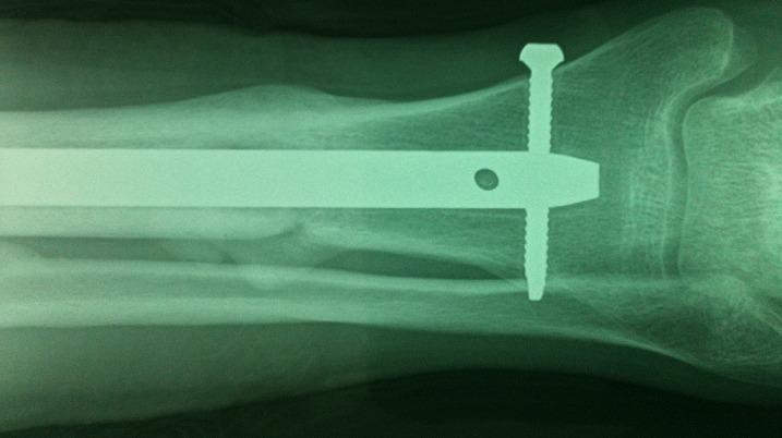
This diagnostic radiograph (X-ray) displays a consolidated spiral fracture of the distal fourth of the tibia and fibula. The tibial fracture, classified as AO/OTA type A1-1, shows significant interval healing with exuberant periosteal callus formation bridging the original fracture site. Alignment is well-maintained with the restoration of the anatomical axis. Internal fixation is demonstrated by a radiopaque intramedullary nail positioned within the tibial canal. A single distal interlocking screw is visible, transfixing the distal tibia and the nail to provide rotational and axial stability. The fibular fracture is also visible in the background with signs of secondary bone healing. The ankle joint space and mortise appear preserved. This image serves as a clinical example of successful surgical management of a distal leg fracture using antegrade intramedullary nailing, emphasizing bone union and alignment preservation in orthopedic trauma.
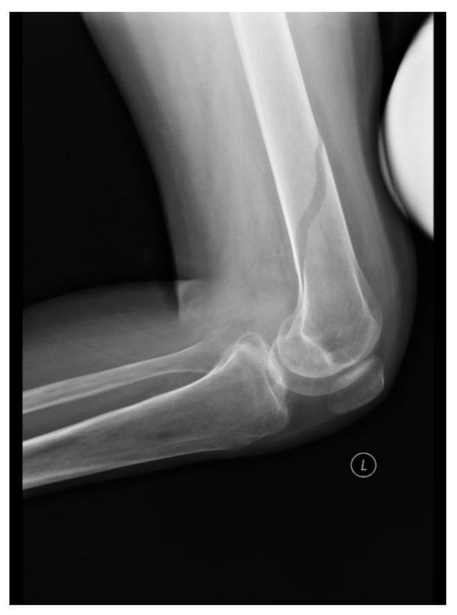
This lateral-view X-ray (radiograph) of the left lower limb demonstrates the distal femur, knee joint, and proximal tibia and fibula. The primary diagnostic finding is a spiral fracture located in the mid-to-distal shaft of the femur. The fracture is characterized by a radiolucent, oblique line that curves around the diaphysis, indicating torsional stress as the mechanism of injury. There is minimal displacement of the femoral fragments, and the cortical alignment is relatively well-preserved despite the discontinuity. The knee joint space appears intact, and the patella is visualized in its expected anterior position. The proximal tibia and fibula show normal bone density and morphology without evidence of additional fractures. This image illustrates a common complication of low-energy trauma in patients with reduced bone mineral density, such as those with chronic spinal cord injuries, where sensory deficits may mask the initial pain of the fracture.
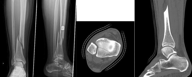
A composite medical diagnostic image including plain X-ray radiography (anteroposterior and lateral views) and Computed Tomography (CT) scans (axial and sagittal planes) of the lower leg and ankle. The imaging demonstrates a spiral fracture of the distal third of the tibial shaft (AO/OTA Type 42A1), characterized by a long, torsional fracture line with moderate displacement and an associated non-comminuted fibular fracture. Accompanying CT images reveal a concomitant posterior malleolus fracture (Volkmann's fragment) involving the articular surface of the distal tibia, which is often occult on initial plain films. The sagittal CT view highlights the longitudinal extent of the tibial shaft fracture and the posterior articular fragment, while the axial view confirms the intra-articular extension at the ankle joint. This case illustrates the high clinical incidence of posterior malleolus involvement in spiral tibial shaft fractures and emphasizes the educational value of CT for preoperative planning and screw osteosynthesis.
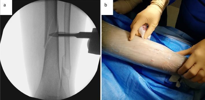
This composite educational image demonstrates the initial steps of a minimally invasive cerclage wiring technique for a tibial spiral fracture. Panel A is an intraoperative fluoroscopic (X-ray) image using an image intensifier. It shows a mid-shaft spiral fracture of the tibia (AO 42A1c) that has undergone anatomical reduction. A radiopaque surgical instrument with a tapered shaft and paddle-like tip is positioned perpendicularly to the tibial shaft to identify the target level for cerclage placement. Panel B is a clinical photograph showing the surgical field preparation of the lower leg. The limb is covered with a transparent sterile adhesive drape. Two small, approximately 1 cm longitudinal incisions are visible: one on the anterolateral aspect and one on the dorsomedial aspect of the tibia. A surgeon’s gloved hand is shown palpating the proximal incision site. The image illustrates the transition from radiological planning to the execution of minimally invasive portals in orthopedic trauma surgery.
Toddler's Fracture - Short Notes
Definition
Epidemiology
- Peak incidence: 9 months to 4 years of age (the walking/toddling stage)
- Occurs in children who have recently started or are learning to walk
- Slightly more common in boys
- One of the few fractures in young children that is NOT suspicious for non-accidental trauma (NAT/child abuse)
Mechanism
- External rotation of the foot with the knee flexed - the classic mechanism
- A minor fall or stumble with the foot planted on the ground while the body twists
- Often the parent reports the child simply "tripped" or "slipped" - the trauma seems trivial
- The spiral/oblique fracture pattern results from the torsional (twisting) force on the tibia
Clinical Features
| Feature | Description |
|---|---|
| Presenting complaint | Limping or refusal to bear weight |
| History | Often vague or absent - parents unsure of any fall |
| Pain | Localized to distal tibia/lower leg |
| Swelling | Minimal or absent - often deceptively mild |
| Deformity | None |
| Tenderness | On palpation and rotation of the distal tibia |
| Neurovascular | Normal |
Key clinical pearl: A toddler who is limping or refuses to weight-bear with no obvious cause should raise suspicion for this fracture.
Radiology
X-ray (standard AP + lateral views)
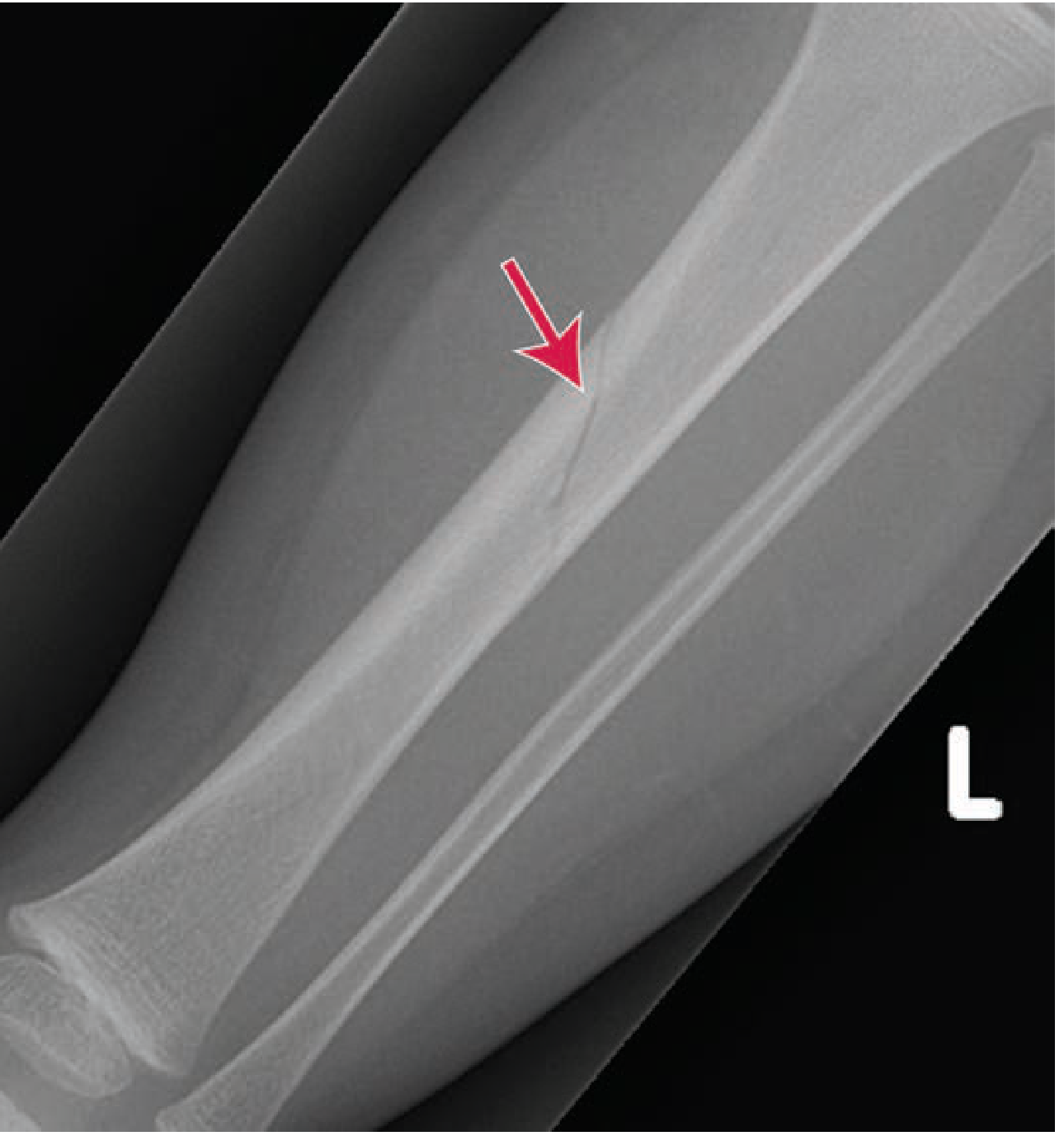
- May show a subtle oblique or spiral lucency through the distal third of the tibial shaft
- The fracture line terminates medially
- Often initially INVISIBLE on standard views - very easy to miss!
- The fibula is intact (no fibular fracture)
Oblique view
- Oblique views are more sensitive and may reveal the fracture when standard views are negative
If X-rays are negative but suspicion remains high
- Periosteal reaction and sclerosis become visible on repeat X-ray after 1-2 weeks due to callus formation
- Ultrasound can detect periosteal elevation before X-ray changes appear
- MRI or bone scan can confirm occult fracture
Differential Diagnosis
| Condition | How to differentiate |
|---|---|
| Non-accidental trauma | Multiple fractures, different healing stages, inconsistent history, unusual fracture patterns |
| Septic arthritis/osteomyelitis | Fever, raised inflammatory markers, joint effusion |
| Transient synovitis | Older age, hip not tibial tenderness |
| Osteogenesis imperfecta | Blue sclerae, recurrent fractures, family history |
| Developmental dysplasia of hip | Hip range of motion abnormality |
Important: Toddler's fracture is a recognized exception among pediatric fractures - it does NOT require a skeletal survey for NAT workup, even without a clear mechanism of trauma.
Management
If X-ray positive
- Immobilization - options include:
- Posterior long-leg splint (ED) followed by above-knee or below-knee cast placed in orthopaedic clinic
- Removable prefabricated device (cast boot)
- No significant difference in outcomes between methods has been shown
- Duration: 3-4 weeks
- Children are allowed to weight-bear as tolerated after immobilization
- Casts should NOT extend above the knee in very young toddlers - risk of cast migration
- No reduction required - fracture is non-displaced
If X-ray negative but clinically suspected
- Two acceptable options:
- Immobilize (splint) and review in 1-2 weeks with repeat X-ray (will show callus)
- No immobilization with close outpatient follow-up
Prognosis
- Excellent - heals completely without growth disturbance
- No risk of malunion as fracture is non-displaced
- Full weight-bearing usually restored within 3-4 weeks of immobilization
- Recurrence is not expected
Key Exam Points
- Toddler's fracture = non-displaced spiral fracture of distal tibia in a child aged 9 months - 4 years
- Classic presentation: limping/refusal to weight-bear, minimal swelling, minimal trauma history
- Mechanism: external rotation of foot with knee flexed (twisting fall with foot planted)
- Fibula is INTACT - distinguishes it from higher-energy fractures
- Initial X-ray may be completely normal - oblique views and/or repeat X-ray at 1-2 weeks are key
- NOT a red flag for child abuse - it is a recognized accidental fracture pattern
- Management: conservative immobilization, no surgery, no reduction needed