Discuss ELISA with its importance in detail as per m pharm
ELISA AND applications
ELISA principles formats direct indirect sandwich competitive official educational resource
https://www.abcam.com/en-us/technical-resources/guides/elisa…
ELISA: Enzyme-Linked Immunosorbent Assay
Definition
Principle
-
Specific antigen-antibody interaction
An antibody binds only to its corresponding antigen or epitope. -
Enzyme-substrate reaction
An enzyme linked to an antibody or antigen converts an added substrate into a detectable product. The signal generated is related to the amount of analyte in the sample.
- Horseradish peroxidase (HRP)
- Alkaline phosphatase (ALP)
- Beta-galactosidase
- Glucose oxidase
| Enzyme | Common substrate | Product/readout |
|---|---|---|
| HRP | TMB, OPD, ABTS | Coloured product |
| Alkaline phosphatase | pNPP | Yellow product |
| HRP | Luminol | Chemiluminescent signal |
Basic components of an ELISA
-
Solid phase
- Usually polystyrene microtitre plate.
- Antigen or capture antibody is adsorbed onto the well surface.
-
Analyte
- The substance to be measured, such as antigen, antibody, cytokine, hormone, or drug.
-
Primary antibody
- Binds specifically to the antigen of interest.
-
Secondary antibody or detection antibody
- Recognizes the primary antibody or antigen.
- May be enzyme-labelled directly.
-
Enzyme conjugate
- Antibody or antigen coupled with HRP, ALP, or another enzyme.
-
Blocking buffer
- Usually BSA, casein, skim milk, or similar protein.
- Occupies uncoated plastic sites and reduces non-specific binding.
-
Wash buffer
- Commonly phosphate-buffered saline or Tris-buffered saline with Tween-20.
- Removes unbound reagents and reduces background signal.
-
Substrate and stop solution
- Substrate produces a measurable signal after enzyme action.
- Stop solution terminates the reaction at a defined time.
-
Controls and standards
- Blank, negative control, positive control, and calibrators/standards are required for valid interpretation.
General ELISA Procedure
-
Coat the microplate
- Add antigen or capture antibody to the wells.
- Incubate to allow adsorption to the solid surface.
-
Wash
- Remove unbound coating material.
-
Block unoccupied sites
- Add blocking buffer to prevent non-specific protein adsorption.
-
Add sample
- Sample may contain the antigen or antibody being measured.
-
Incubate and wash
- Specific antigen-antibody complexes remain attached to the plate.
- Unbound materials are removed by washing.
-
Add enzyme-labelled antibody/conjugate
- This binds to the immunological complex.
-
Wash again
- Essential for reducing false-positive results from residual conjugate.
-
Add substrate
- Enzyme produces colour, fluorescence, or luminescence.
-
Stop reaction
- Used for many colorimetric ELISAs.
-
Read absorbance
- Measure OD using an ELISA microplate reader.
- Interpret results
- Qualitative: positive/negative based on cut-off.
- Semi-quantitative: titre or relative concentration.
- Quantitative: concentration calculated from a standard curve.
Types of ELISA
1. Direct ELISA
Principle
Steps
- Coat plate with antigen.
- Block unoccupied sites.
- Add enzyme-labelled primary antibody.
- Wash.
- Add substrate and measure signal.
Features
- Detects antigen.
- Signal is generally directly proportional to antigen concentration.
Advantages
- Simple and rapid.
- Fewer incubation and washing steps.
- Lower risk of cross-reactivity from a secondary antibody.
- Suitable for screening when a labelled primary antibody is available.
Limitations
- Less signal amplification.
- Usually less sensitive than indirect ELISA.
- Each primary antibody must be individually enzyme-labelled.
- Antigen adsorption may be variable.
2. Indirect ELISA
Principle
Steps
- Coat plate with known antigen.
- Block.
- Add patient serum or test sample containing possible primary antibodies.
- Wash.
- Add enzyme-labelled anti-human immunoglobulin secondary antibody.
- Wash.
- Add substrate and measure OD.
Main use
Examples
- Anti-HIV antibody screening
- Hepatitis B antibody testing
- Anti-HCV antibody testing
- Detection of antibodies against infectious organisms
- Autoantibody measurement
- Vaccine-induced antibody titres
Advantages
- High sensitivity due to signal amplification by several secondary antibodies binding one primary antibody.
- Economical because one labelled secondary antibody can detect many primary antibodies.
- Flexible and widely applicable.
Limitations
- More steps and longer assay time than direct ELISA.
- Secondary antibody cross-reactivity may cause non-specific results.
- Interpretation can be affected by heterophile antibodies, rheumatoid factor, or non-specific serum proteins.
3. Sandwich ELISA
Principle
Steps
- Coat plate with capture antibody.
- Block non-specific sites.
- Add test sample containing antigen.
- Wash.
- Add detection antibody.
- Add enzyme-labelled secondary antibody, if the detection antibody is unlabelled.
- Wash.
- Add substrate and measure signal.
Main use
Examples
- Cytokines: IL-6, TNF-alpha, IL-1 beta
- Hormones: insulin, hCG, TSH
- Tumour markers
- Viral antigens
- Bacterial toxins
- Biomarkers in serum, plasma, culture supernatant, and cell lysates
Advantages
- Very high specificity because two antibodies recognize distinct epitopes.
- High sensitivity.
- Suitable for low-abundance antigens.
- Works well with complex matrices such as serum and plasma.
- Preferred format for protein quantification in research and many diagnostic kits.
Limitations
- Requires a matched antibody pair.
- Not suitable when antigen has only one accessible epitope or is very small.
- High-dose hook effect may occur at very high antigen concentrations, causing falsely low results.
- More expensive and time-consuming to develop.
4. Competitive ELISA
Principle
- More analyte in sample = less labelled material binds = lower colour/signal.
- Less analyte in sample = more labelled material binds = higher colour/signal.
Main use
Examples
- Small hormones
- Steroids
- Drugs and drug metabolites
- Pesticides
- Toxins
- Certain therapeutic drug monitoring assays
Advantages
- Suitable for small molecules and single-epitope antigens.
- Useful when only one antibody is available.
- Can be highly sensitive.
Limitations
- More difficult to design and optimize.
- Signal interpretation is inverse and may confuse inexperienced users.
- Lower signal may represent a higher analyte concentration.
- Reagent standardization is demanding.
Comparison of ELISA Formats
| Feature | Direct ELISA | Indirect ELISA | Sandwich ELISA | Competitive ELISA |
|---|---|---|---|---|
| Main analyte | Antigen | Antibody | Antigen | Small antigen/antibody |
| Labelled reagent | Primary antibody | Secondary antibody | Detection antibody or secondary antibody | Competitor or antibody |
| Signal relationship | Direct | Direct | Direct | Inverse |
| Sensitivity | Moderate | High | Very high | High |
| Specificity | Moderate | Moderate to high | Very high | High |
| Number of steps | Few | Moderate | More | Moderate to high |
| Major use | Rapid antigen detection | Antibody detection | Protein/cytokine quantification | Drugs, hormones, small molecules |
Quantitative Analysis and Standard Curve
Steps
- Prepare serial dilutions of a known standard.
- Measure their OD values.
- Plot concentration on the x-axis and absorbance on the y-axis.
- Use a suitable curve-fitting model, commonly four-parameter logistic (4PL) or five-parameter logistic (5PL) regression.
- Calculate unknown sample concentrations by interpolation from the standard curve.
Important rule
Importance of ELISA in M.Pharm
1. Biopharmaceutical product development
- Monoclonal antibodies
- Recombinant proteins
- Cytokines
- Enzymes
- Growth factors
- Vaccines
- Biosimilars
- Estimation of product concentration
- Detection of residual host-cell proteins
- Measurement of process-related impurities
- Detection of aggregates or degradation-related changes, where assay design permits
- Assessment of antigen-binding activity
- Stability testing during formulation development
2. Immunogenicity assessment
- Binding anti-drug antibodies
- Neutralizing antibodies, with appropriate functional assay designs
- Antibodies against vaccine antigens
- Antibodies against excipients or carrier proteins in selected studies
Importance
- Reduced drug efficacy
- Faster drug clearance
- Hypersensitivity reactions
- Loss of clinical response
- Altered pharmacokinetics
3. Pharmacokinetic and bioanalytical studies
Uses in PK/PD studies
- Measurement of drug concentration over time
- Calculation of pharmacokinetic parameters
- Estimation of exposure-response relationships
- Measurement of biomarkers linked to pharmacodynamic effect
- Monitoring cytokine concentrations after treatment
- Estimation of therapeutic proteins in preclinical animal studies
4. Vaccine development and evaluation
Applications
- Estimation of antigen-specific IgG, IgM, IgA, or IgE
- Measurement of antibody titre after vaccination
- Comparison of immune response between vaccine formulations
- Evaluation of adjuvant effect
- Stability testing of vaccine antigens
- Seroconversion studies
- Screening convalescent or immune sera
5. Drug screening and therapeutic drug monitoring
- Steroid hormones
- Antibiotics
- Antiepileptic drugs
- Digoxin and other narrow-therapeutic-index drugs
- Drugs of abuse
- Pesticides and environmental contaminants
- Mycotoxins
6. Quality control of pharmaceutical and biological products
Examples
- Quantification of active antigen in vaccines
- Detection of residual host-cell proteins
- Detection of residual DNA with suitable assay systems
- Estimation of contaminants or impurities
- Measurement of antigen-antibody binding potency
- Lot-to-lot consistency testing
- Comparative analysis during process development
7. Clinical diagnostics and disease screening
Infectious diseases
- HIV antibodies and p24 antigen
- Hepatitis B surface antigen and antibodies
- Hepatitis C antibodies
- Dengue NS1 antigen and antibodies
- COVID-19 antibody studies
- Tuberculosis and parasitic disease serology
- Lyme disease screening
Autoimmune diseases
- Antinuclear antibodies
- Anti-dsDNA antibodies
- Rheumatoid factor
- Antiphospholipid antibodies
- Autoantibodies in bullous skin disease
Endocrine and metabolic disorders
- Insulin
- hCG
- Thyroid hormones and TSH
- Cortisol
- Vitamin-related binding proteins
- Cytokines and inflammatory biomarkers
Oncology
- PSA
- AFP
- CEA
- CA-125
- Other tumour-associated biomarkers
8. Food, cosmetic, and environmental analysis
Food analysis
- Detection of food allergens such as peanut, milk, egg, soy, and gluten
- Detection of mycotoxins
- Detection of antibiotic residues in milk and meat
- Detection of pesticide residues
- Detection of foodborne microbial toxins
Cosmetic and environmental applications
- Measurement of allergens or proteins in cosmetic products
- Detection of toxins and pollutants
- Monitoring environmental contaminants
- Detection of endocrine-disrupting compounds
Advantages of ELISA
-
High specificity
- Based on antigen-antibody recognition.
-
Good sensitivity
- Enzymatic amplification allows detection of low analyte concentrations.
-
Safe
- Avoids radioactive substances used in radioimmunoassay.
-
Quantitative or qualitative
- Can provide concentration, titre, or positive/negative result.
-
High-throughput
- Many samples can be tested in 96-well or 384-well plates.
-
Cost-effective
- Suitable for routine testing and mass screening.
-
Relatively simple instrumentation
- Requires mainly a plate washer, incubator, and microplate reader.
-
Versatile
- Applicable to proteins, antibodies, hormones, cytokines, pathogens, toxins, and drugs.
-
Good reproducibility
- Achievable with validated reagents, standardized incubation conditions, and appropriate controls.
-
Non-radioactive
- Safer handling and disposal than radioactive immunoassays.
Limitations of ELISA
-
False-positive results
- May result from cross-reactivity, inadequate washing, non-specific binding, heterophile antibodies, rheumatoid factor, or contaminated reagents.
-
False-negative results
- May occur due to low analyte concentration, poor sample collection, degradation, improper timing of test, or antigenic variation.
-
Matrix interference
- Lipemic, hemolyzed, or highly proteinaceous samples may interfere with binding or signal detection.
-
High-dose hook effect
- Very high antigen levels can produce falsely low readings in sandwich assays.
- Suspected samples should be retested after dilution.
-
Need for high-quality antibodies
- Poor antibody specificity produces poor assay specificity.
-
Cross-reactivity
- Structurally related molecules may react with the antibody.
-
Time-consuming manual procedure
- Multiple incubation and wash steps may be needed.
-
Limited multiplexing in conventional ELISA
- A standard well generally measures one analyte at a time, unlike bead-based multiplex immunoassays.
-
Enzyme and substrate instability
- Improper storage, temperature variation, and light exposure can affect results.
-
Assay standardization is essential
- Variation in coating, pipetting, incubation, and washing can affect OD values.
Sources of Error and Troubleshooting
| Problem | Likely cause | Corrective action |
|---|---|---|
| High background | Inadequate washing, poor blocking, excessive conjugate | Increase wash cycles, optimize blocker and antibody dilution |
| Weak signal | Low analyte, inactive enzyme, short incubation | Verify reagents, increase incubation time, check substrate |
| High variability between duplicates | Pipetting error, edge effects, uneven washing | Use calibrated pipettes, avoid edge wells or control temperature |
| Poor standard curve | Incorrect dilution, expired standard, improper curve model | Prepare fresh standards and use validated 4PL/5PL fitting |
| False positive | Cross-reactivity, contamination, non-specific binding | Use controls, optimize blocking and antibody selection |
| False negative | Very low analyte or hook effect | Increase sensitivity or repeat diluted sample |
| No colour development | Omitted reagent, inactive substrate/enzyme | Check assay sequence and reagent storage |
Validation Parameters for an ELISA Method
Key parameters
- Specificity/selectivity: Ability to measure only the intended analyte.
- Accuracy: Closeness of measured value to the true value.
- Precision: Repeatability within a run and reproducibility between runs.
- Linearity: Proportional relationship between concentration and assay response.
- Range: Concentration interval over which accuracy and precision are acceptable.
- Limit of detection (LOD): Lowest amount distinguishable from blank.
- Lower limit of quantification (LLOQ): Lowest concentration that can be quantified reliably.
- Upper limit of quantification (ULOQ): Highest concentration that can be quantified reliably.
- Recovery: Percentage of known analyte recovered from the matrix.
- Dilutional linearity: Reliable quantification after sample dilution.
- Robustness: Resistance to small changes in experimental conditions.
- Stability: Analyte stability during storage, freeze-thaw cycles, and bench-top handling.
Conclusion
- Biopharmaceutical development
- Biosimilar characterization
- Vaccine evaluation
- Immunogenicity and anti-drug antibody testing
- Pharmacokinetic and biomarker studies
- Quality control of biological products
- Clinical diagnostics
- Food and environmental analysis
Discuss ELISA with its importance in detail as per m pharm
"Enzyme-Linked Immunosorbent Assay"
ELISA principle direct indirect sandwich competitive applications validation official guide
ELISA: Principle, Types, Procedure and Importance in M.Pharm
Definition
- Specific antigen-antibody binding
- An enzyme label that produces a measurable signal after addition of a suitable substrate
- Colour formation, by spectrophotometer or ELISA plate reader
- Fluorescence
- Chemiluminescence
Principle of ELISA
Interpretation
- In most direct, indirect, and sandwich ELISAs:
- In competitive ELISA:
Essential Components
| Component | Role |
|---|---|
| Microtitre plate | Solid support for immobilization of antigen or antibody |
| Antigen | Target molecule or coating material |
| Primary antibody | Specifically binds the target antigen |
| Secondary antibody | Detects primary antibody in indirect methods |
| Enzyme conjugate | Produces measurable signal |
| Substrate | Converted by enzyme to coloured, fluorescent, or luminescent product |
| Blocking buffer | Reduces non-specific binding |
| Washing buffer | Removes unbound molecules |
| Standards/calibrators | Used for quantitative estimation |
| Positive and negative controls | Establish assay validity and interpretation |
Common enzyme labels
- Horseradish peroxidase (HRP)
- Alkaline phosphatase (ALP)
- Beta-galactosidase
- Glucose oxidase
Common substrates
| Enzyme | Substrate | Typical signal |
|---|---|---|
| HRP | TMB, OPD, ABTS | Colour |
| ALP | p-nitrophenyl phosphate | Yellow colour |
| HRP | Luminol | Chemiluminescence |
General Procedure of ELISA
-
Coating
Antigen or capture antibody is adsorbed onto the microplate well. -
Blocking
Remaining unoccupied sites are blocked using bovine serum albumin, casein, skim milk, or another blocking protein. -
Addition of sample
The test sample is added. If target analyte is present, it binds specifically. -
Incubation
Allows antigen-antibody complex formation. -
Washing
Removes non-specifically bound and unbound substances. -
Addition of enzyme-labelled antibody or conjugate
The conjugate binds to the formed immune complex. -
Second washing step
Removes free enzyme-labelled reagent. -
Substrate addition
The enzyme produces a measurable colour or light signal. -
Stopping the reaction
In colorimetric assays, an acid or other stop solution may be added. -
Measurement and interpretation
Absorbance is read with an ELISA plate reader and compared with controls or a standard curve.
Types of ELISA
1. Direct ELISA
Principle
Steps
Uses
- Rapid detection of antigen
- Screening of purified antigens
- Research assays where labelled primary antibody is available
Advantages
- Simple procedure
- Shorter assay time
- Fewer reagents
- Less chance of secondary-antibody cross-reactivity
Limitations
- Less sensitive because there is little signal amplification
- Every primary antibody must be individually labelled
- Not ideal for complex samples containing many proteins
2. Indirect ELISA
Principle
Steps
Uses
- HIV antibody detection
- Hepatitis B and hepatitis C antibody tests
- Antibody response after vaccination
- Detection of autoantibodies
- Detection of antibodies in parasitic and bacterial infections
Advantages
- High sensitivity due to signal amplification
- One labelled secondary antibody can detect many primary antibodies
- Economical and flexible
- Useful in serological diagnosis
Limitations
- More incubation steps
- Greater possibility of non-specific binding
- Cross-reactivity of secondary antibody may give false-positive results
3. Sandwich ELISA
Principle
Steps
- Coat plate with capture antibody.
- Add sample containing antigen.
- Antigen binds capture antibody.
- Add detection antibody.
- Add enzyme-linked secondary antibody, if required.
- Add substrate and measure signal.
Uses
- Cytokines: TNF-alpha, IL-6, IL-1 beta
- Hormones: insulin, hCG, TSH
- Tumour markers
- Viral antigens
- Therapeutic proteins
- Biomarkers in serum, plasma, cell lysates, and culture media
Advantages
- Very high specificity because two antibodies recognize the antigen
- High sensitivity
- Suitable for complex samples
- Appropriate for low concentrations of protein analytes
- Commonly used for protein quantification in M.Pharm research
Limitations
- Requires a compatible pair of capture and detection antibodies
- Costlier than direct ELISA
- Not ideal for small antigens with only one available epitope
- Very high antigen concentration can cause the high-dose hook effect, leading to falsely low results
4. Competitive ELISA
Principle
Interpretation
Uses
- Steroid hormones
- Drugs and metabolites
- Pesticides
- Toxins
- Therapeutic drug monitoring analytes
- Small peptides
Advantages
- Suitable for small antigens
- Can be used if only one specific antibody is available
- Useful for analytes that cannot bind two antibodies simultaneously
Limitations
- Interpretation is more complicated
- Requires careful optimization
- Lower signal means higher analyte concentration
- More sensitive to variation in reagent concentration and incubation conditions
Comparison of ELISA Types
| Feature | Direct | Indirect | Sandwich | Competitive |
|---|---|---|---|---|
| Main detection | Antigen | Antibody | Antigen | Small antigen or antibody |
| Labelled component | Primary antibody | Secondary antibody | Detection antibody or secondary antibody | Antigen or antibody |
| Signal relation | Direct | Direct | Direct | Inverse |
| Sensitivity | Moderate | High | Very high | High |
| Specificity | Moderate | High | Very high | High |
| Main use | Rapid antigen detection | Serology | Protein and biomarker quantification | Drugs, hormones, toxins |
Quantitative ELISA and Standard Curve
Steps
- Prepare serial dilutions of a standard with known concentration.
- Perform ELISA for standards and test samples.
- Plot standard concentration against optical density.
- Construct a standard curve.
- Determine unknown concentration by interpolation.
Importance of ELISA in M.Pharm
1. Biopharmaceutical analysis
- Recombinant proteins
- Monoclonal antibodies
- Biosimilars
- Enzymes
- Cytokines
- Growth factors
- Vaccines
Applications
- Quantification of therapeutic proteins
- Measurement of antigen-binding activity
- Detection of product-related impurities
- Analysis of residual host-cell proteins
- Stability studies
- Batch-to-batch consistency testing
- Potency evaluation of biological products
2. Immunogenicity assessment
- Binding anti-drug antibodies
- Antibodies against monoclonal antibodies
- Antibodies against recombinant proteins
- Antibodies generated after enzyme replacement therapy
- Vaccine-induced antibodies
Importance of ADA measurement
- Reduced therapeutic response
- Increased clearance of the drug
- Altered pharmacokinetics
- Hypersensitivity reactions
- Neutralization of drug activity
- Treatment failure
3. Pharmacokinetic and pharmacodynamic studies
Applications
- Measurement of drug concentration at different time points
- Pharmacokinetic profiling of biologics
- Assessment of drug exposure
- Estimation of cytokines and inflammatory mediators
- Evaluation of pharmacodynamic biomarkers
- Correlation of drug concentration with response
4. Vaccine development
Uses
- Determination of antigen-specific IgG, IgA, IgM, and IgE
- Measurement of antibody titre after vaccination
- Assessment of seroconversion
- Comparison of vaccine formulations
- Evaluation of adjuvant activity
- Monitoring vaccine stability
- Studying humoral immune response
5. Clinical diagnosis and disease screening
Infectious diseases
- HIV antibodies and p24 antigen
- Hepatitis B surface antigen
- Hepatitis C antibodies
- Dengue NS1 antigen and antibodies
- COVID-19 antibody studies
- Parasitic infections
- Lyme disease serology
Autoimmune diseases
- Antinuclear antibodies
- Anti-dsDNA antibodies
- Rheumatoid factor
- Antiphospholipid antibodies
- Bullous disease autoantibodies
Hormonal and metabolic disorders
- Insulin
- hCG
- TSH
- Cortisol
- Cytokines
- Vitamin-binding proteins
Cancer biomarkers
- PSA
- AFP
- CEA
- CA-125
6. Quality control of vaccines and biological products
- Estimation of vaccine antigen content
- Assessment of antigen-antibody binding potency
- Detection of residual host-cell proteins
- Monitoring impurities
- Process validation
- Stability testing
- Lot-release testing, where applicable
- Comparability studies after manufacturing changes
7. Drug screening and therapeutic drug monitoring
- Steroids
- Antibiotics
- Antiepileptic drugs
- Drugs of abuse
- Digoxin and similar narrow-therapeutic-index drugs
- Pesticides
- Mycotoxins
- Drug metabolites
8. Food, cosmetic and environmental applications
Food industry
- Detection of allergens such as peanut, milk, egg, soy, and gluten
- Detection of mycotoxins
- Detection of antibiotic residues in milk and meat
- Detection of pesticide residues
- Detection of foodborne toxins
Environmental monitoring
- Detection of pesticides and pollutants
- Detection of microbial toxins
- Detection of endocrine-disrupting chemicals
Cosmetic evaluation
- Measurement of allergens or protein contaminants
- Assessment of biological activity of selected cosmetic ingredients
Advantages of ELISA
- High antigen-antibody specificity
- Good sensitivity
- Non-radioactive and safer than radioimmunoassay
- Can be qualitative, semi-quantitative, or quantitative
- Suitable for high-throughput analysis
- Cost-effective for large numbers of samples
- Relatively simple equipment requirement
- Useful for many sample types, including serum, plasma, saliva, urine, tissue extracts, and cell culture supernatants
- Amenable to automation
- Broad usefulness in research, diagnostics, pharmaceutical development, and quality control
Limitations of ELISA
- False positives due to cross-reactivity, heterophile antibodies, rheumatoid factor, contamination, or inadequate washing.
- False negatives due to low analyte concentration, improper sample timing, poor storage, or antigen degradation.
- Need for high-quality, specific antibodies.
- Results can be affected by sample matrix interference, hemolysis, lipemia, or non-specific proteins.
- Conventional ELISA usually measures one analyte per well.
- Manual assays may be time-consuming due to repeated incubation and washing.
- Plate-to-plate variation may occur.
- Very high antigen concentrations may produce the high-dose hook effect in sandwich assays.
- Enzyme and substrate stability can affect the result.
- Positive results in some screening assays need confirmation by another method.
Validation Parameters for Pharmaceutical ELISA
- Specificity/selectivity
- Accuracy
- Precision
- Intra-assay precision
- Inter-assay precision
- Linearity
- Analytical range
- Limit of detection
- Lower limit of quantification
- Upper limit of quantification
- Recovery
- Dilution linearity
- Robustness
- Stability of analyte and reagents
Conclusion
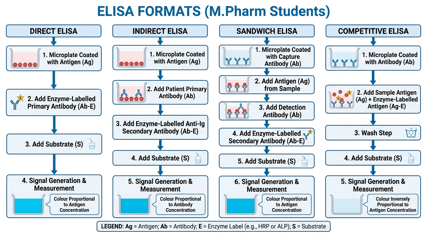
Make a flowchart of the four ELISA formats
A clear educational flowchart of the four ELISA formats for M.Pharm students, showing Direct ELISA, Indirect ELISA, Sandwich ELISA, and Competitive ELISA. Each branch should use labelled step boxes and arrows to show what is coated on the microplate, what binds next, the enzyme-labelled detection reagent, substrate addition, and how the signal relates to analyte concentration. Use scientifically accurate labels: Direct: antigen-coated plate → enzyme-labelled primary antibody → substrate → colour proportional to antigen. Indirect: antigen-coated plate → patient primary antibody → enzyme-labelled anti-Ig secondary antibody → substrate → colour proportional to antibody. Sandwich: capture antibody-coated plate → antigen from sample → detection antibody → enzyme-labelled secondary antibody → substrate → colour proportional to antigen. Competitive: antibody-coated plate + sample antigen competes with enzyme-labelled antigen → wash → substrate → colour inversely proportional to antigen. Include a simple legend: Ag = antigen; Ab = antibody; enzyme label = HRP or ALP. Ensure all text is large, fully legible, and spell-checked.