
Simple diagrams for all types of necrosis and apoptosis
necrosis AND apoptosis
necrosis patterns apoptosis pathology educational resource
A clean, simple labeled medical pathology infographic for students, organized into two panels. Panel 1 title: Types of necrosis. Show six small schematic diagrams with clear labels: coagulative necrosis (preserved tissue outlines with pale dead cells), liquefactive necrosis (liquid cavity with pus-like debris), caseous necrosis (crumbly cheese-like central material in a granuloma), fat necrosis (adipocytes with chalky calcium soap deposits), fibrinoid necrosis (blood vessel wall with bright fibrin-like deposit), and gangrenous necrosis (dry black toe and wet infected swollen foot shown as simple icon illustrations). Panel 2 title: Apoptosis. Show a cell progressing through shrinkage, chromatin condensation, membrane blebs, apoptotic bodies, then phagocyte clearance; include two small pathway arrows: intrinsic mitochondrial pathway and extrinsic death-receptor pathway, both converge on caspases. Use short labels, clear arrows, no dense paragraphs, medically accurate but easy to copy into notes.
https://www.ncbi.nlm.nih.gov/books/NBK557627
Types of necrosis
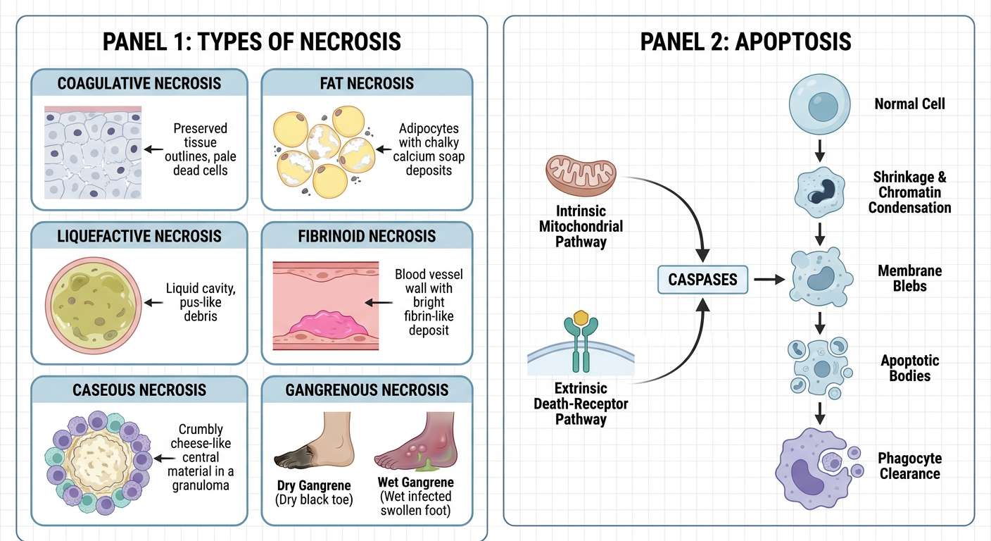
| Type | Simple visual cue | Typical example |
|---|---|---|
| Coagulative | Dead tissue architecture remains as a firm “ghost outline” | Ischemic infarct in solid organs, such as heart or kidney, except brain |
| Liquefactive | Enzymatic digestion turns tissue into a liquid or pus-filled cavity | Brain infarct; bacterial abscess |
| Caseous | Amorphous, crumbly “cheese-like” central material within a granuloma | Tuberculosis |
| Fat | Damaged fat cells plus chalky calcium deposits, called saponification | Acute pancreatitis; traumatic fat injury |
| Fibrinoid | Bright fibrin-like material deposited in a damaged vessel wall | Immune-mediated vasculitis; severe hypertension |
| Gangrenous | Clinical pattern of limb ischemia: dry, black shriveled tissue, or wet infected swollen tissue | Peripheral arterial disease, diabetes-related limb ischemia |
- Dry gangrene = ischemic coagulative necrosis
- Wet gangrene = ischemia with superimposed bacterial infection, producing liquefaction
Apoptosis
Intrinsic pathway                         Extrinsic pathway
DNA damage / growth-factor loss          Fas or TNF death receptor
             ↓                                       ↓
      BAX / BAK activation                       FADD
             ↓                                       ↓
 Cytochrome-c release from mitochondria      Caspase-8
             ↓                                       ↓
        Caspase-9 ───────────────→ Executioner caspases 3, 6, 7
                                             ↓
Cell shrinkage → chromatin condensation → membrane blebs
             → apoptotic bodies → phagocytosis
             → no significant inflammation
Easy contrast diagram
NECROSIS
Cell swelling → membrane rupture → contents spill out → inflammation

APOPTOSIS
Cell shrinkage → membrane blebbing → apoptotic bodies → phagocytosis
                                                    ↓
                                           no inflammation