
Make a sticker with named syndromes in general surgery
A colorful medical sticker sheet featuring named syndromes in general surgery, illustrated in a fun and educational style. Include the following syndromes with small cute icons or symbols next to each name: Mirizzi Syndrome (gallstone blocking bile duct), Boerhaave Syndrome (esophageal rupture), Courvoisier Sign (enlarged gallbladder), Mallory-Weiss Syndrome (esophageal tear), Ogilvie Syndrome (colonic pseudo-obstruction), Meigs Syndrome (ovarian fibroma with ascites and pleural effusion), Budd-Chiari Syndrome (hepatic vein thrombosis), Fitz-Hugh-Curtis Syndrome (perihepatitis), Chilaiditi Syndrome (colon between liver and diaphragm), Leriche Syndrome (aortoiliac occlusion). Bold colorful text labels, sticker-style with white outlines, bright pastel background, clean medical illustration aesthetic, fun educational poster format.
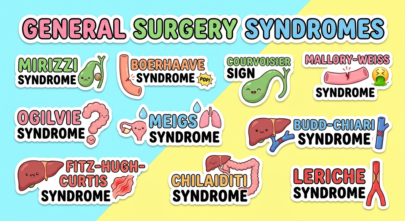
| Syndrome | Key Feature |
|---|---|
| Mirizzi Syndrome | Gallstone compresses the common hepatic duct |
| Boerhaave Syndrome | Spontaneous esophageal rupture (usually from forceful vomiting) |
| Courvoisier Sign | Painless jaundice + palpable gallbladder = periampullary malignancy |
| Mallory-Weiss Syndrome | Mucosal tear at gastroesophageal junction from retching |
| Ogilvie Syndrome | Acute colonic pseudo-obstruction without mechanical cause |
| Meigs Syndrome | Ovarian fibroma + ascites + right pleural effusion |
| Budd-Chiari Syndrome | Hepatic vein obstruction causing congestive hepatopathy |
| Fitz-Hugh-Curtis Syndrome | Perihepatitis (violin-string adhesions) from PID spread |
| Chilaiditi Syndrome | Colon interposition between liver and right hemidiaphragm |
| Leriche Syndrome | Aortoiliac occlusion: claudication + impotence + absent femoral pulses |
Hepatobilliary
A colorful medical sticker sheet featuring named syndromes and signs specific to hepatobiliary surgery, illustrated in a fun educational style with cute icons next to each name. Include: Mirizzi Syndrome (gallstone compressing common hepatic duct, icon: gallbladder with stone), Courvoisier Sign (painless jaundice + palpable gallbladder = pancreatic cancer, icon: distended gallbladder), Budd-Chiari Syndrome (hepatic vein thrombosis, icon: liver with blocked veins), Klatskin Tumor (hilar cholangiocarcinoma at bile duct bifurcation, icon: bile duct fork), Caroli Disease (intrahepatic biliary duct dilatation, icon: dilated bile ducts in liver), Charcot's Triad (fever + jaundice + RUQ pain = cholangitis, icon: three stars), Reynolds' Pentad (Charcot's triad + hypotension + confusion = severe cholangitis, icon: five stars), Kasai Procedure eponym (biliary atresia, icon: small liver), Couinaud Segments (liver anatomy segments I-VIII, icon: segmented liver diagram), Whipple's Triad (insulinoma: hypoglycemia symptoms + low glucose + relief with glucose, icon: pancreas). Bold colorful text labels, sticker-style with white outlines and drop shadows, bright pastel colors, hepatobiliary themed with liver and bile duct motifs, clean fun medical illustration.
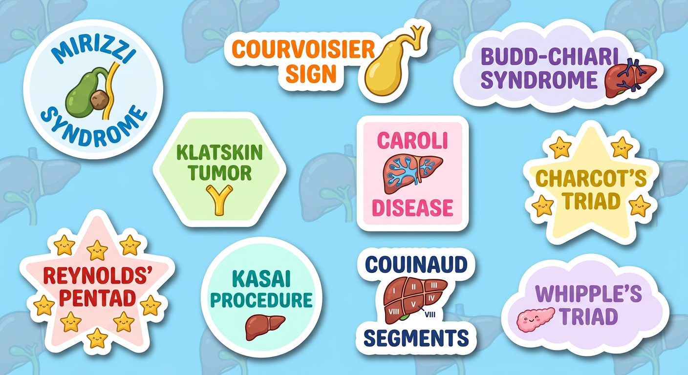
| Name | Key Concept |
|---|---|
| Mirizzi Syndrome | Large gallstone compresses the common hepatic duct causing obstructive jaundice |
| Courvoisier Sign | Painless jaundice + non-tender palpable gallbladder = periampullary malignancy (not stones) |
| Budd-Chiari Syndrome | Hepatic vein outflow obstruction - hepatomegaly, ascites, abdominal pain |
| Klatskin Tumor | Hilar cholangiocarcinoma at the bifurcation of left and right hepatic ducts |
| Caroli Disease | Congenital segmental dilatation of intrahepatic bile ducts - risk of cholangitis and stones |
| Charcot's Triad | Fever + Jaundice + RUQ pain = acute cholangitis |
| Reynolds' Pentad | Charcot's triad + Hypotension + Altered mental status = severe/suppurative cholangitis |
| Kasai Procedure | Portoenterostomy for biliary atresia - named after Morio Kasai |
| Couinaud Segments | Functional liver divided into 8 independent segments (I-VIII) based on portal and hepatic veins |
| Whipple's Triad | Hypoglycemia symptoms + blood glucose <50 mg/dL + relief with glucose = insulinoma |
Git
A colorful medical sticker sheet featuring named syndromes and signs specific to gastrointestinal tract (GIT) surgery, illustrated in a fun educational style with cute icons next to each name. Include: Boerhaave Syndrome (spontaneous esophageal rupture, icon: esophagus bursting), Mallory-Weiss Syndrome (mucosal tear at gastroesophageal junction from retching, icon: torn esophagus), Ogilvie Syndrome (acute colonic pseudo-obstruction, icon: dilated colon), Meckel's Diverticulum Rule of 2s (2 inches long, 2 feet from ileocecal valve, 2% population, icon: small bowel pouch), Hirschsprung Disease (aganglionic megacolon, icon: dilated colon with narrow segment), Peutz-Jeghers Syndrome (hamartomatous polyps + lip pigmentation, icon: spotted lips + polyp), Zollinger-Ellison Syndrome (gastrinoma causing multiple peptic ulcers, icon: stomach with ulcers), Gardner Syndrome (FAP + osteomas + desmoids, icon: colon with polyps), Plummer-Vinson Syndrome (iron deficiency + dysphagia + esophageal web, icon: esophageal web), Cronkhite-Canada Syndrome (GI polyposis + alopecia + nail dystrophy, icon: polyp with hair loss). Bold colorful text labels, sticker-style with white outlines and drop shadows, bright pastel colors, GIT themed with intestine and stomach motifs, clean fun medical illustration.
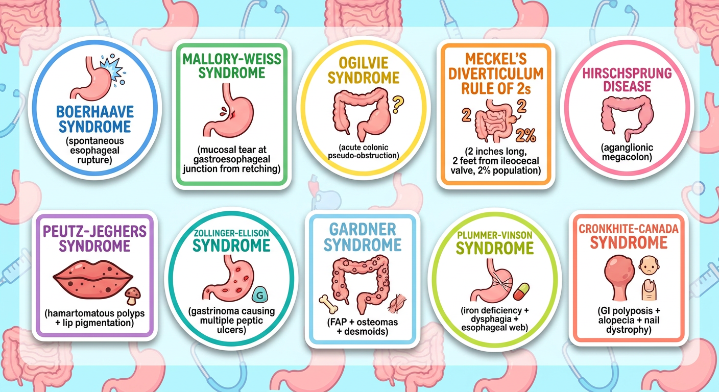
| Name | Key Concept |
|---|---|
| Boerhaave Syndrome | Spontaneous full-thickness esophageal rupture from sudden rise in intraesophageal pressure (forceful vomiting) |
| Mallory-Weiss Syndrome | Partial thickness (mucosal) tear at the gastroesophageal junction - presents with hematemesis after retching |
| Ogilvie Syndrome | Acute colonic pseudo-obstruction - massive colonic dilatation without mechanical cause, often post-op |
| Meckel's Rule of 2s | 2% population, 2 feet from ileocecal valve, 2 inches long, 2 types of ectopic mucosa (gastric/pancreatic), presents in first 2 years |
| Hirschsprung Disease | Congenital absence of ganglion cells (aganglionosis) - narrow aganglionic segment + proximal megacolon |
| Peutz-Jeghers Syndrome | Hamartomatous GI polyps + mucocutaneous melanin pigmentation (lips/fingers) - autosomal dominant, STK11 mutation |
| Zollinger-Ellison Syndrome | Gastrin-secreting tumor (gastrinoma) - refractory peptic ulcers, diarrhea, MEN-1 association |
| Gardner Syndrome | FAP variant: colorectal polyps + osteomas + desmoid tumors + epidermoid cysts - APC gene mutation |
| Plummer-Vinson Syndrome | Iron deficiency anemia + dysphagia + upper esophageal web - risk factor for esophageal/pharyngeal carcinoma |
| Cronkhite-Canada Syndrome | Non-hereditary GI polyposis + ectodermal changes (alopecia, nail dystrophy, skin pigmentation) |
Large bowel pathological and anatomical syndromes
A colorful medical sticker sheet featuring named pathological and anatomical syndromes specific to the large bowel (colon and rectum), illustrated in a fun educational style with cute icons next to each name. Include: Ogilvie Syndrome (acute colonic pseudo-obstruction, icon: massively dilated colon), Hirschsprung Disease (aganglionic megacolon with transition zone, icon: narrow segment + dilated colon), Familial Adenomatous Polyposis FAP (hundreds of colonic polyps, APC gene, icon: colon carpeted with polyps), Lynch Syndrome HNPCC (hereditary non-polyposis colorectal cancer, mismatch repair genes, icon: colon with DNA helix), Sigmoid Volvulus (twisting of sigmoid colon, coffee bean sign on X-ray, icon: twisted sigmoid), Caecal Volvulus (twisting of caecum, icon: twisted caecum), Hartmann's Procedure (sigmoid resection with end colostomy, icon: stoma bag), Anterior Resection Syndrome (bowel dysfunction after low anterior resection, icon: rectum with nerve), Chilaiditi Syndrome (colon interposition between liver and diaphragm, icon: colon between liver and diaphragm), Solitary Rectal Ulcer Syndrome (rectal ulcer with straining and mucus, icon: rectum with ulcer). Bold colorful text labels, sticker-style with white outlines and drop shadows, bright pastel colors, large bowel themed with colon and rectum anatomy motifs, clean fun medical illustration poster.
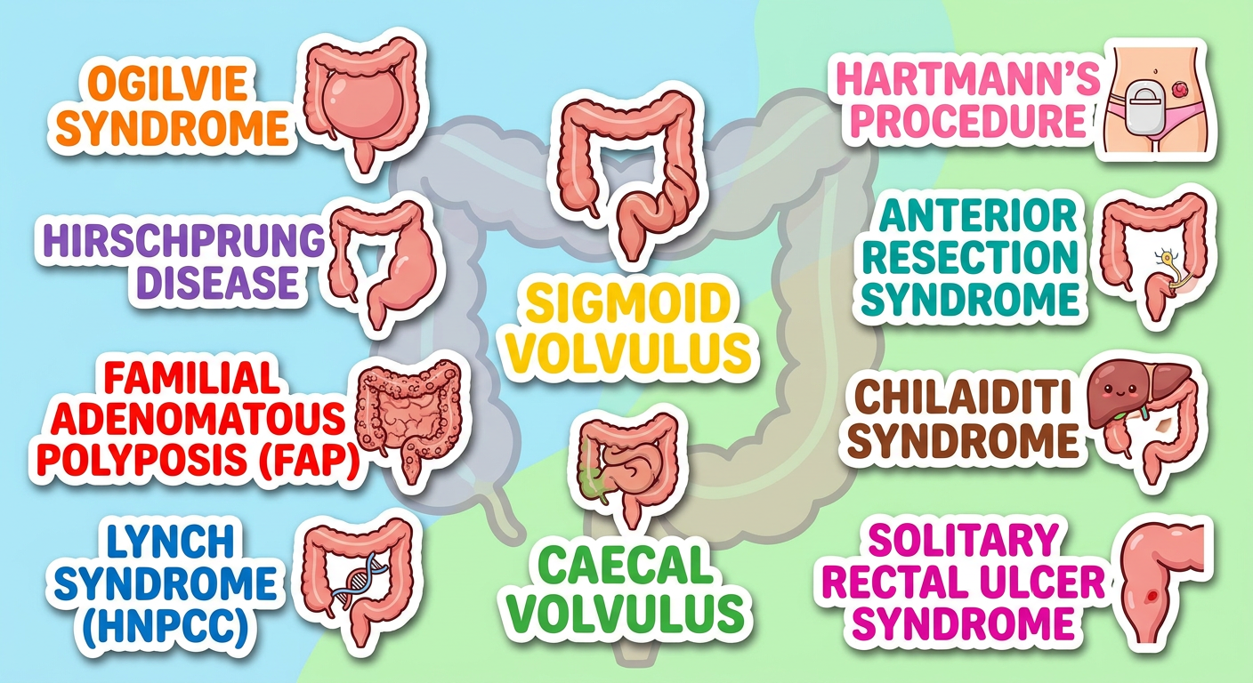
| Name | Key Concept |
|---|---|
| Ogilvie Syndrome | Acute colonic pseudo-obstruction - massive caecal/colonic dilatation without mechanical block; risk of perforation if caecum >12 cm |
| Hirschsprung Disease | Congenital aganglionosis causing a narrow non-peristaltic segment + proximal megacolon; presents with failure to pass meconium in neonates |
| FAP (Familial Adenomatous Polyposis) | Hundreds to thousands of colonic adenomas; APC gene mutation; 100% malignancy risk by age 40 if untreated - prophylactic colectomy required |
| Lynch Syndrome (HNPCC) | Most common hereditary CRC syndrome; mismatch repair gene mutations (MLH1, MSH2, etc.); right-sided colon cancers, extracolonic cancers |
| Sigmoid Volvulus | Twisting of sigmoid on its mesentery - "coffee bean" or "bent inner tube" sign on X-ray; more common in elderly/constipated patients |
| Caecal Volvulus | Axial twist or bend-fold of caecum; less common than sigmoid; "kidney bean" sign; requires surgical fixation or right hemicolectomy |
| Hartmann's Procedure | Sigmoid resection with end colostomy + rectal stump closure; used in emergency (perforated diverticulitis, obstruction) |
| Anterior Resection Syndrome | Cluster of bowel dysfunction after low/ultra-low anterior resection - urgency, frequency, incontinence, incomplete evacuation |
| Chilaiditi Syndrome | Symptomatic interposition of colon between liver and right hemidiaphragm; can mimic pneumoperitoneum on X-ray |
| Solitary Rectal Ulcer Syndrome | Chronic benign condition - rectal ulcer, mucosal prolapse, straining, mucus/blood PR; associated with internal rectal prolapse and dyssynergia |
Abdominal compartment syndrome and its classification and treatment
"abdominal compartment syndrome" AND management
A detailed colorful medical educational sticker/poster about Abdominal Compartment Syndrome (ACS) for surgery students. Include these labeled sections with icons and color coding: TOP SECTION - Title: "Abdominal Compartment Syndrome" with a large illustrated abdomen icon LEFT PANEL - "CLASSIFICATION: Intra-Abdominal Hypertension (IAH) Grades" as a color-coded table: Grade I: 12-15 mmHg (green) Grade II: 16-20 mmHg (yellow) Grade III: 21-25 mmHg (orange) Grade IV: >25 mmHg (red) ACS = sustained IAP >20 mmHg + NEW organ dysfunction MIDDLE PANEL - "TYPES" with icons: PRIMARY ACS: direct abdominal trauma, hemorrhage, surgery (trauma icon) SECONDARY ACS: massive fluid resuscitation, sepsis, burns without abdominal injury (IV drip icon) TERTIARY/RECURRENT ACS: after treated primary or secondary ACS (recycle icon) RIGHT PANEL - "ORGAN EFFECTS" with mini body diagram: Heart: decreased preload, low cardiac output Lungs: high airway pressures, hypoventilation Kidneys: oliguria, renal failure Gut: ischemia, bacterial translocation Brain: raised ICP BOTTOM LEFT - "DIAGNOSIS" box: Bladder pressure via Foley catheter 25 mL sterile saline instilled Measured at end-expiration, supine, muscles relaxed Tense abdomen + oliguria + high airway pressures BOTTOM RIGHT - "TREATMENT LADDER" with upward arrow: Step 1 (mild): Sedation, analgesia, neuromuscular blockade Step 2: Body positioning, NG/colonic decompression Step 3: Percutaneous drainage (if ascites) Step 4 (definitive): DECOMPRESSIVE LAPAROTOMY + open abdomen Monitor: Bladder pressure q4h Bright educational poster style, color coded panels, white outlines, clear fonts, surgical themed.
abdominal compartment syndrome intra-abdominal pressure classification diagram
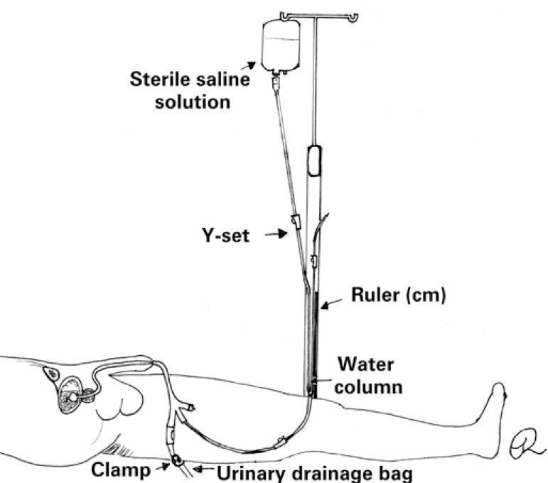
A clinical illustration demonstrating the assembly and components of the Y-set technique for intra-abdominal pressure (IAP) measurement. The diagram features a bag of sterile saline solution suspended from an IV stand, which is connected via tubing to a Y-set connector. One branch of the Y-set is attached to an indwelling urinary catheter (Foley) inserted into the patient to allow for saline instillation into the bladder. The second branch of the Y-set is positioned vertically against a centimeter ruler, creating a manometer setup with a visible water column to measure hydrostatic pressure. A clamp is clearly indicated on the tubing distal to the patient, leading toward the urinary drainage bag, which must be closed during the measurement process to ensure an accurate pressure reading. This setup illustrates a common bedside method used in critical care and surgical settings to monitor for intra-abdominal hypertension or abdominal compartment syndrome.
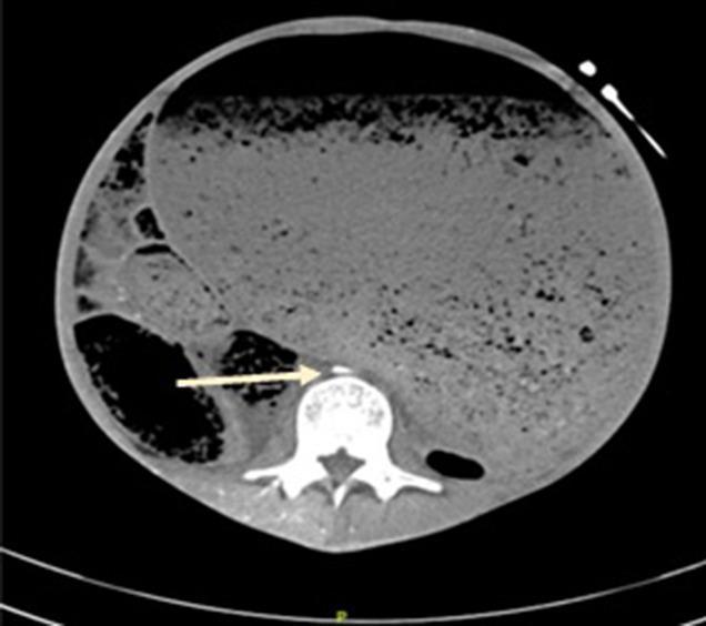
This axial computed tomography (CT) scan of the abdomen demonstrates severe abdominal compartment syndrome (ACS) resulting from massive gastric distension. The most prominent feature is a grossly dilated stomach filled with heterogeneous material and air pockets, occupying nearly the entire cross-section of the abdominal cavity. This extreme distension creates high intra-abdominal pressure, leading to significant compression of retroperitoneal vascular structures. A white arrow points to the abdominal aorta, which is severely compressed into a flattened, slit-like configuration anterior to the lumbar vertebral body. The inferior vena cava (IVC) is not visible, suggesting complete collapse due to the high extrinsic pressure. The image serves as a clinical illustration of the radiological signs of ACS, focusing on vascular compromise and visceral displacement. The anatomical relationship between the distended viscus, the spine, and the compressed major vessels is a key educational focus for understanding the pathophysiology of abdominal hypertension.
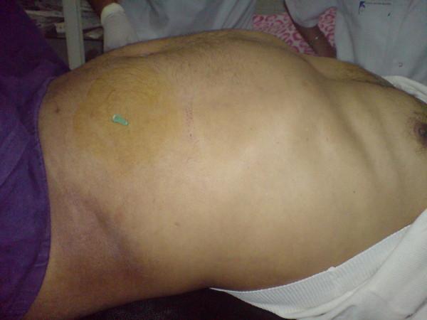
This clinical photograph displays a significantly distended and tense abdomen of a patient in a critical care setting. The abdomen shows marked protrusion, consistent with high intra-abdominal pressure seen in Abdominal Compartment Syndrome (ACS). A large, circular area of yellowish-brown skin discoloration is present on the left flank/abdominal wall, likely representing a localized antiseptic preparation or ecchymosis. Centrally within this discolored area, a 14-gauge needle (indicated by the green hub) is seen inserted percutaneously. This image illustrates the emergency bedside technique of fine-needle decompression used as a salvage procedure to relieve intra-abdominal hypertension. The photograph demonstrates the severe physical findings of abdominal tension and the immediate interventional approach to restoring hemodynamic stability in the context of critical intra-abdominal pathology.
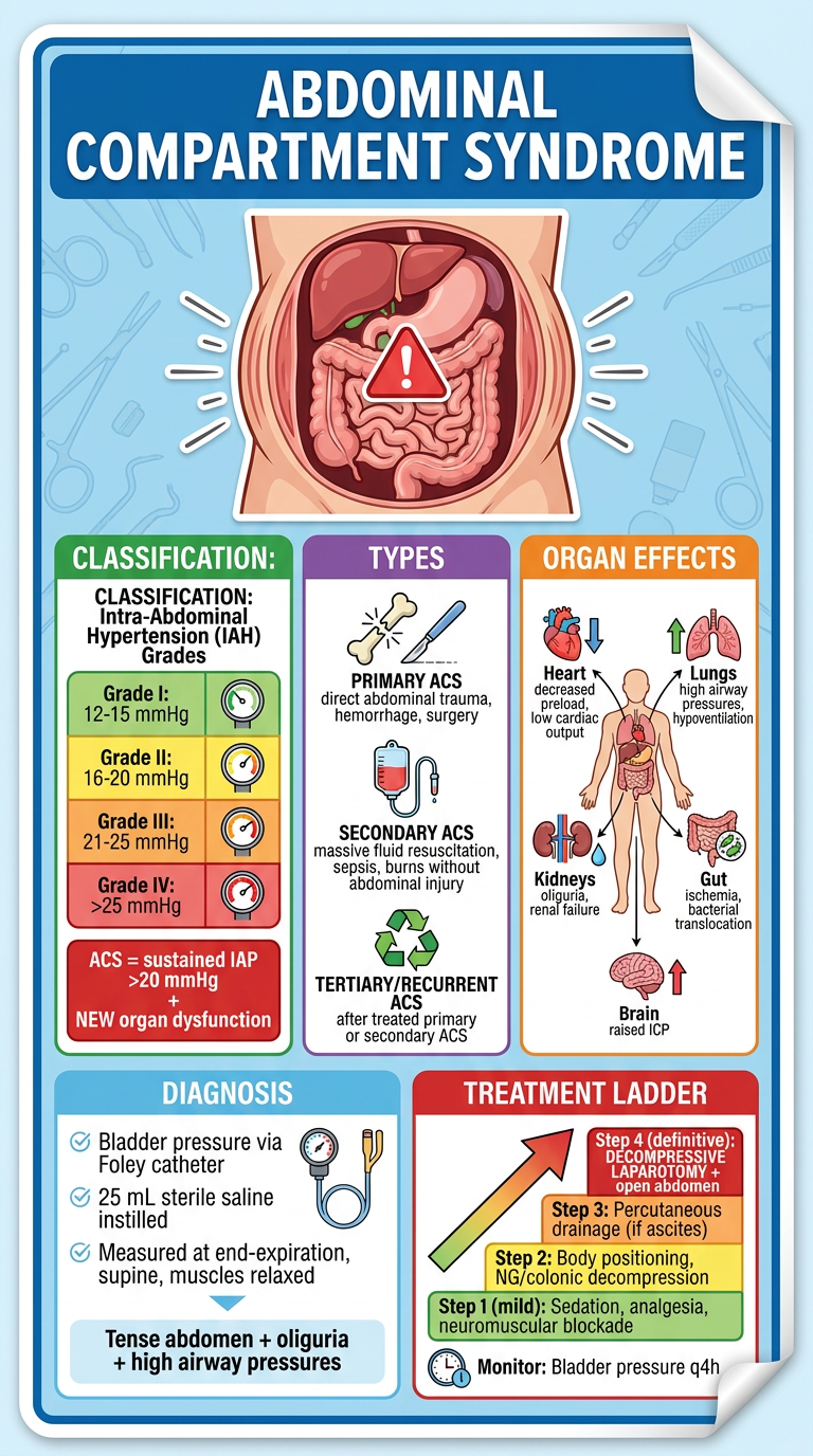
Abdominal Compartment Syndrome - Full Reference
Definition
- Intra-Abdominal Hypertension (IAH): Sustained IAP >12 mmHg
- ACS: Sustained IAP >20 mmHg with new-onset organ dysfunction or failure
- Abdominal Perfusion Pressure (APP): MAP - IAP; APP <60 mmHg signals critical ischemia
WSACS Grading of Intra-Abdominal Hypertension
| Grade | IAP (mmHg) | Clinical Action |
|---|---|---|
| I | 12-15 | Vigilant monitoring, optimize volume |
| II | 16-20 | Active non-operative treatment |
| III | 21-25 | Consider decompression |
| IV | >25 | Urgent decompressive laparotomy |
Classification by Cause
| Type | Mechanism | Examples |
|---|---|---|
| Primary ACS | Direct abdominal pathology | Trauma with hemorrhage, damage control surgery, acute pancreatitis, ruptured AAA |
| Secondary ACS | No direct abdominal injury | Massive crystalloid resuscitation, burns >60% TBSA, sepsis |
| Tertiary (Recurrent) ACS | Recurrence after treated ACS | Following previously decompressed primary or secondary ACS |
Pathophysiology - Organ Effects
| System | Effect |
|---|---|
| Cardiovascular | Impedes venous return → reduced preload and cardiac output; raised SVR |
| Respiratory | Diaphragm elevation → high airway pressures, hypoventilation, shunting |
| Renal | Direct compression of renal parenchyma + renal veins → oliguria, AKI |
| GI | Mesenteric ischemia, bacterial translocation, mucosal breakdown |
| CNS | Raised intracranial pressure (ICP) via impaired cerebral venous drainage |
Diagnosis
- Gold standard: Intravesical (bladder) pressure via Foley catheter
- Instill 25 mL sterile saline, clamp catheter, measure at end-expiration, patient supine, muscles relaxed
- Monitor every 4 hours in at-risk patients
- Unreliable in: bladder trauma, pelvic packing, neurogenic bladder, adhesions
Treatment Ladder
- Sedation and analgesia (reduces abdominal wall tension)
- Neuromuscular blockade (brief trial as temporizing measure)
- Body positioning (reverse Trendelenburg, avoid prone)
- Nasogastric and/or colonic decompression
- Diuresis / albumin-driven fluid removal once resuscitation complete
- Percutaneous drainage - if ascites/fluid is the primary cause (identified by bedside USS)
- Decompressive laparotomy - open the abdomen, release fascial tension
- Can be done at bedside in ICU to avoid transporting unstable patients
- Temporary abdominal closure (TAC) - fenestrated sterile drape + Ioban (Bogota bag, vacuum-assisted closure)
- Monitor bladder pressure q4h post-op - recurrent ACS is possible even with open abdomen
- Goal: close abdomen as fast as possible - delayed closure raises risk of SSI, fistula, ventral hernia
Prevention
- Avoid aggressive crystalloid resuscitation - use Damage Control Resuscitation (DCR): blood products early, balanced ratios
- Consider open abdomen prophylactically if >10 units PRBCs or >15 L crystalloid given at index laparotomy (Sabiston, 21e)
Compartment syndrome of upper and lower limbs and detailed theoretical description and various diagnostic and therapeutic techniques
"compartment syndrome" AND fasciotomy AND extremity
compartment syndrome leg fasciotomy incision anatomy diagram
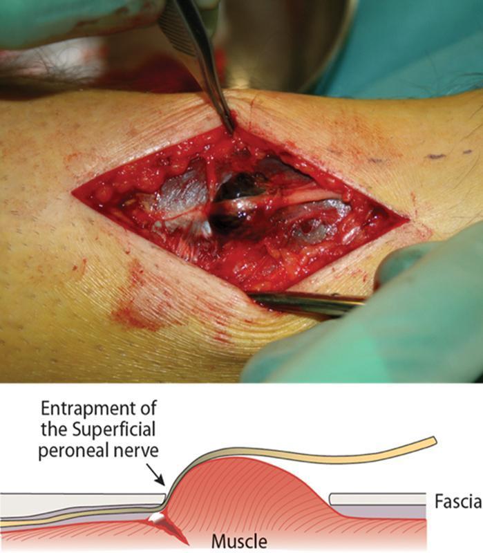
This clinical photograph and accompanying anatomical diagram illustrate an intraoperative view of the distal lateral leg during a fasciotomy for compartment syndrome. The clinical image shows a longitudinal surgical incision exposing the underlying muscle fascia and neurovascular structures. A prominent localized dark-red hematoma is visible within the surgical field, indicating acute trauma. The superficial peroneal nerve (SPN) is seen coursing through the incision site, appearing compressed and entrapped against the fascial opening. The underlying peroneus tertius muscle demonstrates significant disruption, consistent with rupture and herniation through the fascia. The anatomical diagram below the photograph provides a cross-sectional schematic of the pathology, highlighting how muscle herniation causes mechanical entrapment of the SPN against the rigid edge of the fascia. This visual material is used in surgical education to demonstrate the mechanical etiology of nerve entrapment secondary to muscle trauma and the subsequent need for surgical decompression and fasciotomy.
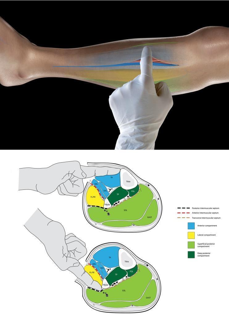
This educational graphic consists of a clinical photograph and two anatomical cross-sections illustrating the surgical technique for identifying leg compartments during a fasciotomy. The top photograph shows a gloved surgeon's finger inserted into an incision in the lower leg's fascia to palpate internal structures. Below, two schematic transverse cross-sections of the mid-leg categorize the four compartments: anterior (blue), lateral (yellow), superficial posterior (light green), and deep posterior (dark green). The top diagram demonstrates the 'tibia test': the finger enters the anterior compartment and palpates the medial tibia to confirm correct localization. The bottom diagram demonstrates the identification of the lateral (peroneal) compartment by palpating the fibula. The diagrams highlight the role of the intermuscular septa (anterior, posterior, and transverse) as physical barriers that prevent cross-compartment palpation, serving as a vital intraoperative check to ensure all compartments are adequately decompressed in cases of acute compartment syndrome.
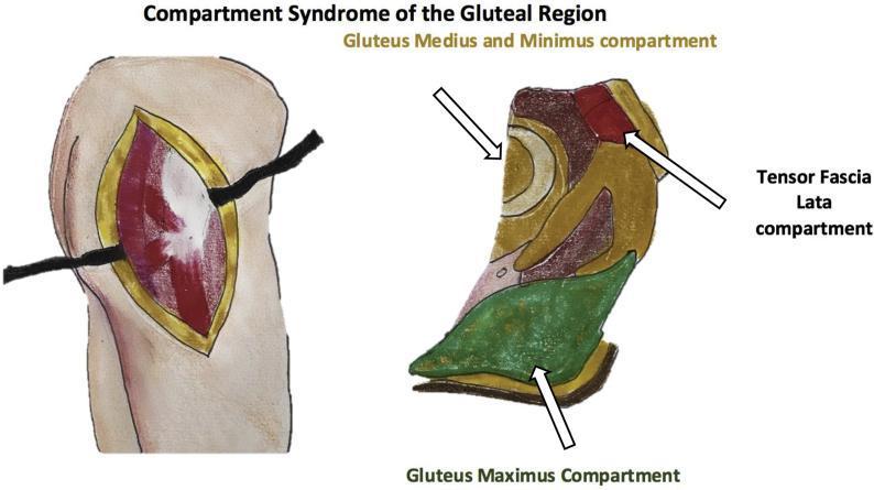
This composite medical illustration depicts the surgical and anatomical aspects of compartment syndrome in the gluteal region. The image is divided into two primary sections: a surgical clinical illustration and an anatomical diagram. On the left, a surgical illustration shows a decompressive fasciotomy of the right buttock with the patient in a lateral decubitus position. A large, linear incision through the skin and fascia exposes bulging muscle tissue, which appears edematous and reddish with focal pale areas, suggesting pressure-induced ischemia or muscle fiber disruption. On the right, a color-coded anatomical diagram identifies the three non-distensible fascial compartments of the gluteal region: the tensor fascia lata compartment (positioned anteriorly in red), the gluteus medius and minimus compartment (located centrally with a layered appearance), and the gluteus maximus compartment (positioned posteriorly in green). White arrows indicate the specific spatial boundaries and contents of each compartment. This visual serves as an educational tool for orthopedic surgery and emergency medicine, demonstrating the surgical management (fasciotomy) and the underlying regional anatomy relevant to diagnosing and treating acute gluteal compartment syndrome.
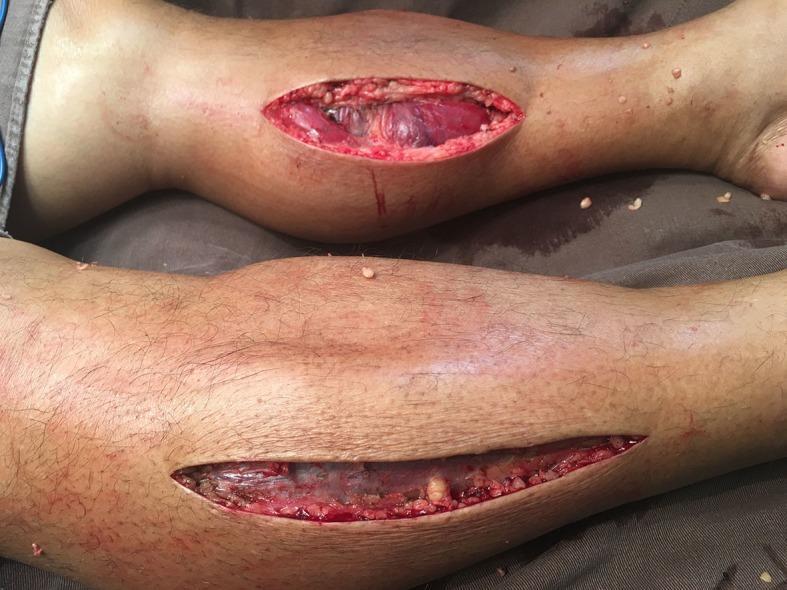
This clinical photograph displays a bilateral lower limb fasciotomy performed to treat acute compartment syndrome. The image shows two lower legs with longitudinal surgical incisions. The upper leg exhibits a wide, elliptical incision over the anterolateral aspect, revealing viable, bulging muscle tissue with a deep reddish-purple hue and no evidence of necrosis. The lower leg shows a more linear, elongated incision along the medial aspect, exposing underlying fascial planes and muscle fibers which appear paler and slightly translucent. These open incisions illustrate the surgical decompression of the four anatomical compartments of the lower leg (anterior, lateral, superficial posterior, and deep posterior). The visible extrusion of the muscle through the skin openings demonstrates the release of high intracompartmental pressure. This visual is significant for teaching emergency surgical management of compartment syndrome and recognizing healthy versus ischemic muscle tissue post-decompression.
forearm compartment syndrome fasciotomy upper limb anatomy
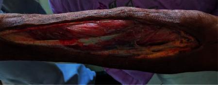
This clinical photograph displays a longitudinal fasciotomy on the left upper limb, performed for the management of compartment syndrome following brachial artery thrombosis. The large, linear surgical incision extends along the forearm, revealing underlying musculoskeletal structures. The exposed muscle tissue appears predominantly viable with a beefy red, striated texture, though some areas exhibit darker, dusky discoloration suggestive of evolving ischemia or injury. Anatomical structures visible within the wound bed include deep fascia, muscular compartments, and white longitudinal structures consistent with tendons or nerve fibers. The surrounding skin is dark and shows a somewhat dry, leathery texture with signs of underlying edema. This image serves as an educational example of emergency surgical decompression used to prevent muscle necrosis and limb loss in the setting of acute vascular compromise and subsequent compartment syndrome.
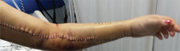
This clinical photograph displays the left upper limb of a patient on the 5th postoperative day following surgical treatment for compartment syndrome secondary to a Crotalinae (pit viper) snakebite. A long, linear fasciotomy incision is visible, extending from the distal upper arm, across the antecubital fossa, and down the length of the anterior forearm to the wrist. The wound is closed with multiple interrupted non-absorbable sutures, showing well-apposed edges with no evidence of dehiscence or purulent discharge. Significant perilesional ecchymosis and bruising are noted, with dark purple and reddish skin discoloration concentrated around the elbow and proximal forearm. The skin appears slightly shiny, suggesting resolving edema. Two small, darkened puncture marks proximal to the elbow likely represent the initial bite site. This image serves as a clinical example of postoperative recovery following emergency surgical decompression and the local dermatologic manifestations of envenomation-induced tissue injury and deep vein thrombosis.
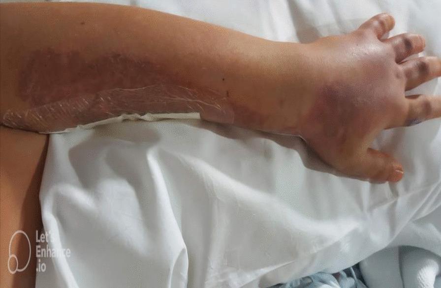
Clinical photograph of a human left upper extremity demonstrating significant postoperative skin changes following a fasciotomy for compartment syndrome. The forearm, dorsum of the hand, and fingers exhibit diffuse, pronounced edema, resulting in a tense, swollen appearance. Extensive ecchymotic patches are visible, particularly on the hand and fingers, with colors ranging from deep reddish-purple to blue-gray, indicating subcutaneous hemorrhage. A large, erythematous, and possibly necrotic-appearing lesion is present on the dorsal aspect of the proximal forearm, consistent with the site of a jellyfish sting and subsequent tissue reaction. A transparent occlusive dressing is applied over a surgical incision site on the medial aspect of the forearm. The image serves as a clinical example of the local manifestations of jellyfish venom-induced compartment syndrome and the immediate postoperative appearance after limb-saving surgical intervention.
A detailed colorful medical educational poster/sticker about Compartment Syndrome of Upper and Lower Limbs. Divided into clear color-coded sections: TITLE at top: "COMPARTMENT SYNDROME OF THE LIMBS" with a dramatic surgical icon LEFT COLUMN - UPPER LIMB (blue theme): ARM COMPARTMENTS: - Anterior arm: biceps brachii, brachialis, coracobrachialis (musculocutaneous nerve) - Posterior arm: triceps (radial nerve) FOREARM COMPARTMENTS: - Volar (flexor) compartment: superficial + deep flexors, pronators (median + ulnar nerve) - Dorsal (extensor) compartment: wrist + finger extensors (radial nerve) - Mobile wad: brachioradialis, ECRL, ECRB HAND COMPARTMENTS: 10 total (4 dorsal interosseous, 3 volar interosseous, thenar, hypothenar, adductor pollicis) FASCIOTOMY TECHNIQUE upper limb: S-shaped or curvilinear incision from antecubital fossa to wrist + carpal tunnel release MIDDLE COLUMN - DIAGNOSIS (yellow/gold theme): CLINICAL SIGNS - "6 Ps": Pain out of proportion (earliest) Pressure (tense compartment) Paresthesia (numbness/tingling) Paralysis (late sign) Pallor (late) Pulselessness (very late) PRESSURE MEASUREMENT: Normal: <10 mmHg IAH threshold: >30 mmHg FASCIOTOMY threshold: Delta P ≤30 mmHg (Diastolic BP - Compartment P) Absolute threshold: >30 mmHg DEVICES shown: Stryker handheld monitor Whitesides 3-way stopcock method Arterial line manometer Wick catheter RIGHT COLUMN - LOWER LIMB (red/orange theme): LEG COMPARTMENTS (cross-section diagram): - Anterior: tibialis anterior, EHL, EHD, EDL, deep peroneal nerve, anterior tibial artery - Lateral (peroneal): peroneus longus + brevis, superficial peroneal nerve - Superficial posterior: gastrocnemius, soleus, plantaris, sural nerve - Deep posterior: FHL, FDL, tibialis posterior, tibial nerve, posterior tibial + peroneal arteries TWO-INCISION FASCIOTOMY technique: LATERAL incision: 2cm lateral to anterior tibial border - releases anterior + lateral compartments MEDIAL incision: 1-2cm posterior to medial tibial border - releases superficial + deep posterior compartments THIGH COMPARTMENTS: - Anterior: quads, femoral nerve - Posterior: hamstrings, sciatic nerve - Medial: adductors, obturator nerve Single lateral incision through iliotibial band BOTTOM PANEL - TREATMENT ALGORITHM: Suspected ACS → Clinical exam → Delta P ≤30? → YES: IMMEDIATE FASCIOTOMY → NO: Equivocal → Serial monitoring q1h → Not improving → FASCIOTOMY Key: Leave wound OPEN, return to OR in 48-72h, delayed primary closure or split-thickness skin graft COMPLICATIONS box: Rhabdomyolysis → AKI → Volkmann contracture → amputation Bright educational poster, color coded, anatomical cross-sections, surgical diagrams, clean medical illustration style.
Compartment Syndrome of the Limbs - Detailed Reference
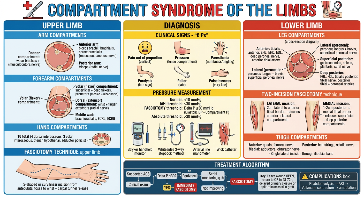
Definition & Pathophysiology
- Normal compartment pressure: <10 mmHg
- Significant elevation: >30 mmHg
- Fasciotomy threshold: Delta P ≤ 30 mmHg (Diastolic BP - Compartment Pressure)
- Muscle necrosis begins at >30 mmHg sustained for >8 hours
Classification
| Type | Features |
|---|---|
| Acute | Fractures, trauma, vascular injury, burns, revascularisation, prolonged compression |
| Chronic Exertional (CECS) | Exercise-induced; resolves with rest; common in runners, military recruits; forearm in weightlifters |
| Subacute | Gradual onset, e.g. from tight casts, circumferential dressings |
Anatomical Compartments
Lower Limb
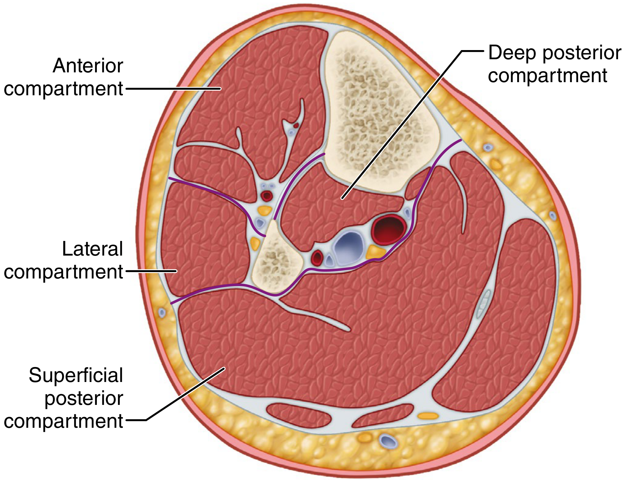
| Compartment | Muscles | Nerve | Artery |
|---|---|---|---|
| Anterior | Tibialis anterior, EHL, EDL, peroneus tertius | Deep peroneal nerve | Anterior tibial artery |
| Lateral (Peroneal) | Peroneus longus, peroneus brevis | Superficial peroneal nerve | Peroneal artery perforators |
| Superficial Posterior | Gastrocnemius, soleus, plantaris | Sural nerve | - |
| Deep Posterior | FHL, FDL, tibialis posterior | Tibial nerve | Posterior tibial + peroneal arteries |
- Anterior: quadriceps, sartorius; femoral nerve + femoral artery
- Posterior: biceps femoris, semimembranosus, semitendinosus; sciatic nerve
- Medial (adductor): adductor group; obturator nerve + profunda femoris artery
Upper Limb
- Volar (flexor): superficial flexors (FCR, FCU, FDS, PL) + deep flexors (FDP, FPL) + pronators; median + ulnar nerves; ulnar + radial arteries
- Dorsal (extensor): wrist and finger extensors; posterior interosseous nerve
- Mobile wad: brachioradialis, ECRL, ECRB; radial nerve
- Anterior: biceps brachii, brachialis, coracobrachialis; musculocutaneous nerve
- Posterior: triceps; radial nerve
Clinical Diagnosis - The "6 Ps"
| Sign | Notes |
|---|---|
| Pain out of proportion | Earliest and most important - worse on passive stretch of compartment muscles |
| Pressure (tense compartment) | Hard, wooden feel on palpation |
| Paresthesia | Type C (non-myelinated) sensory fibres are most hypoxia-sensitive; earliest neurological sign |
| Paralysis | Late sign - motor loss |
| Pallor | Late sign |
| Pulselessness | Very late sign - do NOT wait for this |
Key: Palpable pulses do NOT rule out compartment syndrome. The diagnosis should be made clinically before pulses are lost. (Bailey & Love, 28e)
Pressure Measurement Techniques
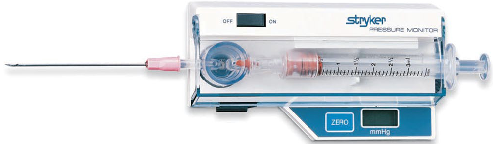
| Device | Method | Notes |
|---|---|---|
| Stryker handheld monitor | Side-port needle into compartment; digital readout in mmHg | Most widely used bedside device |
| Whitesides 3-way stopcock | Mercury manometer connected via IV tubing and saline-filled needle | Simple, low-cost bedside technique |
| Arterial line manometer | Standard arterial transducer connected to intramuscular needle | Accurate, available in ICU |
| Wick catheter / slit catheter | Soft catheter left in compartment for continuous monitoring | Best for continuous monitoring post-op or in high-risk fractures |
| Ultrasonography (emerging) | Dual-probe ultrasound for simultaneous pressure and imaging | High sensitivity/specificity in recent studies but not yet standard |
Pressure measurements are erroneous in up to 30-35% of cases and should not be used as the sole determinant for fasciotomy. Clinical examination + pressure measurement combined raises positive predictive value to 69%. (Campbell's 2026)
Diagnostic Algorithm
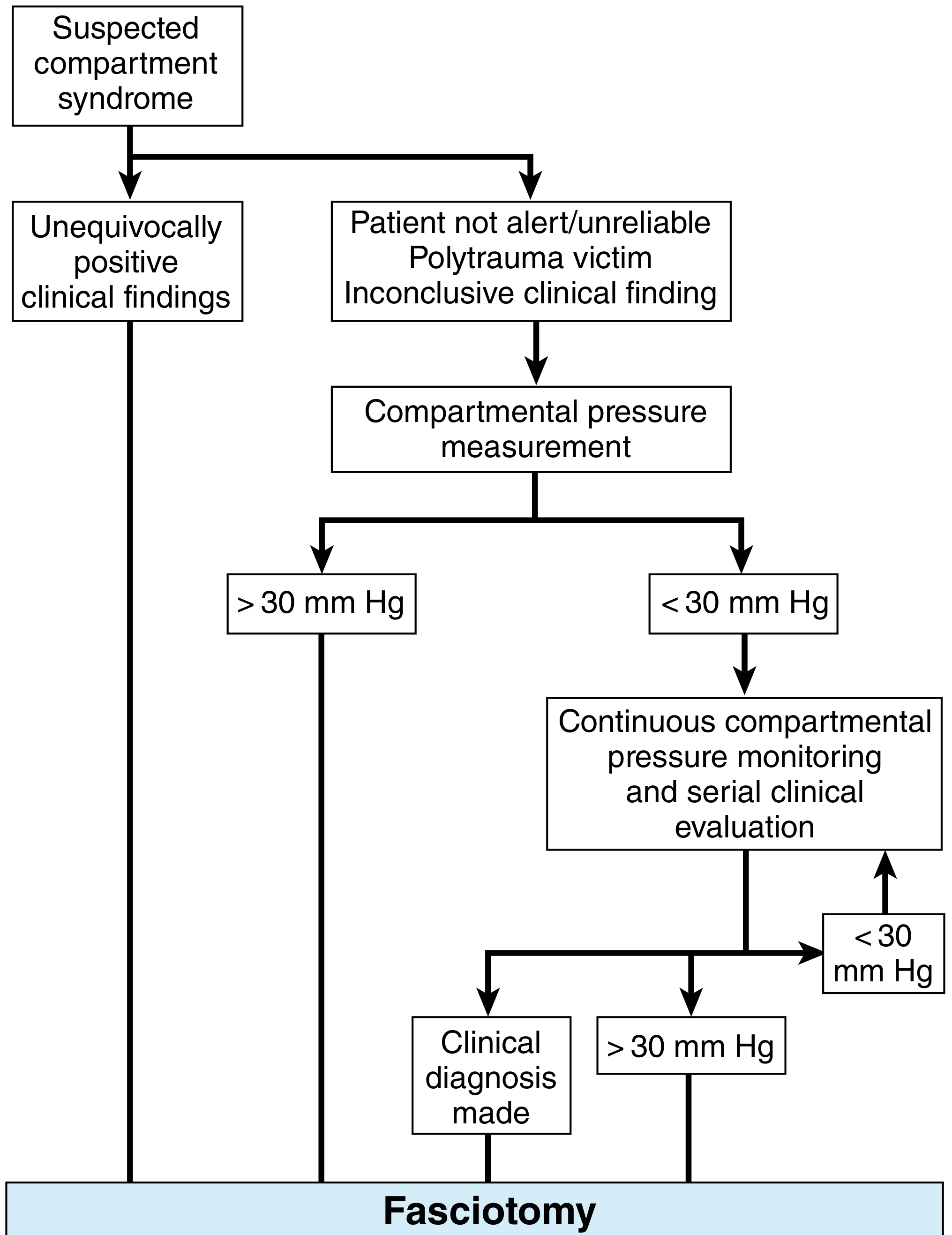
Treatment
Non-operative (First Steps)
- Remove all constricting dressings, casts, splints - can reduce compartment pressure by 50-85%
- Position limb at the level of the heart - do NOT elevate (elevation decreases arterial inflow without meaningfully improving venous outflow, worsening ischemia)
- Correct hypotension and hypoxia
- IV fluids to prevent rhabdomyolysis-related AKI
- Serial neurovascular examination every 30-60 minutes
- If no improvement within 30-60 minutes and Delta P remains ≤30 mmHg → proceed to fasciotomy
Operative: Fasciotomy
Lower Leg - Two-Incision Four-Compartment Fasciotomy
- Releases anterior and lateral (peroneal) compartments
- Incise the intermuscular septum between the two compartments using a long scissors
- Protect the superficial peroneal nerve (exits fascia ~12 cm above the lateral malleolus)
- Releases superficial posterior and deep posterior compartments
- Must incise the deep posterior fascia behind the soleus bridge (most commonly missed step)
- Protect the saphenous vein and nerve anteriorly
Thigh - Lateral Single-Incision Fasciotomy (Tarlow Technique)
- Lateral incision from just distal to the intertrochanteric line to the lateral epicondyle
- Incise the iliotibial band in line with the skin incision
- Reflect vastus lateralis off the lateral intermuscular septum - coagulate perforators
- Make a 1.5 cm incision in the lateral intermuscular septum; extend proximally and distally with Metzenbaum scissors
- This releases the anterior and posterior compartments
- Measure medial (adductor) compartment - if elevated, add a separate medial incision
Forearm - Volar + Dorsal Fasciotomy
- Volar (Henry) incision: S-shaped or curvilinear from antecubital fossa, across the wrist, continuing distally to include carpal tunnel release - releases volar compartment and mobile wad
- Dorsal incision: Straight longitudinal incision over dorsal forearm - releases dorsal extensor compartment
- Extend proximally into anterior arm compartment if biceps/brachialis are involved
Wound Management After Fasciotomy
- Wounds are left open - pack loosely with saline-soaked gauze or negative pressure wound therapy (NPWT/VAC)
- Return to operating room in 48-72 hours for reassessment and debridement
- Delayed primary closure once swelling resolves (usually day 5-7)
- If fasciotomy wounds cannot be closed primarily: split-thickness skin graft (STSG)
- Vacuum-assisted closure (VAC) devices help reduce edema and facilitate skin edge approximation
Postoperative Management & Complications
| System | Complication | Management |
|---|---|---|
| Renal | Rhabdomyolysis → AKI (myoglobinuria) | IVF to urine output >100 mL/hr until CPK <5000 U/L; urine alkalinisation with NaHCO₃ |
| Metabolic | Hyperkalemia, acidosis, hypocalcemia | Correct electrolytes; monitor ECG |
| Infection | Wound infection, osteomyelitis | Debridement, antibiotics |
| Neurological | Persistent paresthesia, motor deficit | Nerve decompression; physio/OT |
| Contracture | Volkmann's ischemic contracture (forearm) | If missed - tendon lengthening, muscle slide procedures, eventual reconstruction |
| Amputation | Delayed or missed diagnosis | Avoid by early fasciotomy |
Key Rules to Remember
- Do not wait for pulselessness - this is a pre-amputation finding
- Do not elevate the limb - keep at heart level
- Delta P ≤ 30 mmHg = fasciotomy now (AAOS guideline)
- Always decompress the deep posterior compartment of the leg - it is the most missed
- Include carpal tunnel release in all forearm fasciotomies
- Fasciotomy after 12 hours carries a dramatically worse prognosis
- Bilateral fasciotomy should be performed liberally after prolonged limb ischemia/revascularization
Retro peritoneal necrotising fasciitis
"retroperitoneal necrotizing fasciitis"
necrotising fasciitis retroperitoneal CT scan gas fascial planes
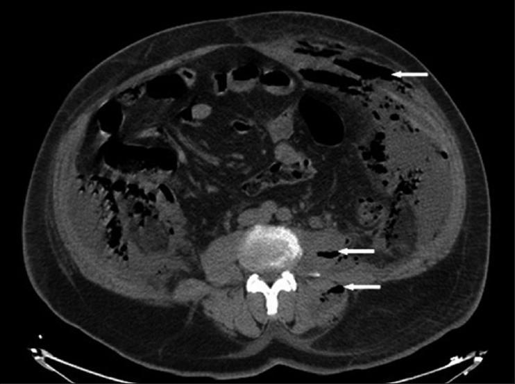
This diagnostic axial computed tomography (CT) scan of the lower abdomen and retroperitoneum demonstrates extensive scattered gas collections (indicated by white arrows) characteristic of necrotizing fasciitis. The hypodense (dark) gas pockets are visible within multiple anatomical compartments, including the left psoas muscle, latero-vertebral muscles, and the fascial planes. Significant subcutaneous emphysema is also noted in the lower abdominal wall. The gas appears to dissect through muscle layers and fascial boundaries, causing architectural distortion and tissue separation. These findings are clinically significant as they indicate a fulminant soft-tissue infection or retroperitoneal-necrotizing fasciitis, often secondary to intra-abdominal or urological suppuration like pyonephrosis. The presence of gas along the retroperitoneal structures and abdominal wall highlights the invasive nature of the disease and serves as a critical radiological marker for urgent surgical debridement.
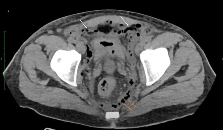
Axial non-contrast computed tomography (CT) scan of the pelvis demonstrating extensive gas collections in multiple compartments. Intraperitoneal gas (pneumoperitoneum) is visible as small, well-defined black bubbles (white arrows) located predominantly along the anterior abdominal wall and between visceral structures. Substantial retroperitoneal gas (orange arrows) is also evident, dissecting along the fascial planes and posterior pelvic structures. Key anatomical landmarks include the iliac wings and the distended pelvic cavity. These findings represent a significant cephalad extension of necrotizing fasciitis (Fournier's gangrene), where gas-producing organisms have tracked from the perineum into the abdominal and retroperitoneal spaces. This diagnostic image is used to illustrate the severity of soft-tissue infections and the importance of cross-sectional imaging in assessing surgical debridement margins.
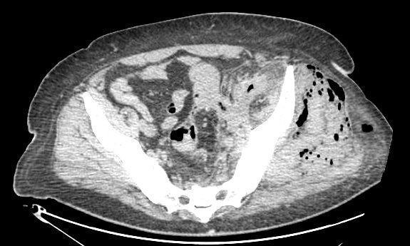
This diagnostic image is an axial computed tomography (CT) scan of the pelvis demonstrating significant pathology within the soft tissues. The most prominent finding is the presence of extensive gas collections (emphysema) within the left gluteal and left psoas musculature, appearing as multiple low-attenuation (dark) punctate foci and larger confluent pockets. The gas is seen dissecting through the muscle fibers and along the fascial planes, which is a hallmark finding of necrotizing fasciitis or gas-forming infections. There is associated swelling and distortion of the normal muscular architecture on the left side compared to the right. The scan also shows evidence of retroperitoneal involvement, with gas extending along the psoas muscle. This visual information is critical for diagnosing surgical emergencies like necrotizing soft tissue infections (NSTI) and determining the extent of required debridement. The imaging provides anatomical context for the spread of sepsis from retroperitoneal sources, such as perforated diverticulitis, into the musculoskeletal compartments of the pelvis and lower limb.
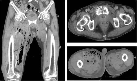
Diagnostic Image: This composite shows a computerized tomography (CT) scan in coronal (left) and axial (right top and bottom) views, focusing on the pelvis and lower extremities. The images demonstrate extensive subcutaneous and deep soft tissue gas (emphysema), characterized by multiple low-attenuation (black) bubbles and streaks. These gas pockets are primarily localized to the right femoral region, tracking along fascial planes, and extending into the retroperitoneal space. A significant skeletal abnormality is visible in the upper axial slice, showing a fracture and cortical disruption of the pubic bone/symphysis. The soft tissues exhibit marked swelling, distortion, and loss of normal muscle planes, with surrounding increased density suggestive of edema and inflammation. These findings are highly characteristic of a severe, necrotizing soft tissue infection (necrotizing fasciitis) potentially secondary to osteomyelitis or urinary fistulation. Key educational concepts include the identification of gas-forming infections, recognition of fascial plane disruption, and the evaluation of pelvic fractures in the context of surrounding soft tissue pathology.
PMID: 38382145
PMID: 34664780
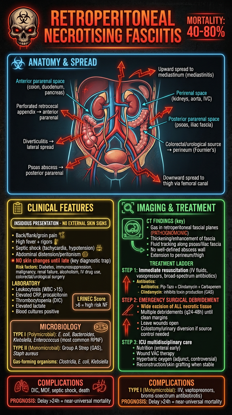
A dramatic, detailed medical educational poster about Retroperitoneal Necrotising Fasciitis (RPNF) for surgery students. Dark background with urgent red/orange color scheme conveying danger and urgency. TITLE: "RETROPERITONEAL NECROTISING FASCIITIS" with a skull-warning icon and "MORTALITY: 40-80%" in red LEFT PANEL - ANATOMY & SPREAD (blue outline, anatomy diagram): Retroperitoneal spaces labeled: - Anterior pararenal space (colon, duodenum, pancreas) - Perirenal space (kidneys, aorta, IVC) - Posterior pararenal space (psoas, iliac fascia) Spread pathways shown with red arrows: - Perforated retrocecal appendix → anterior pararenal - Diverticulitis → lateral spread - Psoas abscess → posterior pararenal - Colorectal/urological source → perineum (Fournier's) - Upward spread to mediastinum (mediastinitis) - Downward spread to thigh via femoral canal MIDDLE PANEL - CLINICAL FEATURES (yellow): "INSIDIOUS PRESENTATION - NO EXTERNAL SKIN SIGNS" Symptoms: - Back/flank/groin pain - High fever + rigors - Septic shock (tachycardia, hypotension) - Abdominal distension/peritonism - NO skin changes until late (key diagnostic trap) Risk factors: Diabetes, immunosuppression, malignancy, renal failure, alcoholism, IV drug use, colorectal/urological surgery LABORATORY: - Leukocytosis (WBC >15) - Elevated CRP, procalcitonin - Thrombocytopenia (DIC) - Elevated lactate - Blood cultures positive LRINEC Score box: >6 = high risk NF MICROBIOLOGY box: TYPE I (Polymicrobial): E. coli, Bacteroides, Klebsiella, Enterococcus (most common RPNF) TYPE II (Monomicrobial): Group A Strep (GAS), Staph aureus Gas-forming organisms: Clostridia, E. coli, Klebsiella RIGHT PANEL - IMAGING & TREATMENT (green/red): CT FINDINGS (key): - Gas in retroperitoneal fascial planes (PATHOGNOMONIC) - Thickening/enhancement of fascia - Fluid tracking along psoas/iliac fascia - No well-defined abscess wall - Extension to perineum/thigh TREATMENT LADDER (urgent upward arrows): STEP 1: Immediate resuscitation (IV fluids, vasopressors, broad-spectrum antibiotics) Antibiotics: Pip-Tazo + Clindamycin + Carbapenem Clindamycin: inhibits toxin production (GAS) STEP 2: EMERGENCY SURGICAL DEBRIDEMENT - Wide excision of ALL necrotic tissue - Multiple debridements (q24-48h) until clean margins - Leave wounds open - Colostomy/urinary diversion if source control needed STEP 3: ICU multidisciplinary care - Nutrition (enteral early) - Wound VAC therapy - Hyperbaric oxygen (adjunct, controversial) - Reconstruction/skin grafting when stable BOTTOM: COMPLICATIONS box - DIC, MOF, septic shock, death PROGNOSIS: Delay >24h = near-universal mortality Bright urgent poster style, anatomical retroperitoneal diagram, CT scan icon, clear fonts, surgical emergency theme.
retroperitoneal necrotizing fasciitis management LRINEC score CT diagnosis 2023 2024
Retroperitoneal Necrotising Fasciitis (RPNF)
Definition
"Rarely, patients may develop necrotizing fasciitis of the retroperitoneum, a condition with high mortality." - Schwartz's Principles of Surgery, 11e
Anatomy of the Retroperitoneum - Why It Matters
| Space | Contents | Infection source |
|---|---|---|
| Anterior pararenal | Ascending/descending colon, duodenum, pancreas | Perforated retrocecal appendix, diverticulitis, duodenal perforation, pancreatitis |
| Perirenal | Kidneys, adrenal glands, aorta, IVC | Pyelonephritis, pyonephrosis, perinephric abscess |
| Posterior pararenal | Psoas, iliac fascia, preperitoneal fat | Psoas abscess, vertebral osteomyelitis, iliopsoas spread |
- The mediastinum superiorly (via aortic/esophageal hiatus) - allowing upward spread causing mediastinitis
- The perineum and thigh inferiorly via the femoral canal and inguinal ligament - giving rise to Fournier's gangrene
- The anterior abdominal wall via preperitoneal fat continuity
Aetiology and Sources
| Source | Mechanism |
|---|---|
| Colorectal (most common) | Perforated retrocecal appendix, diverticulitis, anastomotic leak, colorectal carcinoma |
| Urological | Pyonephrosis, perinephric abscess, urinary fistula, Foley trauma |
| Vertebral/spinal | Discitis, vertebral osteomyelitis with psoas abscess |
| Pancreatic | Infected pancreatic necrosis, pancreatic abscess |
| Post-surgical | Contamination after retroperitoneal or laparoscopic surgery |
| Haematogenous | Primary bacteraemia in immunocompromised patients |
| Cryptogenic | No clear source in ~20% of cases |
Risk Factors
- Diabetes mellitus (most important - impaired neutrophil function, microvascular disease)
- Immunosuppression (steroids, chemotherapy, transplant)
- Malignancy
- Chronic renal failure
- Alcoholism / liver cirrhosis
- Obesity
- Peripheral vascular disease
- IV drug use
- Recent abdominal/urological surgery
Microbiology
| Type | Organisms | Notes |
|---|---|---|
| Type I - Polymicrobial (most common in RPNF) | E. coli, Bacteroides, Klebsiella, Enterococcus, Pseudomonas, Clostridia | Synergistic infection; anaerobes + aerobes; gas-forming |
| Type II - Monomicrobial | Group A Streptococcus (GAS), Staphylococcus aureus | Associated with exotoxin production; toxic shock syndrome |
| Type III | Vibrio vulnificus, Aeromonas (water exposure) | Rare; particularly virulent; high mortality |
Clinical Features
| Feature | Detail |
|---|---|
| Severe back, flank, or groin pain | Often the presenting complaint; may mimic renal colic |
| Fever and rigors | High-grade, rapidly progressive |
| Septic shock | Tachycardia, hypotension, altered mental status (often presents in shock) |
| Abdominal distension/peritonism | May mimic perforated viscus |
| NO skin erythema/crepitus until very late | The hallmark that makes this diagnosis so treacherous |
| Generalised peritonitis | Can mimic perforated appendicitis (as seen in the 2024 case report, PMID: 38382145) |
Diagnosis
Laboratory - LRINEC Score
| Parameter | Score |
|---|---|
| CRP ≥150 mg/L | +4 |
| WBC 15-25 ×10³/mm³ | +1; WBC >25: +2 |
| Haemoglobin 11-13.5 g/dL | +1; Hb <11: +2 |
| Sodium <135 mmol/L | +2 |
| Creatinine >141 µmol/L | +2 |
| Glucose >10 mmol/L (180 mg/dL) | +1 |
- Score ≤5: Low risk (<50%)
- Score 6-7: Moderate risk (50-75%)
- Score ≥8: High risk (>75%)
- PPV 92%, NPV 96% at cutoff of ≥6
- Critical: A score <6 does NOT rule out NF - 10% of confirmed cases score <6
Imaging - CT Abdomen/Pelvis with IV Contrast (Gold Standard)
| CT Finding | Significance |
|---|---|
| Gas in retroperitoneal fascial planes | Pathognomonic - presence of gas-forming organisms; seen along psoas, iliac fascia, pararenal spaces |
| Asymmetric fascial thickening | Inflammation tracking along fascia |
| Lack of fascial enhancement post-contrast | Most specific finding - indicates fascial necrosis and absent perfusion |
| Fluid tracking along psoas/iliac fascia | Extensive spread; correlates with extent of debridement needed |
| No well-defined abscess wall | Distinguishes from simple abscess - there is no capsule |
| Extension to perineum/thigh | Indicates Fournier's gangrene component |
| Pneumoperitoneum + retroperitoneal gas | Suggests bowel perforation as source |
Operative Findings (Intraoperative Diagnosis)
- Easy finger dissection along fascial planes - the "finger test"; the fascia separates readily with minimal resistance (hallmark of NF)
- Grey/brown/black necrotic fascia and fat - no bleeding when cut
- Absence of bleeding from fascial edges
- Foul-smelling grey "dishwater" fluid in the retroperitoneal space
- Bowel/visceral wall still viable initially but surrounded by necrotic fascia
Treatment
1. Immediate Resuscitation (Simultaneous with Surgical Preparation)
- IV fluid resuscitation; vasopressors if in shock
- Blood cultures x2 before antibiotics (do NOT delay antibiotics for this)
- Urinary catheter and strict monitoring
- ICU admission/HDU monitoring
- Correct coagulopathy (FFP, platelets if DIC)
2. Empirical Broad-Spectrum Antibiotics - Start IMMEDIATELY
| Regimen | Rationale |
|---|---|
| Piperacillin-tazobactam (4.5g IV q6-8h) | Gram-negative + anaerobic cover |
| Clindamycin (900mg IV q8h) | Toxin suppression - inhibits exotoxin production by GAS; maintains efficacy even in high bacterial loads |
| Carbapenem (meropenem/imipenem) | For resistant organisms; add if septic shock or recent healthcare contact |
| Add Vancomycin/Linezolid | If MRSA suspected |
3. EMERGENCY SURGICAL DEBRIDEMENT (Definitive Treatment)
- Midline laparotomy (most common) - allows full retroperitoneal exploration
- Retroperitoneal access via medial visceral rotation (Mattox or Cattel-Braasch) to access all retroperitoneal spaces
- Assess source and perform source control first: right hemicolectomy, Hartmann's, urostomy as needed
- Radical excision of ALL necrotic tissue - until healthy, bleeding tissue is reached at all margins
- Extension of debridement as far as the infection tracks - if it reaches the thigh, perform thigh fasciotomy and debridement
- Leave all wounds completely open - no primary closure
- Peritoneal lavage with warm saline
- Temporary abdominal closure (TAC/Bogota bag) for open abdomen management
4. Post-operative ICU Management
- Ventilatory support if needed
- Vasopressors (noradrenaline is first line)
- Early enteral nutrition within 24-48h (massive catabolic state)
- Wound negative pressure therapy (VAC) once wounds are clean
- Serial CRP, WBC, LRINEC score follow-up to guide re-look timing
- Renal replacement therapy if AKI develops
5. Adjuncts
| Therapy | Evidence |
|---|---|
| Hyperbaric oxygen (HBO) | Controversial; some benefit in Type I infections; impractical in unstable patients; used as adjunct only in stable survivors |
| IVIG | May benefit Type II (GAS) by neutralising superantigens; insufficient evidence for routine use |
| Maggot therapy | For wound bed preparation in clean chronic wounds post-debridement |
6. Reconstruction
- Once infection controlled and wounds clean (typically 2-4 weeks)
- Split-thickness skin grafting (STSG) for large defects
- Tissue flaps for complex reconstruction if perineum/groin involved
- Stoma reversal considered 3-6 months later if clinically stable
Complications
| Complication | Mechanism |
|---|---|
| Septic shock / Multi-organ failure | Endotoxin + exotoxin release, microcirculatory failure |
| DIC | Massive tissue destruction, endothelial activation |
| Mediastinitis | Upward spread via aortic/diaphragmatic hiatus |
| Fournier's gangrene | Downward perineal spread |
| Enteroatmospheric fistula | Open abdomen management complication |
| Massive hernias | After open abdomen; requires delayed reconstruction |
| AKI/renal failure | Sepsis + rhabdomyolysis |
| Death | 40-80% mortality despite optimal treatment |
Prognosis
- Overall mortality: 40-80% (higher in retroperitoneal vs. peripheral NF)
- Delay of >24 hours to surgery: near-universal mortality
- Independent predictors of death: age >60, diabetes, delayed surgery, polymicrobial infection, shock on presentation, renal failure, extent of involvement
- Survival is directly linked to speed and completeness of debridement and multidisciplinary ICU management (Fairweather et al., ANZ J Surg 2022, PMID: 34664780)
Key Teaching Points
- No skin signs ≠ no NF - retroperitoneal NF is invisible from outside
- CT with IV contrast is mandatory in any septic patient with back/flank pain + risk factors
- Gas in retroperitoneal fascial planes on CT = emergency laparotomy NOW
- LRINEC ≥6 in the right clinical context = high suspicion; act surgically
- Antibiotics alone will not cure this - surgery is the only definitive treatment
- Multiple re-look debridements are the standard, not just one operation
- Source control (bowel/urinary diversion) is as important as debridement
- Clindamycin must be included for toxin suppression even if GAS not confirmed