
Flowchart of pathophysiology of psoriasis
psoriasis pathophysiology diagram keratinocyte T cell immune
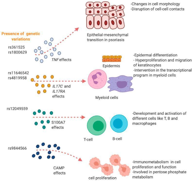
A pathophysiology diagram illustrating the impact of specific regulatory single nucleotide polymorphisms (rSNPs) on cellular processes involved in psoriasis. The diagram maps genetic variations (rs361525, rs1800629, rs11646542, rs4819958, rs12049559, and rs9844566) to downstream clinical effects across four major biological axes. 1) TNF effects lead to epithelial-mesenchymal transition (EMT), characterized by altered cell morphology and disrupted cell-cell contacts. 2) IL17C and IL17RA effects impact the epidermis through epidermal differentiation, keratinocyte hyperproliferation/migration, and transcriptional changes in myeloid cells. 3) S100A7 effects influence the development and activation of T-cells, B-cells, and macrophages. 4) CAMP effects modulate immunometabolism in proliferating cells and the pentose phosphate pathway. Anatomical structures shown include a stratified epidermis and various immune cell types (T-cell, B-cell, myeloid cells). This educational illustration serves to link dermatological genomics with observed cellular pathology and immune dysregulation in psoriatic disease.
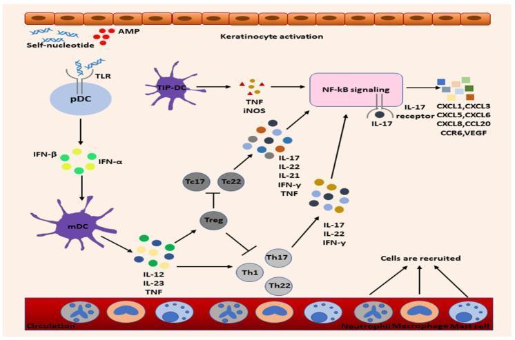
This pathophysiology diagram illustrates the complex immune signaling pathways in psoriasis. The process begins with keratinocyte activation and the release of antimicrobial peptides (AMPs) and self-nucleotides, which trigger plasmacytoid dendritic cells (pDC) via Toll-like receptors (TLR). These pDCs secrete IFN-α/β, activating myeloid dendritic cells (mDC). Activated mDCs release pro-inflammatory cytokines, including IL-12, IL-23, and TNF, which drive the differentiation of T helper cells (Th1, Th17, Th22) and cytotoxic T cells (Tc17, Tc22). These T cells produce IL-17, IL-22, and IFN-γ, which activate NF-κB signaling pathways. NF-κB activation further induces keratinocytes to release a broad spectrum of chemokines (CXCL1, CXCL3, CXCL5, CXCL6, CXCL8, CCL20, CCR6) and VEGF. This molecular cascade leads to the recruitment of neutrophils, macrophages, and mast cells from systemic circulation into the skin. Regulatory T cells (Treg) are shown providing inhibitory feedback on the T cell populations. The diagram serves as an educational summary of the cytokine-driven feed-forward loop that sustains chronic skin inflammation.
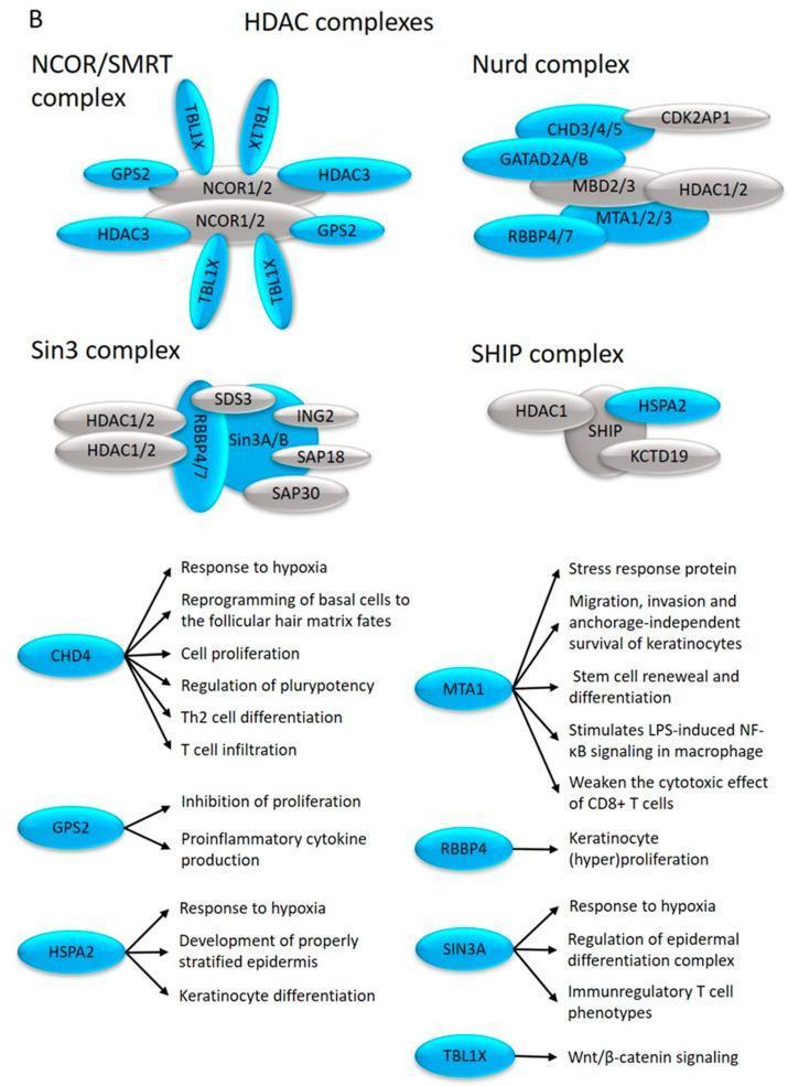
This medical pathophysiology diagram illustrates the molecular architecture and functional pathways of Class I Histone Deacetylase (HDAC) complexes relevant to skin pathology, particularly psoriasis. The top section depicts four multi-protein complexes: the NCOR/SMRT complex (composed of NCOR1/2, GPS2, HDAC3, and TBL1X), the Nurd complex (CHD3/4/5, GATAD2A/B, MBD2/3, MTA1/2/3, and RBBP4/7), the Sin3 complex (Sin3A/B, HDAC1/2, RBBP4/7, and various SAP/SDS3 subunits), and the SHIP complex (HDAC1, SHIP, HSPA2, and KCTD19). The bottom section functions as a signaling flowchart, mapping individual subunits to specific clinical and cellular processes. Key pathways include CHD4 regulating keratinocyte proliferation and Th2 differentiation; MTA1 influencing CD8+ T cell effects and macrophage NF-̄kB signaling; and SIN3A modulating epidermal differentiation and immune phenotypes. The diagram highlights the clinical significance of these epigenetic regulators in modulating inflammation, hypoxia responses, and epidermal homeostasis.
psoriasis pathogenesis
Pathophysiology of Psoriasis - Flowchart
1. TRIGGER
   Genetic susceptibility (HLA-Cw6, PSORS1 locus, >85 non-MHC risk loci)
   + Environmental trigger (skin trauma/Koebner phenomenon, streptococcal
     infection, drugs, stress)
        |
        v
2. KERATINOCYTE/INNATE ACTIVATION
   Injured or stressed keratinocytes release self-DNA/RNA and
   antimicrobial peptides (e.g., LL-37/cathelicidin)
        |
        v
3. PLASMACYTOID DENDRITIC CELL (pDC) ACTIVATION
   Self-nucleic acid-LL37 complexes engage Toll-like receptors (TLR7/9) on pDCs
        |
        v
4. TYPE I INTERFERON RELEASE
   pDCs secrete IFN-alpha/beta -> activates dermal myeloid dendritic cells (mDCs)
        |
        v
5. DENDRITIC CELL CYTOKINE OUTPUT
   Activated mDCs produce IL-23, IL-12, and TNF-alpha
        |
        v
6. T-CELL DIFFERENTIATION (adaptive amplification)
   - IL-23 + TGF-beta -> drives Th17 and Tc17 cell development
   - IL-12 -> drives Th1 cells (IFN-gamma)
   - Net effect: expansion of Th1/Th17/Th22 and cytotoxic Tc17/Tc22 cells
        |
        v
7. EFFECTOR CYTOKINE RELEASE
   T cells secrete IL-17A, IL-22, IFN-gamma, TNF-alpha
        |
        v
8. KERATINOCYTE RESPONSE (the "feed-forward loop")
   IL-17 (TNF-alpha-augmented) activates NF-kB and STAT3 signaling in
   keratinocytes, causing:
     - Hyperproliferation and abnormal/accelerated differentiation
       (parakeratosis, thickened epidermis - acanthosis)
     - Release of antimicrobial peptides, chemokines (CXCL1, CXCL3, CXCL5,
       CXCL8, CCL20) and VEGF
        |
        v
9. INFLAMMATORY CELL RECRUITMENT
   Chemokines/VEGF recruit neutrophils (-> Munro microabscesses),
   macrophages, mast cells, and more T cells into the dermis/epidermis;
   VEGF drives dermal angiogenesis (visible as pinpoint bleeding -
   Auspitz sign)
        |
        v
10. SELF-PERPETUATING CYCLE
    Recruited neutrophils and activated keratinocytes further stimulate
    dendritic cells and T cells, sustaining chronic plaque formation
    (regulatory T cells normally provide inhibitory feedback but this is
    defective/overwhelmed in psoriasis)
        |
        v
CLINICAL RESULT: erythematous plaques with silvery scale, epidermal
thickening, and (in a subset) psoriatic arthritis via similar
IL-23/IL-17/TNF-driven inflammation in entheses/joints
- Goldman-Cecil Medicine, "Pathobiology" (psoriasis), p. 3261
- Fitzpatrick's Dermatology, "Psoriasis" pathogenesis discussion, pp. 2789-2797