
Erytropisis 10 mark physiology
I'll look up erythropoiesis from the medical library for a comprehensive 10-mark answer.
Reading File
Finding Sources
Finding Sources
Good - I found relevant sections. Let me read the most relevant physiology-focused content from Histology and Biochemistry books, plus any Guyton/physiology textbook content.
Reading File
Reading File
Finding Sources
Now let me read the histology textbook's dedicated erythropoiesis section and fetch the diagram:
Reading File
Loading Image
Finding Sources
Now let me get the dedicated histology erythropoiesis section and the Kinetics of Erythropoiesis:
Reading File
Finding Sources
Let me find the actual erythropoiesis development section in the histology textbook:
Finding Sources
Finding Sources
Finding Sources
Excellent! Found the key histology plate. Let me read the full erythropoiesis section:
Reading File
Reading File
I now have all the information needed for a thorough 10-mark answer. Let me also fetch the erythropoiesis plate image:
Loading Image
I now have comprehensive information to write a detailed 10-mark physiology answer on erythropoiesis.
Erythropoiesis (10 Marks)
Definition
Erythropoiesis is the process by which red blood cells (erythrocytes) are formed in a steady state to maintain their normal concentration in the peripheral blood. It begins with pluripotent stem cells in the bone marrow and ends with the release of mature erythrocytes into the circulation.
- Histology: A Text and Atlas, Plate 10.3
- Basic Medical Biochemistry, Ch. 42
Site of Erythropoiesis
- Embryonic life (yolk sac): Primitive erythropoiesis begins in the yolk sac during the first weeks of gestation.
- Fetal life: Liver and spleen are the main sites (hepatic phase).
- After birth and in adults: Red bone marrow of flat bones (sternum, ribs, vertebrae, iliac crest) and the epiphyses of long bones.
- In severe hemolytic anemias, the liver and spleen may resume erythropoiesis (extramedullary erythropoiesis).
Stages of Development (Erythroid Maturation Series)
The progression is from stem cells through committed progenitors to recognizable morphological precursors:
Stem Cell Hierarchy:
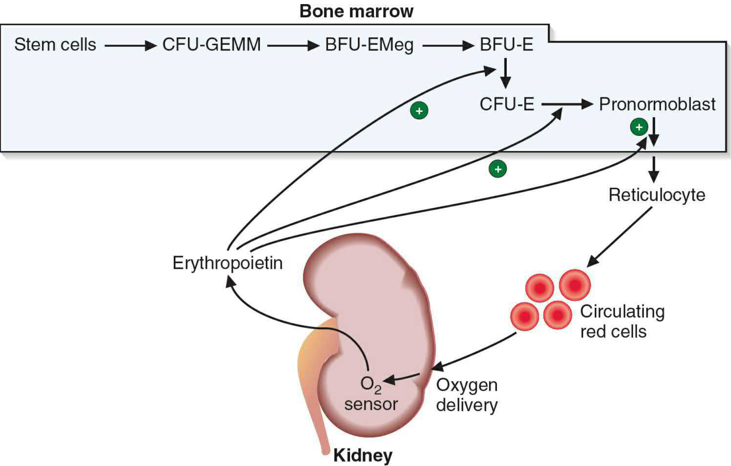
Pluripotent Stem Cell → CFU-GEMM → BFU-EMeg → BFU-E → CFU-E → Pronormoblast
Recognizable Morphological Stages (in bone marrow smear):
| Stage | Size | Nucleus | Cytoplasm | Mitosis |
|---|---|---|---|---|
| 1. Proerythroblast (Pronormoblast) | Largest (~20 µm) | Large, pale, 1-2 nucleoli | Basophilic (deep blue), no Hb | Yes |
| 2. Basophilic erythroblast | Smaller | Smaller, dense chromatin | Strong basophilia (many ribosomes synthesizing Hb) | Yes |
| 3. Polychromatophilic erythroblast | Smaller | Smaller, coarser chromatin | Blue-gray (mix of ribosomes + Hb accumulating) | Yes (last stage of active mitosis) |
| 4. Orthochromatic erythroblast (Normoblast) | ~8-10 µm | Very small, pyknotic/condensed | Pink/eosinophilic (lots of Hb, fewer ribosomes) | NO - cannot divide |
| 5. Reticulocyte (Polychromatophilic erythrocyte) | Near mature size | Nucleus extruded | Slight basophilia (residual ribosomes, still synthesizes Hb) | No |
| 6. Mature Erythrocyte | 6-8 µm, biconcave | Absent | Eosinophilic, full of Hb | No |
Key facts:
- Each normoblast undergoes 4 mitotic divisions before nucleus extrusion.
- Nuclear extrusion occurs at the orthochromatic normoblast stage.
- Reticulocytes retain ribosomes and mRNA and can still synthesize hemoglobin.
- Reticulocytes circulate for 1-2 days, maturing in the spleen where ribosomes and mRNA are lost.
- It takes approximately 1 week for a basophilic erythroblast progeny to reach the circulation.
Changes During Maturation (Progressive Trends)
- Cell size decreases progressively from proerythroblast to mature RBC.
- Nuclear size decreases and chromatin condenses - nucleus is finally extruded.
- Cytoplasm changes color from deep blue (basophilic) → blue-gray (polychromatic) → pink (orthochromatic) as hemoglobin accumulates and ribosomes decrease.
- Hemoglobin content increases progressively with each division.
- Ribosomes decrease as hemoglobin synthesis nears completion.
Regulation: Role of Erythropoietin (EPO)
Erythropoietin is the primary regulator of erythropoiesis. It is a 34 kDa glycoprotein hormone synthesized and secreted predominantly by peritubular interstitial cells of the kidney (90%) and to a lesser extent by the liver (10%).
Stimulus: Decreased oxygen delivery to the kidney (hypoxia) triggers EPO release via HIF-1α (Hypoxia-Inducible Factor).
Mechanism:
- Hypoxia/anemia → kidney O₂ sensor detects low O₂ delivery
- Kidney releases erythropoietin
- EPO acts on specific receptors (EPO-R, a cytokine receptor) on erythroid progenitors (CFU-E and BFU-E) in bone marrow
- Activates JAK2/STAT5 signaling pathway
- Promotes proliferation, differentiation, and survival of erythroid precursors
- Accelerates Hb synthesis and reticulocyte release
- Increased RBC mass → improved O₂ delivery → negative feedback reduces EPO secretion
Clinical note: In chronic kidney disease, EPO production falls - causing anemia of CKD. Recombinant EPO (epoetin alfa, darbepoetin) is used therapeutically.
Requirements for Erythropoiesis
| Requirement | Role |
|---|---|
| Iron | Core of heme in hemoglobin; deficiency causes microcytic hypochromic anemia |
| Vitamin B12 (Cobalamin) | DNA synthesis (thymidine); deficiency causes megaloblastic anemia |
| Folic acid | DNA synthesis; deficiency causes megaloblastic anemia |
| Erythropoietin | Proliferation and differentiation stimulus |
| Proteins/amino acids | Globin chain synthesis |
| Vitamin C | Iron absorption from gut |
| Vitamin B6 (Pyridoxine) | Heme synthesis (ALA synthase cofactor) |
| Copper | Iron mobilization from stores |
Fate of Old Erythrocytes
- RBC lifespan = 120 days in humans.
- Senescent RBCs are phagocytosed by macrophages in the spleen, bone marrow, and liver (reticuloendothelial system).
- Heme and globin dissociate:
- Globin → hydrolyzed to amino acids → re-enter metabolic pool
- Iron → released, stored as ferritin/hemosiderin in spleen, reused for new Hb synthesis
- Porphyrin ring → converted to biliverdin → bilirubin → bound to albumin → transported to liver → conjugated as bilirubin glucuronide → excreted in bile
Normal Values
| Parameter | Value |
|---|---|
| RBC count (male) | 4.5 - 5.5 × 10⁶/µL |
| RBC count (female) | 3.8 - 5.0 × 10⁶/µL |
| Hemoglobin (male) | 13.5 - 17.5 g/dL |
| Hemoglobin (female) | 12 - 15.5 g/dL |
| Reticulocyte count | 0.5 - 1.5% of RBCs |
| RBC lifespan | ~120 days |
Summary Diagram (Pathway)
Pluripotent Stem Cell
        ↓
    CFU-GEMM
        ↓
     BFU-E  ←──── Erythropoietin (EPO)
        ↓
     CFU-E  ←──── EPO
        ↓
  Proerythroblast  ←── EPO
        ↓
Basophilic Erythroblast  [mitosis ×4]
        ↓
Polychromatophilic Erythroblast
        ↓
Orthochromatic Erythroblast (Normoblast) → Nucleus extruded
        ↓
    Reticulocyte (1-2 days in circulation)
        ↓
  Mature Erythrocyte (120-day lifespan)
Sources: Histology: A Text and Atlas with Correlated Cell and Molecular Biology (Plate 10.3, pp. 746-777); Basic Medical Biochemistry: A Clinical Approach, 6e (Fig. 42.13, pp. 1535-1536).
This is a shared conversation. Sign in to Orris to start your own chat.