
Classify Neuroendocrine Neoplasms according to the latest WHO classification. Discuss the pathology, grading, immunohistochemistry, molecular alterations, and prognostic factors of Neuroendocrine Tumours with suitable examples.
Reading File
Now let me run parallel searches across the library for all aspects of this question.
Finding Sources
Finding Sources
Searching PubMed
"neuroendocrine neoplasms" AND "WHO classification"
Let me try broader heading searches and paragraph searches:
Finding Sources
Finding Sources
Reading Article
PMID: 40335070
Now let me read the key textbook sections in parallel:
Reading File
Reading File
Reading File
Excellent - very useful content found. Let me continue reading deeper into the Goldman-Cecil NEN section and also get the surgical therapy and GI sections:
Reading File
Reading File
Let me continue reading the Goldman-Cecil section on NEN pathology and grading, and also search for specific IHC and molecular alteration content:
Reading File
Finding Sources
Finding Sources
Excellent - found Robbins and Sabiston with key molecular content. Let me read those critical sections now:
Reading File
Reading File
Reading File
I now have excellent content from Robbins, Goldman-Cecil, Sabiston, and others. Let me get the Yamada Gastroenterology IHC section and the Fishman Pulmonary section for lung NETs, plus the Sabiston DAXX/ATRX molecular section and prognosis content:
Reading File
Reading File
Reading File
Excellent - I now have outstanding content from multiple textbooks. Let me also fetch the key histology images from Yamada's and check for the Harrison's/Fischer's content on IHC and prognosis:
Reading File
Reading File
I now have comprehensive content from multiple authoritative textbooks. Let me fetch the key histology images and compile the response:
Loading Image
Loading Image
Loading Image
I now have all the information needed to compile a comprehensive, well-referenced answer. Let me write it up:
Neuroendocrine Neoplasms (NENs): Classification, Pathology, Grading, IHC, Molecular Alterations, and Prognosis
1. Definition and Origin
Neuroendocrine neoplasms (NENs) originate from the diffuse neuroendocrine system, which is distributed throughout the body. All NENs share cytologic features of endodermal origin. Ultrastructurally, they contain electron-dense secretory granules that store multiple peptides, biogenic amines, neuron-specific enolase (NSE), synaptophysin, and chromogranins. Historically, extrapancreatic NENs in the gut and lung were called "carcinoid tumors," but this term is now discouraged in favour of the site-specific NEN terminology. The annual incidence in the US is approximately 12,000 new cases, with a prevalence of ~175,000. About 50% occur in the GI tract, 20% in the lung, and 12% have an unknown primary.
- Goldman-Cecil Medicine, p. 2754
2. WHO Classification of NENs (2022, 5th Edition)
The 2018 IARC/WHO framework, incorporated into the 2022 WHO 5th Edition classification, introduced a unified, site-agnostic conceptual framework for all NENs. The WHO 5th Edition update (2025 review) confirms the following overarching structure:
2.1 Top-Level Division: Differentiation
NENs are primarily divided into two biologically and behaviourally distinct groups based on histologic differentiation:
| Category | Differentiation | Behaviour |
|---|---|---|
| Neuroendocrine Tumour (NET) | Well-differentiated | Indolent to intermediate |
| Neuroendocrine Carcinoma (NEC) | Poorly differentiated | Highly aggressive |
| Mixed Neuroendocrine - Non-Neuroendocrine Neoplasm (MiNEN) | Mixed | Variable |
This distinction is fundamental because NETs and NECs have different molecular pathogeneses, respond to different therapies, and have radically different prognoses. A high-grade NEC is NOT simply a "high-grade NET."
- Henry's Clinical Diagnosis and Management by Laboratory Methods, p. 425
- Fischer's Mastery of Surgery, p. 5113
2.2 WHO Grading System for Well-Differentiated NETs
Grading applies to NETs only (all are considered potentially malignant) and is based on two proliferative parameters - the Ki-67 labeling index and the mitotic count per 10 high-power fields (HPF). When Ki-67 and mitotic index give discordant grades, the higher grade takes precedence (NANETS 2013).
| Grade | Ki-67 Index | Mitoses per 10 HPF |
|---|---|---|
| G1 (Low) | ≤ 3% | < 2 |
| G2 (Intermediate) | 3-20% | 2-20 |
| G3 (High) | > 20% | > 20 |
Neuroendocrine Carcinomas (NEC) are by definition poorly differentiated and G3. They are further subtyped as:
- Small Cell NEC (SC-NEC) - analogous to SCLC
- Large Cell NEC (LC-NEC)
Both have Ki-67 typically > 55-80% and very high mitotic rates.
An important distinction: ~10% of well-differentiated NETs can have Ki-67 > 20% ("G3 NET") with relatively better outcomes than poorly differentiated NECs with the same Ki-67. This distinction is made on morphology and confirmed by molecular markers.
- Sabiston Textbook of Surgery, p. 1591 (Table 76.2)
- Yamada's Textbook of Gastroenterology, p. 511
3. Pathology of NETs
3.1 Gross Pathology
NETs are typically firm, well-circumscribed masses, often hypervascular. Small bowel NETs may produce a striking desmoplastic reaction in the mesentery, causing foreshortening. Insulinomas are usually small (< 2 cm), encapsulated, and solitary. Gastrinomas are often small (< 1 cm) in the duodenal wall and may be multiple in MEN1.
3.2 Histopathology of Well-Differentiated NETs
H&E features:
- Uniform, small to medium-sized cells growing in organoid patterns: nests, trabeculae (ribbons), or gyriform (pseudoglandular) arrangements
- Cells have eosinophilic, finely granular cytoplasm (reflecting abundant neurosecretory granules)
- Round to oval nuclei with "salt and pepper" chromatin (finely stippled) - the hallmark of neuroendocrine differentiation
- Nucleoli are absent or inconspicuous
- Mitotic figures are rare; necrosis is absent in low-grade tumors
H&E staining of an insulinoma (well-differentiated G1 pancreatic NET) - organoid nesting pattern:
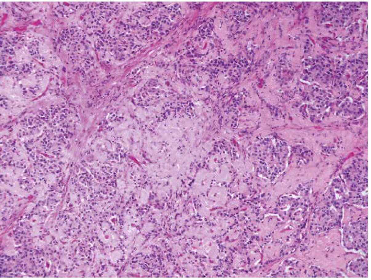
Figure: H&E staining of an insulinoma. Well-differentiated NETs grow in an organoid pattern with nesting and trabecular arrangements. Cells are uniform with eosinophilic cytoplasm and salt-and-pepper chromatin (Yamada's Textbook of Gastroenterology, Fig. 80.3, courtesy Dr Vikram Deshpande)
3.3 Histopathology of Poorly Differentiated NECs
-
Small cell NEC: sheets of densely packed small cells (< 3 lymphocyte diameters, ~20 µm), scant cytoplasm, poorly defined cell borders, granular "salt and pepper" chromatin, absent/inconspicuous nucleoli, frequent nuclear molding, extensive necrosis, and very high mitotic rate (~60 mitoses per 2 mm²). Appears blue on H&E at low power due to high N:C ratio.
-
Large cell NEC: nests of cells with moderate-to-abundant cytoplasm, large vesicular nuclei with prominent nucleoli, frequent necrosis, and high mitotic count (must be ≥ 11 per 2 mm² to distinguish from low-grade carcinoids).
-
Fishman's Pulmonary Diseases & Disorders, p. 3618-3623
4. Immunohistochemistry (IHC) of NETs
IHC is indispensable for: (1) confirming neuroendocrine differentiation, (2) determining site of origin in metastatic disease, (3) assessing grade/prognosis, and (4) guiding treatment selection.
4.1 Pan-Neuroendocrine Markers
| Marker | Nature | Utility | Notes |
|---|---|---|---|
| Synaptophysin | Integral membrane protein of small synaptic vesicles | Most sensitive pan-NE marker | Positive in ALL well-differentiated and poorly differentiated NETs/NECs; most reliable |
| Chromogranin A | Protein integral to membrane of neurosecretory granules | Highly specific for NE differentiation | Absent in poorly differentiated NECs (few granules) and in well-differentiated rectal NETs |
| CD56 (NCAM) | Neural cell adhesion molecule | Sensitive (>90% in SCLC) | Less specific; also expressed in skeletal muscle, leukocyte subsets, some NSCLC |
| INSM1 | Transcription factor (Insulinoma-associated protein 1); nuclear localisation | Emerging gold standard; high sensitivity AND specificity | Expressed in developing neuroendocrine tissue; nuclear staining aids diagnosis; may outperform other markers in SCLC |
| NSE (Neuron-specific enolase) | Cytosolic enzyme | Less specific | Not routinely recommended |
| PGP9.5, chromogranin B, CD57 | Various | Less specific | Rarely used in routine practice |
Synaptophysin immunostaining (strong diffuse brown cytoplasmic staining in a NET):
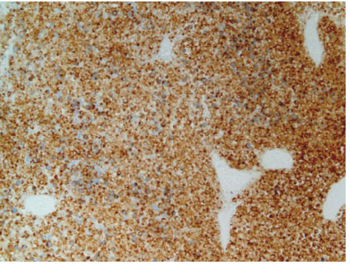
Figure: Synaptophysin immunostaining of a NET. Synaptophysin is expressed in all NE cells and stains positively in both well-differentiated and poorly differentiated NETs (Yamada's Textbook of Gastroenterology, Fig. 80.4, courtesy Dr Vikram Deshpande)
4.2 Proliferative Marker
- Ki-67 (MIB-1 clone): The single most important prognostic IHC marker. Nuclear brown staining quantified as the % of positive neoplastic nuclei. The foundation of the WHO grading system. Ki-67 > 20% with poorly differentiated morphology defines NEC (G3).
Ki-67 IHC in a poorly differentiated NET (high Ki-67 labeling):
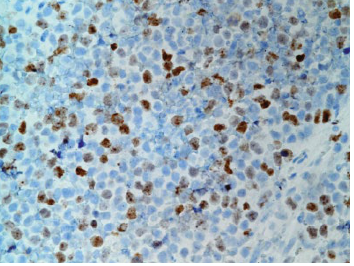
Figure: Ki-67 immunostaining of a poorly differentiated NET. A Ki-67 index > 20% nuclei or mitotic count > 20/hpf defines a poorly differentiated NEC and predicts poor prognosis (Yamada's Textbook of Gastroenterology, Fig. 80.2, courtesy Dr Vikram Deshpande)
4.3 Site-Specific and Lineage-Specific IHC Markers
| Marker | Tumour/Site | Notes |
|---|---|---|
| Insulin | Insulinoma (pancreatic β-cell) | Confirms functional β-cell NET; nonfunctional NETs can also stain positive |
| Gastrin | Gastrinoma | Duodenal/pancreatic origin |
| Glucagon | Glucagonoma | Pancreatic α-cell |
| Somatostatin | Somatostatinoma | Pancreatic δ-cell; also duodenal |
| PDX-1 | Pancreatic NET | Up to 93% specificity, 72% sensitivity for pNET |
| NESP-55 | pNET and phaeochromocytoma | Chromogranin polypeptide; specific |
| CDX-2 | Midgut/GI NET | Marker of intestinal lineage |
| Somatostatin receptor 2 (SSTR2) | NETs (>80% express SSTR2) | Used to predict response to somatostatin analogue therapy and PRRT |
Critical note: Functionality is defined by the presence of clinical symptoms, not IHC staining alone. Nonfunctional NETs frequently stain for multiple hormones but fail to secrete them at symptom-causing levels.
- Yamada's Textbook of Gastroenterology, p. 539-551; Fischer's Mastery of Surgery, p. 4237-4246
5. Molecular Alterations
Unlike most carcinomas, NENs rarely harbour mutations in common oncogenes (RAS, MYC, FOS) or common tumour suppressors (TP53, RB1) in the well-differentiated subtype. Instead, their molecular pathogenesis differs by site and differentiation status.
5.1 Pancreatic NETs (pNETs) - Molecular Profile
Genomic sequencing has identified recurrent somatic alterations in four main pathways:
1. MEN1 (Menin gene, chromosome 11q13)
- Inactivating mutations in 44% of sporadic pNETs
- MEN1 encodes menin, a scaffold protein in a histone methyltransferase complex (chromatin remodelling)
- Also the germline mutation causing MEN1 syndrome (multiple endocrine neoplasia type 1: parathyroid hyperplasia, pituitary tumours, pNETs)
2. DAXX/ATRX pathway (chromatin remodelling / telomere maintenance)
- Inactivating mutations in DAXX (death domain-associated protein) or ATRX (α-thalassaemia/intellectual disability syndrome X-linked chromatin remodeller) in 43% of pNETs
- DAXX and ATRX form a chromatin remodelling complex involved in telomere maintenance
- pNETs with DAXX or ATRX mutations show Alternative Lengthening of Telomeres (ALT) - a telomerase-independent mechanism of telomere maintenance
- Nearly half of pNETs have a somatic mutation in DAXX or ATRX (but not both), consistent with their function in the same pathway
- DAXX/ATRX loss is an independent adverse prognostic marker: 5-year survival ~45% in ALT-positive vs ~85-86% in ALT-negative/DAXX/ATRX-intact tumours
- Routine IHC for DAXX/ATRX loss (loss of nuclear staining) and ALT assessment are now recommended in practice
3. mTOR pathway (PI3K/PTEN/TSC/mTOR)
- Loss-of-function mutations in PTEN and TSC2 tumour suppressors in ~14% of pNETs
- Results in oncogenic activation of the mTOR signalling pathway
- This is the therapeutic target for everolimus (mTOR inhibitor), an approved treatment for advanced pNETs
- Loss of PTEN protein (with increased pAKT expression) may further identify more aggressive GI NETs
4. Chromosomal alterations
-
Allelic losses at 1p, 1q, 3p, 11p (MEN1 locus), and 22p
-
Robbins, Cotran & Kumar Pathologic Basis of Disease, p. 2353-2359; Goldman-Cecil Medicine, p. 2793-2795
5.2 GI (Midgut/Carcinoid) NETs - Molecular Profile
- Chromosomal losses at 18q, 18p, 9p, and 16q are characteristic
- Small intestinal NETs have a low overall mutation rate (one of the lowest of any cancer)
- Recurrent somatic mutations in CDKN1B (encoding the cell cycle inhibitor p27) in ~8% of tumours
- Epigenetic dysregulation (e.g., promoter methylation, histone modification) plays an emerging role
5.3 Neuroendocrine Carcinomas (NECs) - Molecular Profile (Distinctly Different)
- NECs resemble high-grade carcinomas molecularly and share features with SCLC
- TP53 mutations - hallmark of NEC
- RB1 inactivation - common in SCLC and large cell NEC
- Loss of both TP53 and RB1 is characteristic of small cell NEC
- These mutations are rare in well-differentiated G3 NETs, which is a key distinguishing feature
5.4 Hereditary Syndromes Associated with NENs
| Syndrome | Gene | Associated NENs |
|---|---|---|
| MEN1 | MEN1 (11q13) | pNETs in 80-100% (gastrinoma, insulinoma, NF-pNET), anterior pituitary, parathyroid |
| VHL Disease | VHL (3p25) | pNETs in 10-17%; clear cell variant; often multiple |
| Tuberous Sclerosis | TSC1/TSC2 | pNETs in ~0.5%; mTOR pathway driven |
| Neurofibromatosis-1 | NF1 | Duodenal somatostatinomas in ~12% (periampullary) |
| MEN2A/2B | RET | Medullary thyroid carcinoma, phaeochromocytoma |
- Goldman-Cecil Medicine, p. 2805
6. Clinical Examples of NETs
6.1 Insulinoma (Pancreatic β-cell NET)
- Most common functional pNET (incidence: 1-4 per million/year)
- Nearly 90% are benign (G1), solitary, < 2 cm
- Whipple's triad: hypoglycaemic symptoms + blood glucose < 50 mg/dL + relief with glucose
- IHC: positive for insulin, synaptophysin, chromogranin A
- Morphology: well-differentiated, trabecular/nesting pattern; amyloid deposits in stroma (unique)
- Molecular: sporadic insulinomas rarely carry MEN1 mutations; mostly carry YY1 and EIF1AX mutations
6.2 Gastrinoma (Zollinger-Ellison Syndrome)
- 60-90% are malignant (nodal and liver metastases)
- 80-100% located in the duodenum (small, < 1 cm); 20% pancreatic (larger)
- 20-25% occur in MEN1 context (MEN1/ZES): 80-95% are duodenal in this setting
- Gastrin excess drives parietal cell hyperplasia → elevated basal acid output → peptic ulcers, diarrhea, steatorrhoea
- Diagnosis: elevated fasting gastrin (> 1000 pg/mL diagnostic; secretin stimulation test for borderline)
6.3 Small Bowel (Midgut) NET / "Carcinoid"
- Arise from enterochromaffin (Kulchitsky) cells in crypts of Lieberkühn
- Most common small bowel malignancy (>20% of malignant small bowel tumours)
- Located within 60 cm of the ileocecal valve (35-45% of all GI NETs)
- At presentation: 40% have multifocal tumours > 2 cm; 70% invade muscularis propria/lymph nodes; 50% have liver metastases
- Carcinoid syndrome (occurs only with liver metastases, as hepatic passage normally inactivates serotonin): diarrhoea, flushing, bronchospasm, carcinoid heart disease (right-sided valvular lesions from serotonin)
- On CT: hyperenhancing (vs. adenocarcinoma which is moderately enhancing); desmoplastic mesenteric reaction
- IHC: serotonin-positive; chromogranin A strongly positive; CDX-2 positive
6.4 Small Cell Lung Cancer (SC-NEC)
- Poorly differentiated, G3 NEC; highly aggressive
- Nuclear molding, crush artefact, 60 mitoses/2 mm² on average
- IHC: synaptophysin+, chromogranin A+, CD56+ (>90%), INSM1+, Ki-67 80-100%
- TTF-1 positive in ~80%; CK (dot-like pattern)
- Molecular: TP53 and RB1 co-mutation; almost no MEN1/DAXX/ATRX mutations
7. Prognostic Factors in NETs
7.1 Most Important Prognostic Factors
-
Histologic grade (Ki-67 and mitotic index) - the single most powerful determinant
- G1 pNETs: 5-year survival 75-90%
- G2 pNETs: 5-year survival 62-63%
- G3 NET/NEC: 5-year survival 7-12%
-
Differentiation - well-differentiated (NET) vs poorly differentiated (NEC) - independent of grade
-
Stage (AJCC 8th edition):
- Local small bowel NET: 5-year cancer-specific survival ~95%
- Nodal disease: 84%
- Distant (liver) metastases: 51%; overall survival 32% with distant disease
-
DAXX/ATRX mutational status / ALT phenotype - in pNETs: ALT-positive tumours have ~45% 5-year survival vs ~86% in ALT-negative tumours; routine molecular assessment now recommended
-
Functional status: ~90% of insulinomas are benign; 60-90% of other functioning and nonfunctioning pNETs are malignant
-
Site of origin: pNETs generally more aggressive than midgut NETs of the same grade; rectal NETs tend to be less aggressive
-
SSTR2 expression: 90% of well-differentiated NETs overexpress SSTR2; SSTR2-negative tumours (more common in G3 and insulinomas) cannot benefit from somatostatin analogue therapy or PRRT, and carry a worse prognosis in high-grade tumours
-
Tumour size: > 2 cm strongly associated with malignant behaviour in pNETs; small bowel tumours > 2 cm have high rates of nodal metastasis
-
Lymphovascular and perineural invasion - adverse prognostic feature
-
MEN1 context: pNETs in MEN1 tend to be multiple but often lower grade; however, non-insulinoma pNETs in MEN1 still carry significant malignant potential
- Sabiston Textbook of Surgery, p. 1591; Current Surgical Therapy, p. 1675; Goldman-Cecil Medicine, p. 2795-2803
Summary Table: NET vs NEC
| Feature | Well-differentiated NET | Poorly differentiated NEC |
|---|---|---|
| Differentiation | Well | Poor |
| Grade | G1, G2, or G3 (rare) | G3 only |
| Ki-67 | ≤ 3% (G1), 3-20% (G2), >20% (G3-NET) | Usually >55-80% |
| Mitoses | Low to moderate | Very high (>10-60/mm²) |
| Histology | Organoid nests/trabeculae, salt-and-pepper chromatin | Sheets, nuclear molding, necrosis |
| Synaptophysin | Positive | Positive |
| Chromogranin A | Positive (strong) | Weak or absent |
| SSTR2 | Positive (>80%) | Often negative |
| Key mutations | MEN1, DAXX/ATRX, mTOR pathway (pNET); CDKN1B (midgut) | TP53, RB1 |
| Treatment | SSA, everolimus, PRRT, surgery | Platinum/etoposide |
| Prognosis | Variable, often indolent | Very poor (median survival <1 year for stage IV) |
Sources consulted:
- Goldman-Cecil Medicine (International Edition) - Wolin & Jensen, Chapter 213
- Robbins, Cotran & Kumar Pathologic Basis of Disease (10th ed.), Chapter on Pancreatic NENs
- Sabiston Textbook of Surgery (22nd ed.), Chapter 76
- Yamada's Textbook of Gastroenterology (7th ed.), Chapter 80
- Fischer's Mastery of Surgery (8th ed.)
- Fishman's Pulmonary Diseases and Disorders, Chapter on Lung NENs
- Current Surgical Therapy (14th ed.)
- Henry's Clinical Diagnosis and Management by Laboratory Methods
- Hernandez-Prera et al. (2025) - WHO 5th Edition Endocrine/Neuroendocrine Updates, Arch Pathol Lab Med [PMID: 40335070]
This is a shared conversation. Sign in to Orris to start your own chat.