
Ild
"interstitial lung disease"[MeSH Terms] AND "diagnosis"[MeSH Terms]
interstitial lung disease HRCT high resolution CT pattern UIP
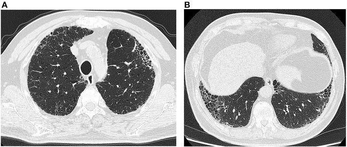
High-resolution computed tomography (HRCT) of the chest in axial view at two different levels (A: upper/mid-thorax, B: lung bases). The images demonstrate bilateral, symmetric pulmonary abnormalities characteristic of a usual interstitial pneumonia (UIP) pattern. Key findings include prominent reticular opacities and peripheral, subpleural honeycombing, particularly evident in the subpleural regions of both lungs. There is a clear predilection for the proximal pleural parts and the lung bases, with relative sparing of the more central lung parenchyma. No significant ground-glass opacities, consolidations, or exudative changes are observed. The presence of these structural alterations—specifically honeycombing and architectural distortion—is highly suggestive of chronic fibrosing interstitial lung disease, such as idiopathic pulmonary fibrosis (IPF). These findings are essential for medical learners to recognize the diagnostic hallmarks of UIP on CT imaging.
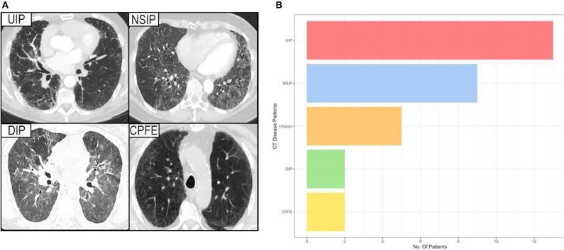
This composite educational resource consists of a diagnostic imaging panel (A) and a comparison bar chart (B) illustrating interstitial lung disease (ILD) patterns in patients with Sjögren's syndrome. Panel A displays axial High-Resolution Computed Tomography (HRCT) slices of four distinct ILD patterns: Usual Interstitial Pneumonia (UIP) characterized by peripheral reticulation and honeycombing; Non-Specific Interstitial Pneumonia (NSIP) showing more uniform ground-glass opacities; Desquamative Interstitial Pneumonia (DIP) with diffuse ground-glass attenuation; and Combined Pulmonary Fibrosis and Emphysema (CPFE) demonstrating upper-lobe emphysematous lucencies alongside fibrotic changes. Panel B is a horizontal bar chart quantifying the prevalence of these CT disease patterns among the study cohort. The chart indicates that UIP is the most frequent pattern, followed by NSIP, unspecific changes (Unspez), and lastly DIP and CPFE, which show equal, lower prevalence. This visual aid is intended for medical education regarding the radiologic classification and epidemiological distribution of pulmonary manifestations in systemic autoimmune diseases.
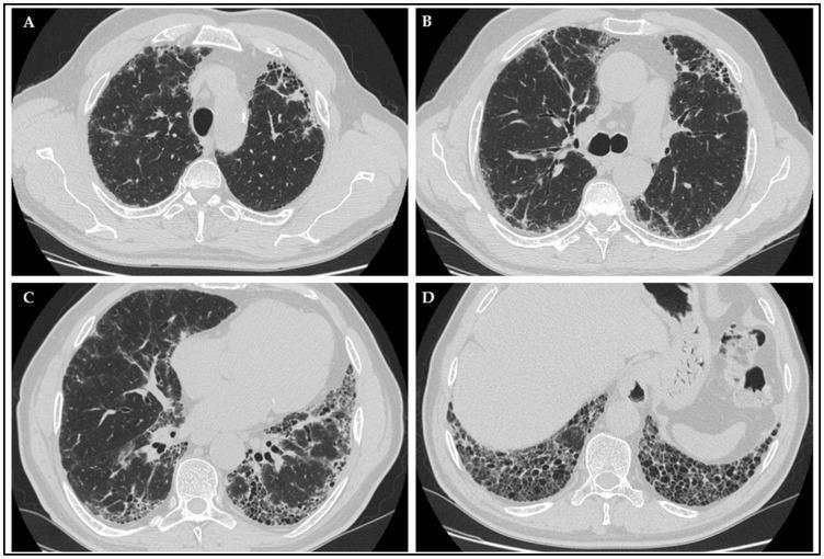
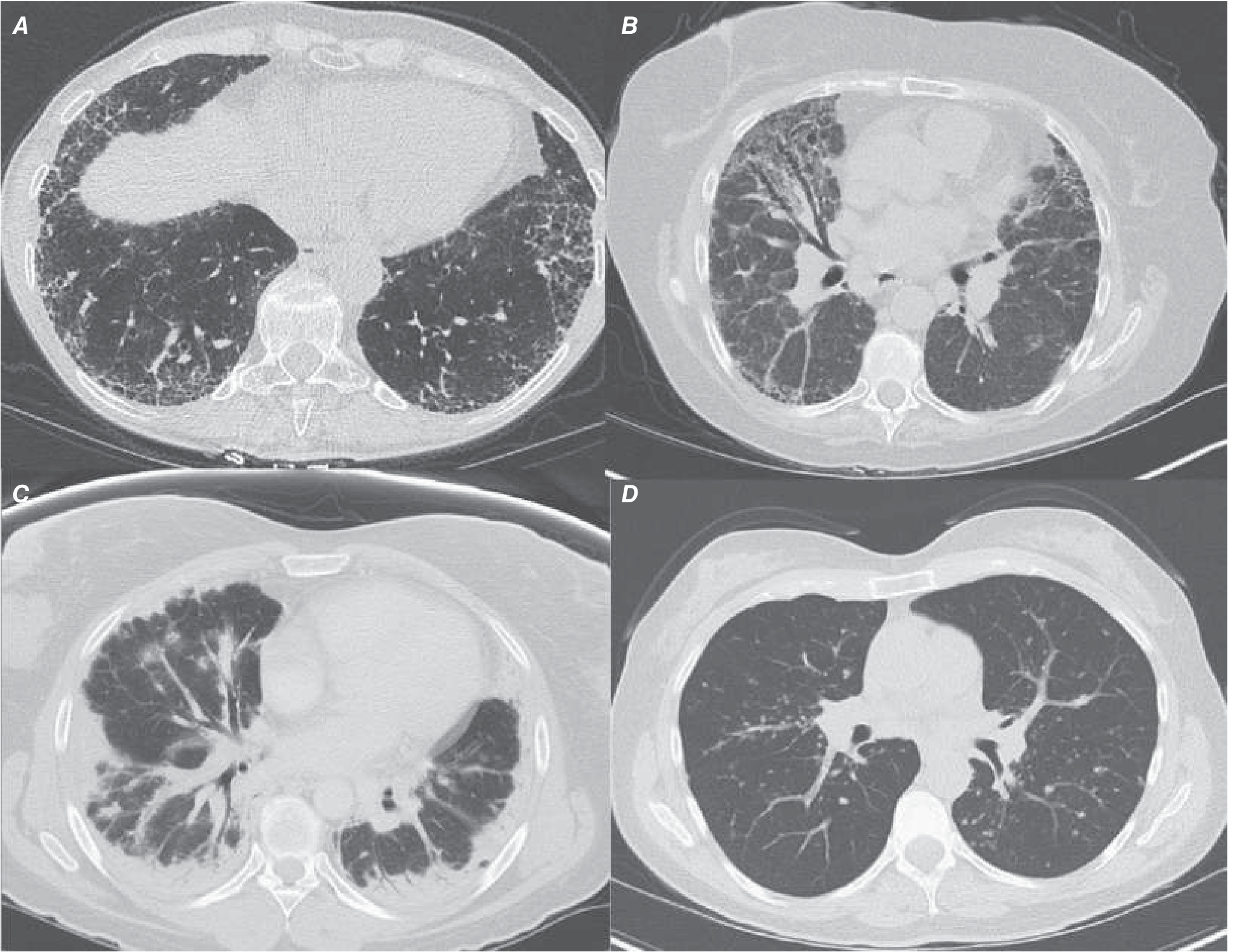
This composite diagnostic image displays four axial slices (A, B, C, D) of a high-resolution computerized tomography (HRCT) scan of the chest, illustrating a Usual Interstitial Pneumonia (UIP) pattern in a patient with Idiopathic Pulmonary Fibrosis (IPF). The scans demonstrate a classic subpleural and basal predominant distribution of interstitial lung disease. Key radiological features include: (A) early peripheral reticular opacities; (B and C) prominent traction bronchiectasis, characterized by the irregular dilation of bronchioles due to surrounding parenchymal fibrosis; and (D) extensive honeycombing, visible as clustered, thick-walled cystic airspaces in the subpleural regions of the lung bases. These findings collectively indicate advanced pulmonary fibrosis with architectural distortion. The image serves as an educational tool for identifying the hallmark CT characteristics of a UIP pattern, essential for the diagnosis of IPF and its differentiation from other interstitial lung diseases in a clinical pulmonology and radiology context.
idiopathic pulmonary fibrosis classification ILD flowchart diagram
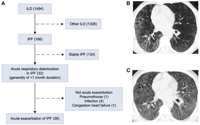
This composite educational image includes a clinical flowchart and two high-resolution computed tomography (HRCT) axial slices of the chest, illustrating the progression from stable Idiopathic Pulmonary Fibrosis (IPF) to an Acute Exacerbation (AE-IPF). Panel A presents a study flowchart detailing the selection process of 26 patients with AE-IPF from a cohort of 1,494 patients with interstitial lung disease (ILD). Panel B displays an axial HRCT scan showing classic features of Usual Interstitial Pneumonia (UIP) associated with stable IPF, including peripheral-predominant reticular opacities, traction bronchiectasis, and subpleural honeycombing. Panel C shows the same patient during an acute exacerbation. In addition to the pre-existing fibrotic changes seen in Panel B, there is now diffuse, bilateral ground-glass opacities (GGO), representing the hallmark radiological sign of acute alveolar damage superimposed on chronic fibrosis. This comparison highlights the key diagnostic imaging criteria for AE-IPF, essential for pulmonology and radiology education.
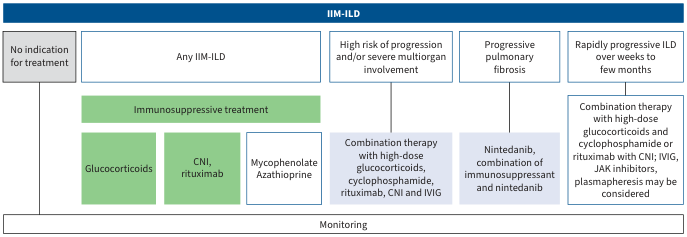
Summary : This flowchart outlines the treatment algorithm for idiopathic inflammatory myopathy-associated interstitial lung disease (IIM-ILD), specifying treatment options based on disease severity and progression risk. flowchart: # Nodes : • No indication for treatment (rectangle) • Any IIM-ILD (rectangle) • High risk of progression and/or severe multiorgan involvement (rectangle) • Progressive pulmonary fibrosis (rectangle) • Rapidly progressive ILD over weeks to few months (rectangle) • Immunosuppressive treatment (rectangle, green background) • Glucocorticoids (rectangle, green background) • CNI, rituximab (rectangle, green background) • Mycophenolate, Azathioprine (rectangle, white background) • Combination therapy with high-dose glucocorticoids, cyclophosphamide, rituximab, CNI and IVIG (rectangle, blue background) • Nintedanib, combination of immunosuppressant and nintedanib (rectangle, blue background) • Combination therapy with high-dose glucocorticoids and cyclophosphamide or rituximab with CNI; IVIG, JAK inhibitors, plasmapheresis may be considered (rectangle, blue background) • Monitoring (rectangle, bottom row) # Connectors : • Flow is left to right, starting from "No indication for treatment" and "Any IIM-ILD" to more severe categories. • "Any IIM-ILD" leads to "Immunosuppressive treatment," which branches to "Glucocorticoids," "CNI, rituximab," and "Mycophenolate, Azathioprine." • "High risk of progression and/or severe multiorgan involvement" leads to "Combination therapy with high-dose glucocorticoids, cyclophosphamide, rituximab, CNI and IVIG." • "Progressive pulmonary fibrosis" leads to "Nintedanib, combination of immunosuppressant and nintedanib." • "Rapidly progressive ILD over weeks to few months" leads to "Combination therapy with high-dose glucocorticoids and cyclophosphamide or rituximab with CNI; IVIG, JAK inhibitors, plasmapheresis may be considered." • All treatment pathways converge to "Monitoring" at the bottom. # Layout : • Horizontal arrangement of disease severity categories from left (mild/no indication) to right (rapidly progressive). • Treatment options are layered below each severity category. • Monitoring is a unified bar at the bottom, spanning all categories. # Analysis : • The flowchart provides a stepwise escalation of therapy based on disease severity, starting with immunosuppressive agents for all IIM-ILD, progressing to combination and advanced therapies for high-risk or rapidly progressive cases. • Monitoring is emphasized as a universal step regardless of treatment pathway. • The use of color highlights the escalation from standard immunosuppressive therapy (green) to more aggressive or adjunctive therapies (blue) as disease severity increases.
Interstitial Lung Disease (ILD)
Definition & Overview
- Inflammation and/or fibrosis of the lung parenchyma
- Dyspnea on exertion
- Bilateral infiltrates on chest imaging
- Restrictive physiology with diffusion impairment on PFTs
Classification
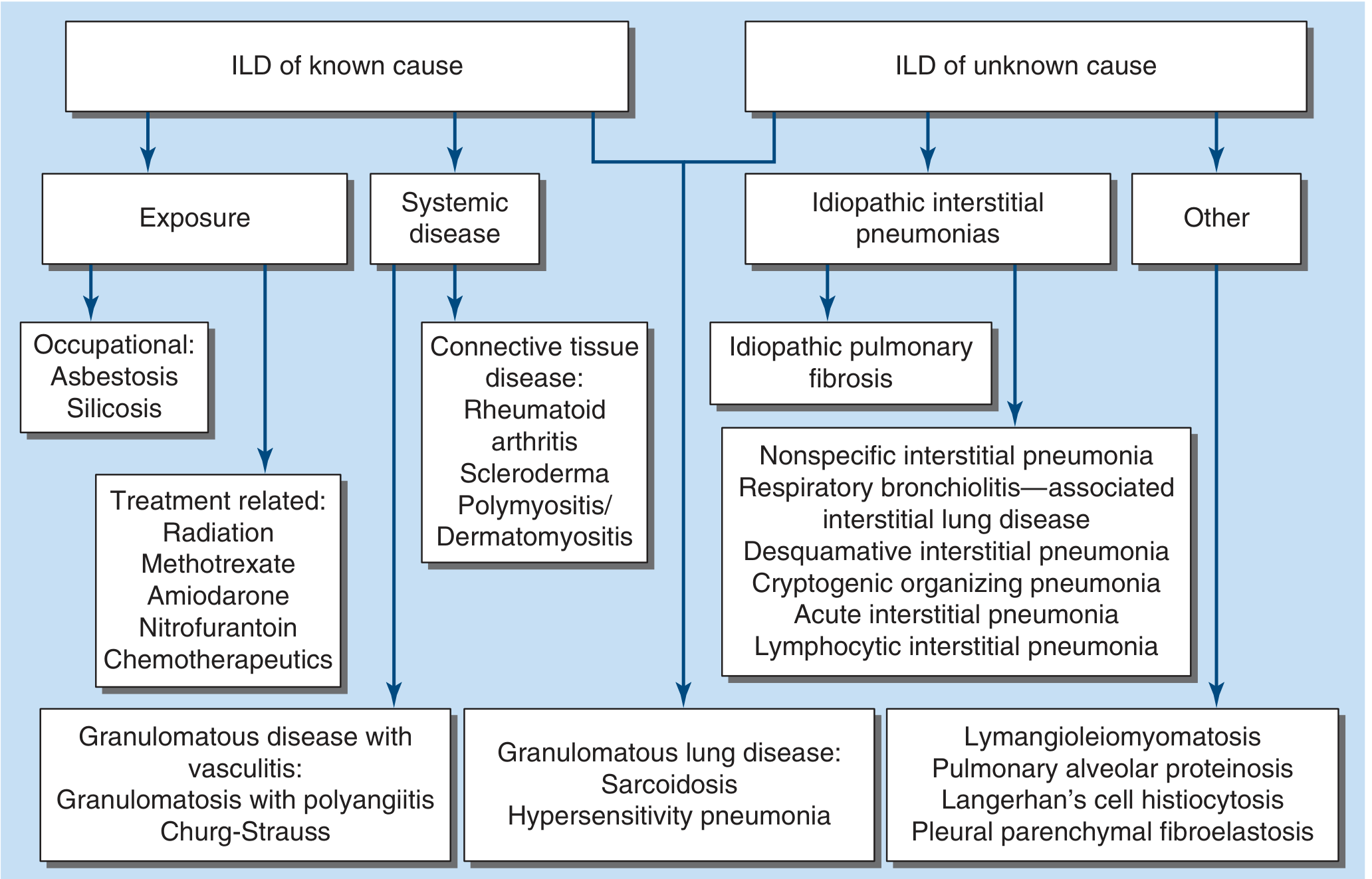
Known Causes
| Category | Examples |
|---|---|
| Connective tissue diseases (CTD) | RA, SLE, Sjogren's, polymyositis/dermatomyositis, systemic sclerosis |
| Occupational/environmental exposures | Asbestos (asbestosis), silica (silicosis), hypersensitivity pneumonitis (bird fancier's lung, farmer's lung) |
| Drug-induced | Nitrofurantoin, amiodarone, bleomycin, methotrexate, immunotherapy |
| Granulomatous | Sarcoidosis, chronic beryllium disease |
| Miscellaneous | Pulmonary alveolar proteinosis, eosinophilic pneumonia, LAM |
Unknown Causes - Idiopathic Interstitial Pneumonias (IIPs)
- Idiopathic Pulmonary Fibrosis (IPF) - UIP pattern
- Idiopathic NSIP (Non-specific interstitial pneumonia)
- Respiratory Bronchiolitis-ILD (RB-ILD)
- Desquamative Interstitial Pneumonia (DIP)
- Cryptogenic Organizing Pneumonia (COP)
- Acute Interstitial Pneumonia (AIP / Hamman-Rich syndrome)
- Lymphoid Interstitial Pneumonia (LIP)
- Pleuroparenchymal Fibroelastosis (PPFE)
Clinical Features
Symptoms
- Progressive exertional dyspnea - most common presenting complaint
- Dry cough - especially prominent in IPF
- Fatigue - common to all ILDs
- Hemoptysis - rare; suggests DAH (Goodpasture's, GPA), LAM, or infection
- Chest pain - rare; may occur in sarcoidosis
- Acuity of onset is diagnostically important:
- Acute (days-weeks): AIP, eosinophilic pneumonia, HP, GPA
- Subacute (weeks-months): Sarcoidosis, CTD-ILD, drug-induced, COP
- Chronic (months-years): IPF (most typical)
Physical Examination
- Fine end-inspiratory crackles (Velcro rales) at the lung bases - earliest sign, most common in IPF
- Wheezing - uncommon; seen in sarcoidosis, HP, eosinophilic GPA
- Signs of advanced disease: cyanosis, digital clubbing, cor pulmonale
Patient Characteristics That Guide Diagnosis
| Feature | ILD suggested |
|---|---|
| Age >60, male, smoker | IPF |
| Female, 5th decade, non-smoker | NSIP |
| Age 20-40 | Sarcoidosis, CTD-ILD, LAM, PLCH |
| Young woman | LAM |
| Male | RA-ILD, occupational ILDs |
History - Key Elements to Elicit
- Occupational/Environmental exposures - asbestos, silica, birds, mold, hot tubs, humidifiers, wind instruments, farming
- Medications - nitrofurantoin, amiodarone, methotrexate, bleomycin, NSAIDs, immunotherapy (check pneumotox.com for full list)
- Smoking history - associated with RB-ILD, DIP, and ~75% of IPF patients
- Family history - up to 20% of pulmonary fibrosis is familial; MUC5B gene variant and telomerase gene (TERT) variants are key genetic risk factors
- CTD symptoms - Raynaud's phenomenon, arthritis, skin changes, sicca, myalgia
- Vaping/EVALI - vitamin E acetate in vaping products causes acute lung injury
Investigations
Laboratory Studies
- ANA, anti-dsDNA, anti-Ro/La, anti-Scl-70, anti-Jo-1, anti-CCP, RF - for CTD screen
- CBC, LFTs, renal function - baseline
- Serum precipitins - for HP (bird/mold antigens)
- Serum ACE - elevated in sarcoidosis
Pulmonary Function Tests (PFTs)
- Restrictive pattern: reduced TLC, FRC, RV; FEV1/FVC normal or increased
- Reduced DLCO - correlates with fibrosis extent and predicts outcome
- GAP index (Gender, Age, Physiology) uses FVC and DLCO to estimate mortality in IPF
- 6-minute walk test - valuable for severity, oxygen requirements, and prognosis
High-Resolution CT (HRCT)
- Subpleural, basal predominant reticular opacities
- Honeycombing (with or without traction bronchiectasis)
- No features suggesting an alternative diagnosis
- Subpleural, basal reticular pattern with traction bronchiectasis
- No honeycombing
- Cysts, marked mosaic attenuation, predominant GGO
- Profuse micronodules, centrilobular nodules, consolidation
- Upper/mid-lung predominance, peribronchovascular distribution
- Pleural plaques (asbestos), dilated esophagus (CTD/scleroderma)
Bronchoscopy
- BAL cellular analysis:
- Eosinophils >25% → virtually diagnostic of eosinophilic pneumonia
- Lymphocytes >25% → sarcoidosis, HP, NSIP, drug reaction, LIP, COP
- CD4+/CD8+ ratio >4 → supports sarcoidosis
- Bloody return → diffuse alveolar hemorrhage
- Milky white fluid → pulmonary alveolar proteinosis (confirm with PAS stain)
- Transbronchial biopsy - useful for granulomatous disease (sarcoidosis, HP)
- Transbronchial cryobiopsy - larger samples, increasing diagnostic yield
Surgical Lung Biopsy
- Reserved for when diagnosis remains uncertain after HRCT + BAL
- VATS (video-assisted thoracoscopic surgery) preferred
- Essential to distinguish UIP from NSIP, DIP, LIP, COP histologically
Management
General Principles
- Multidisciplinary team (MDT) review - pulmonologist, radiologist, pathologist - is considered best practice and can significantly impact diagnostic and management decisions
- Remove or treat the underlying cause where possible (drugs, antigens)
- Smoking cessation
- Supplemental oxygen when indicated - reduces pulmonary hypertension risk, improves exercise tolerance
- Pulmonary rehabilitation
IPF - Specific Treatment
- Antifibrotic agents (2014 onwards - game-changing trials):
- Pirfenidone - inhibits TGF-β-mediated fibrosis
- Nintedanib - tyrosine kinase inhibitor; also approved for progressive pulmonary fibrosis (PPF) beyond IPF
- Immunosuppression is NOT indicated in IPF - associated with increased morbidity and mortality
- Lung transplantation - considered for eligible patients; extends survival and improves quality of life
CTD-ILD & Other Inflammatory ILDs
- Corticosteroids - mainstay for COP, acute HP, sarcoidosis, CTD-ILD
- Azathioprine, mycophenolate mofetil - steroid-sparing agents
- Cyclophosphamide - severe rapidly progressive CTD-ILD
- Rituximab - for refractory cases (especially anti-MDA5+ myositis-ILD, RA-ILD)
- Calcineurin inhibitors (CNI) - IIM-ILD
COP
- Corticosteroids produce substantial improvement; continue for at least 6 months (high relapse rate)
- Evidence growing for mycophenolate, cyclophosphamide, rituximab in refractory cases
AIP (Hamman-Rich)
- Supportive care, mechanical ventilation
- Glucocorticoids often given but not clearly effective (>50% mortality at 6 months)
Key Contrasts Between Major IIPs
| Feature | IPF (UIP) | NSIP | COP | AIP |
|---|---|---|---|---|
| Demographics | >60, male, smoker | Female, 5th decade | Any | Any |
| Onset | Chronic | Subacute-chronic | Subacute | Acute |
| HRCT | Subpleural honeycombing, traction bronchiectasis, basal | Bilateral GGO, subpleural sparing, symmetric | Peribronchovascular consolidation, reversed halo | Bilateral GGO, dependent consolidation |
| Histology | Temporal heterogeneity, fibroblast foci | Temporal uniformity, inflammation or fibrosis | Granulation plugs in airways/alveoli | Diffuse alveolar damage (DAD) |
| Response to steroids | None/harmful | Good | Excellent | Poor |
| Prognosis | Poor (median survival 3-5 yr) | Better | Good (if treated) | Very poor (>50% mortality) |
Take-Home Points
- ILD is a broad category - always establish the underlying cause (known vs idiopathic)
- HRCT is central to diagnosis - a typical UIP pattern on HRCT in the right clinical context can diagnose IPF without biopsy
- MDT discussion is the gold standard approach for diagnosis
- Antifibrotics (pirfenidone, nintedanib) are the treatment of choice for IPF; immunosuppression is harmful in IPF
- Nintedanib is now also approved for progressive pulmonary fibrosis (PPF) regardless of etiology
- Always check medications (pneumotox.com), occupational history, and CTD serology before labeling any ILD as "idiopathic"