
Abdominal discomfort after eating meals
"functional dyspepsia" AND postprandial
common causes of abdominal discomfort after eating meals
Abdominal Discomfort After Eating: Overview
1. Functional dyspepsia (most common non-structural cause)
- Postprandial distress syndrome - bothersome fullness or early satiation that starts during/after a normal-sized meal
- Epigastric pain syndrome - burning or pain in the upper abdomen, not necessarily tied to meals
2. Gastritis / peptic ulcer disease
3. GERD (acid reflux)
4. Biliary colic (gallstones)
5. Chronic mesenteric ischemia ("intestinal angina")
6. Simpler/benign triggers
7. Other considerations
When it needs prompt medical evaluation ("alarm features")
- New onset after age 50
- Unintentional weight loss
- Persistent vomiting
- Difficulty/pain swallowing (dysphagia/odynophagia)
- GI bleeding or iron-deficiency anemia
- Palpable abdominal mass or lymphadenopathy
- Family history of GI cancer
Practical takeaway
- Occasional, mild discomfort after a large or fatty meal that settles within a few hours is usually benign (overeating, mild indigestion).
- Recurrent pain, especially tied specifically to fatty foods and in the right upper quadrant, raises suspicion for gallstones.
- Pain with weight loss and "fear of eating" in someone with vascular risk factors warrants urgent workup for mesenteric ischemia.
- Any alarm feature above should prompt a medical visit rather than self-management.
Duodenal ulcer
"peptic ulcer" AND "Helicobacter pylori" AND treatment
Duodenal ulcer
Duodenal ulcer
"peptic ulcer disease" management guideline
duodenal ulcer endoscopic image
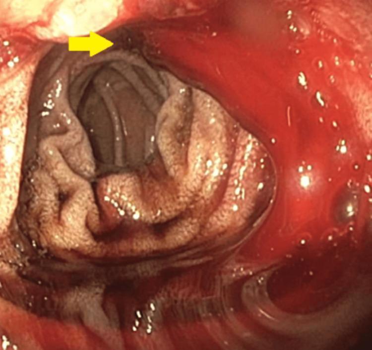
This endoscopic image (Esophagogastroduodenoscopy) provides a view of the duodenal bulb and proximal duodenum. A yellow arrow points to a cratered duodenal ulcer located on the mucosal wall. The ulcer exhibits stigmata of active hemorrhage, specifically a slow, consistent oozing of liquid, dark red blood into the duodenal lumen, characteristic of a Forrest Class Ib ulcer. The surrounding duodenal mucosa appears erythematous and edematous. This visual serves as a clinical example of an upper gastrointestinal bleed (UGIB) originating from peptic ulcer disease. The image is intended for medical training in gastroenterology, focusing on the endoscopic identification, classification, and assessment of bleeding risk in peptic ulcers.
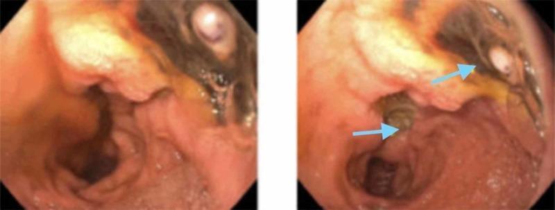
This diagnostic image consists of two endoscopic views of the duodenal bulb, demonstrating a large, cratered duodenal ulcer on the posterior wall. The ulcer measures approximately 35 mm and is characterized by irregular, raised borders and a deep central depression. Within the base of the ulcer, a prominent, non-bleeding visible vessel is identified (indicated by the lower blue arrow in the right panel), which represents a high-risk stigmata of recent hemorrhage (Forrest IIa classification). The surrounding duodenal mucosa appears erythematous, edematous, and friable, consistent with active inflammation. Additional features include yellowish fibrinous exudate and minor areas of dark pigmentation within the ulcer bed. This image serves as a clinical example of severe peptic ulcer disease and the endoscopic identification of vascular structures that necessitate close monitoring or therapeutic intervention for gastrointestinal bleeding.
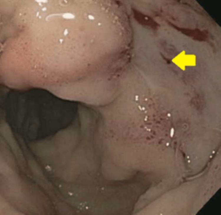
This endoscopic clinical photograph displays the internal mucosal surface of the duodenal bulb during an esophagogastroduodenoscopy (EGD). The primary finding is a large, cratered duodenal ulcer characterized by mucosal architectural disruption and erythema. A yellow arrow points to a prominent, non-bleeding visible vessel (Forrest Class IIa) located within the ulcer base, which represents a high-risk stigmata of recent hemorrhage. The surrounding duodenal mucosa is diffusely erythematous and edematous, showing scattered petechiae and small spots of oozing blood, indicative of active inflammation and recent bleeding. Bubbles of gastric/duodenal secretions are visible on the mucosal surface, and the distal lumen is partially visualized in the background. This image is an essential educational resource for identifying endoscopic risk factors for recurrent peptic ulcer bleeding and for training in Forrest classification for gastrointestinal pathology.
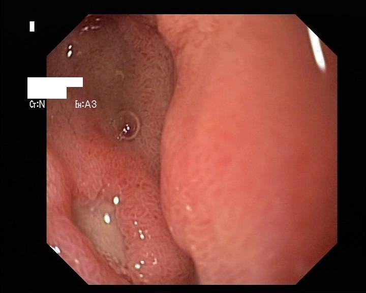
This endoscopic clinical photograph displays a peptic ulcer located within the duodenal mucosa. The diagnostic image reveals a well-circumscribed, depressed ulcer bed characterized by a yellowish-white fibrin base, which indicates tissue necrosis or inflammatory exudate. The surrounding duodenal wall exhibits significant erythema and hyperemia, suggesting localized inflammation of the mucosal lining. The adjacent mucosa displays a granular, slightly cobblestone texture typical of the small intestine, though with visible reactive changes. Notable incidental findings include a small, translucent circular air bubble or mucous collection near the ulcer site. This visual illustrates a classic complication of non-steroidal anti-inflammatory drug (NSAID) therapy in an elderly patient. The educational focus is on the endoscopic identification of upper gastrointestinal pathology, specifically the morphological characteristics of a duodenal ulcer in the context of drug-induced mucosal injury.
Duodenal Ulcer
Epidemiology
Etiology / Pathophysiology
- Helicobacter pylori infection - present in over 70% of PUD cases; produces chronic gastritis and disrupts mucosal defense and acid-gastrin feedback. Only 5-10% of infected individuals actually develop an ulcer, so host and bacterial-strain factors matter.
- NSAID/aspirin use - impairs prostaglandin-mediated mucosal protection; increasingly the leading cause as H. pylori rates fall.
- Zollinger-Ellison syndrome (gastrinoma) - causes multiple, often refractory ulcers via constitutive gastrin-driven acid hypersecretion
- Smoking (reduces mucosal blood flow and healing), high-dose corticosteroids, alcohol-related cirrhosis, COPD, chronic renal failure, hyperparathyroidism (hypercalcemia stimulates gastrin)
- Physiologic mechanisms: duodenal ulcer patients as a group have higher basal and maximal acid output, increased gastric emptying delivering more acid to the duodenum, and reduced duodenal bicarbonate secretion/duodenal gastric metaplasia that impairs buffering - Schwartz's Principles of Surgery.
Clinical Features
- Classic symptom: mid-epigastric pain, well localized, generally tolerable, and characteristically relieved by food or antacids, recurring 2-3 hours after eating or waking the patient at night (contrasts with gastric ulcer pain, which is often worsened by eating).
- Pain becoming constant suggests deeper penetration.
- Pain radiating to the back suggests penetration into the pancreas.
- Sudden, diffuse peritoneal pain signals free perforation - a surgical emergency (Sabiston Textbook of Surgery).
Diagnosis
- Upper endoscopy (EGD) is the test of choice - direct visualization, biopsy capability (mainly for gastric ulcers to exclude malignancy; duodenal ulcer biopsy is not routinely needed) - Yamada's Textbook of Gastroenterology; Current Surgical Therapy.
- Upper GI barium radiography has similar diagnostic accuracy for duodenal ulcer but is inferior to endoscopy for gastric ulcers - Harrison's Principles of Internal Medicine.
- H. pylori testing: urea breath test (good for initial diagnosis and confirming eradication), stool antigen, or biopsy-based testing (rapid urease test/histology) during endoscopy.
- Serum gastrin level if refractory to therapy or surgery is being considered (screens for Zollinger-Ellison syndrome).
Complications
- Hemorrhage - the most common complication (15-20% of PUD patients); posterior duodenal ulcers can erode into the gastroduodenal artery causing major bleeding - Rosen's Emergency Medicine; Schwartz's Principles of Surgery.
- Perforation - the second most common complication and now the most frequent indication for surgery; presents as sudden severe pain and an acute abdomen - Current Surgical Therapy.
- Penetration (e.g., into the pancreas, causing back pain and possibly pancreatitis)
- Gastric outlet obstruction from scarring/edema in the duodenal bulb
Treatment
- Medical management (first line for uncomplicated ulcers):
- Acid suppression with a proton pump inhibitor (PPI)
- H. pylori eradication if positive - standard regimens combine a PPI with antibiotics (e.g., clarithromycin-based or bismuth quadruple therapy) for 10-14 days - Current Surgical Therapy. A 2024 systematic review supports potassium-competitive acid blockers (e.g., vonoprazan) as an emerging alternative to PPI-based regimens for H. pylori eradication (PMID 38577188).
- Discontinue NSAIDs/aspirin where possible; stop smoking
- Long-term PPI is reasonable for patients who cannot avoid NSAIDs/aspirin or who had a complicated ulcer
- Endoscopic therapy for active bleeding (injection, thermal coagulation, clipping) based on Forrest classification and rebleeding risk.
- Surgery is now reserved for complications refractory to medical/endoscopic management: perforation (omental/Graham patch closure with peritoneal washout is the treatment of choice for most perforations), massive or recurrent bleeding, or intractable/obstructing disease. Vagotomy and drainage procedures, once mainstays, are used far less often since H. pylori eradication and PPIs became available - Current Surgical Therapy; Fischer's Mastery of Surgery.
- After surgery, recurrence is minimized by confirming H. pylori eradication and strict avoidance of NSAIDs.