
Muscles of thenar eminence
The thenar eminence is the fleshy mound at the base of the thumb, formed by four muscles that control thumb movement.
Searching sources...
Reading File
Finding Sources
Finding Sources
Reading File
Reading File
Finding Sources
Now let me fetch the Thieme Atlas image of the thenar muscles and grab the detailed table from Gray's:
Reading File
Finding Sources
Now let me fetch the Thieme Atlas image and get the thenar muscle table from the Thieme Atlas:
Loading Image
Reading File
I now have comprehensive data from Gray's Anatomy for Students and the Thieme Atlas of Anatomy. Here is a complete, well-structured answer:
Muscles of the Thenar Eminence
The thenar eminence is the prominent fleshy mound on the lateral (radial) side of the palm at the base of the thumb. It is formed by three thenar muscles proper, plus the nearby adductor pollicis (which some authors group with the thenar eminence).
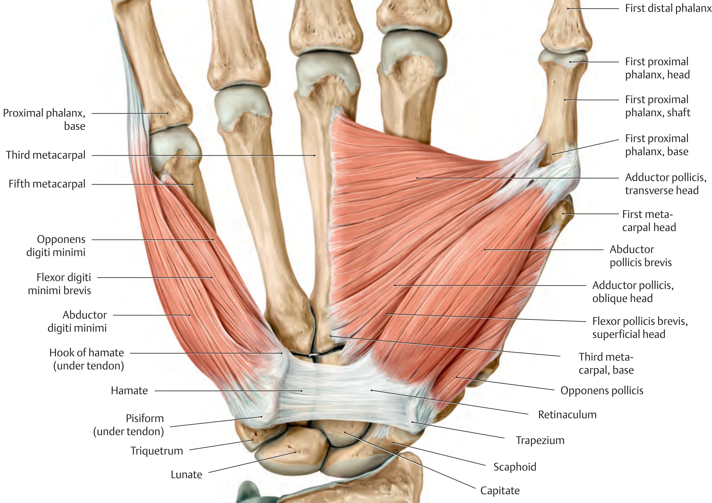
The Three True Thenar Muscles
(all innervated by the recurrent branch of the median nerve, C8, T1)
1. Abductor Pollicis Brevis (APB)
- Position: Most superficial and proximal of the three; overlies the opponens pollicis
- Origin: Tubercles of the scaphoid and trapezium + adjacent flexor retinaculum
- Insertion: Lateral side of the base of the proximal phalanx of the thumb + extensor hood
- Action: Abducts the thumb (principally at the MCP joint); moves the thumb away from the palm in a plane perpendicular to it
- Nerve: Recurrent branch of median nerve (C8, T1)
- Clinical note: The APB is the key muscle tested when assessing median nerve function at the wrist (e.g., in carpal tunnel syndrome)
2. Opponens Pollicis
- Position: Deepest of the three thenar muscles (lies beneath APB and FPB)
- Origin: Tubercle of the trapezium + adjacent flexor retinaculum
- Insertion: Entire length of the lateral margin and adjacent palmar surface of metacarpal I
- Action: Opposition of the thumb - rotates and flexes metacarpal I at the CMC joint, bringing the thumb pad to face the finger pads; the most important muscle for precision grip
- Nerve: Recurrent branch of median nerve (C8, T1)
3. Flexor Pollicis Brevis (FPB)
- Position: Distal to abductor pollicis brevis; lies lateral to opponens pollicis
- Origin:
- Superficial head: Tubercle of trapezium + adjacent flexor retinaculum
- Deep head: Capitate, trapezium (variable - may attach to other carpal bones)
- Insertion: Lateral side of the base of the proximal phalanx of the thumb (tendon often contains a sesamoid bone)
- Action: Flexes the MCP joint of the thumb
- Nerve: Superficial head - recurrent branch of median nerve (C8, T1); deep head - often ulnar nerve (C8, T1)
4. Adductor Pollicis
(deep to the thenar muscles; forms the deep plane of the palm; NOT in the thenar eminence proper)
- Position: Deep, not part of thenar eminence but functionally grouped with thumb muscles
- Origin:
- Transverse head: Anterior aspect of the shaft of metacarpal III
- Oblique head: Capitate + adjacent bases of metacarpals II and III
- Insertion: Medial side of the base of the proximal phalanx of the thumb + extensor hood (tendon often contains a sesamoid bone)
- Action: Adducts the thumb; powerful muscle for gripping
- Nerve: Deep branch of the ulnar nerve (C8, T1)
- Note: The radial artery passes between its two heads to form the deep palmar arch
Summary Table
| Muscle | Origin | Insertion | Action | Nerve |
|---|---|---|---|---|
| Abductor pollicis brevis | Scaphoid, trapezium, flexor retinaculum | Base of proximal phalanx (lateral) | Abducts thumb | Median (recurrent branch) |
| Opponens pollicis | Trapezium, flexor retinaculum | Lateral border of metacarpal I (full length) | Opposition of thumb | Median (recurrent branch) |
| Flexor pollicis brevis | Trapezium, flexor retinaculum (+ deep head: capitate) | Base of proximal phalanx (lateral) | Flexes thumb MCP joint | Median (superficial head); Ulnar (deep head) |
| Adductor pollicis | Metacarpal III (transverse); Capitate + MC II/III (oblique) | Base of proximal phalanx (medial) | Adducts thumb, gripping | Ulnar (deep branch) |
Key Clinical Points
- Median nerve palsy / Carpal tunnel syndrome: Loss of the recurrent branch of the median nerve causes wasting of the APB, opponens pollicis, and FPB (superficial head), leading to flattening of the thenar eminence and loss of opposition. The hand adopts an "ape hand" deformity.
- Ulnar nerve palsy: Adductor pollicis is paralyzed - leads to Froment's sign (patient flexes the IP joint of the thumb using flexor pollicis longus to compensate when trying to pinch paper).
- Recurrent branch of median nerve ("the million dollar nerve"): It hooks around the distal edge of the flexor retinaculum to enter the thenar muscles. Its superficial position makes it vulnerable to lacerations at the base of the thumb.
- Opposition is the combination of flexion, abduction, and rotation of the thumb - this uniquely human movement is largely dependent on the opponens pollicis.
Sources: Gray's Anatomy for Students, pp. 928-931; Thieme Atlas of Anatomy (General Anatomy and Musculoskeletal System), pp. 340-342
This is a shared conversation. Sign in to Orris to start your own chat.