
UNIT I - Introduction to Physiology: The Cell and General Physiology 1. Functional Organization of the Human Body and Control of the "Internal Environment" 2. The Cell and Its Functions 3. Genetic Control of Protein Synthesis, cell function, and cell reproduction In detail from any standard book
Reading File
Now let me search for all three topics in parallel from the medical library.
Finding Sources
Finding Sources
Finding Sources
All three topics are in Guyton & Hall, block 1. Let me read that file in large sections to cover all three chapters.
Finding Sources
Reading File
Good - I can see the structure. Now let me read the three major sections in parallel - Chapter 1 (lines 13-317), Chapter 2 (lines 318-910), and Chapter 3 (lines 911+).
Reading File
Reading File
Reading File
Excellent content. Now let me read more of each chapter to get complete coverage.
Reading File
Reading File
Reading File
Great content is coming through. Let me now read the remaining sections I haven't covered yet - the homeostasis/control systems part of Ch1, the later parts of Ch2 (cell organelles), and cell reproduction from Ch3.
Reading File
Reading File
Reading File
I now have comprehensive content from all three chapters of Guyton & Hall. Let me compile the full, detailed answer.
UNIT I - Introduction to Physiology: The Cell and General Physiology
Source: Guyton and Hall Textbook of Medical Physiology (14th Edition)
CHAPTER 1: Functional Organization of the Human Body and Control of the "Internal Environment"
What Is Physiology?
Physiology is the branch of biology that explains the physical and chemical mechanisms responsible for the origin, development, and progression of life in living organisms. Human physiology specifically explains the characteristics and mechanisms of the human body that make it a living being. Survival is the result of complex control systems - hunger, fear, sensations of cold, and the drives for fellowship and reproduction are all part of this automatic sequence of life.
Human physiology links the basic life sciences with medicine and integrates the multiple functions of cells, tissues, and organs into the functions of the living body. This integration requires communication and coordination by a vast array of control systems operating at every level - from genes that program molecular synthesis to the complex nervous and hormonal systems coordinating entire organ systems.
Cells as the Living Units of the Body
Each tissue or organ is an aggregate of many different cells held together by intercellular supporting structures. Key facts:
- The entire human body contains about 35 to 40 trillion cells
- Red blood cells alone number about 25 trillion per person - the most abundant cell type, transporting oxygen from the lungs to tissues
- All cells share basic characteristics: oxygen reacts with carbohydrate, fat, and protein to release energy; chemical mechanisms for converting nutrients to energy are similar across all cell types
- Almost all cells can reproduce additional cells of their own type
Microbiota: In addition to human cells, trillions of microbes (microbiota) inhabit the body - on skin, mouth, gut, and nose. The gastrointestinal tract contains 400 to 1000 species of microorganisms, actually outnumbering human cells. These microbes have known roles in food digestion but additional roles in nutrition and immunity continue to be discovered.
Extracellular Fluid - The "Internal Environment"
About 50% to 70% of the adult human body is fluid, mainly a water solution of ions and other substances. This fluid exists in two main compartments:
- Intracellular fluid (~28 litres in a 70-kg adult; ~40% of body weight)
- Extracellular fluid (~14 litres; ~20% of body weight)
The extracellular fluid is subdivided into:
- Interstitial fluid (~11 litres) - fluid that bathes the cells directly
- Plasma (~3 litres) - the non-cellular portion of blood
The extracellular fluid is often called the "internal environment" (Claude Bernard's concept of the milieu intérieur). Cells of the body live and function within this internal environment, and as long as it remains normal, the cells continue to live and function properly.
Key constituents of extracellular fluid maintained by homeostasis:
- Oxygen and carbon dioxide
- Glucose, fatty acids, amino acids (nutrients)
- Sodium, potassium, calcium, chloride, bicarbonate ions
- Waste products: urea, creatinine, uric acid
Homeostasis - Maintenance of a Nearly Constant Internal Environment
The term homeostasis describes the maintenance of nearly constant conditions in the internal environment. Almost every organ and tissue in the body performs functions that help maintain these constant conditions.
Transport of Nutrients to Cells
The Circulatory System: Blood is pumped through all tissues by the heart. It delivers oxygen absorbed by the lungs and nutrients absorbed from the gastrointestinal tract, and carries away carbon dioxide and metabolic waste products. The lymphatic system carries proteins and large particulate matter from the interstitium back to blood.
The Respiratory System: Oxygen is absorbed into blood from inspired air, and carbon dioxide is excreted. Approximately 250 mL of oxygen is absorbed per minute and ~200 mL of CO2 excreted.
The Gastrointestinal Tract: Ingested food is broken down into small molecules - glucose, fatty acids, amino acids - then absorbed through the intestinal mucosa into the blood.
The Liver: Changes the chemical compositions of many absorbed substances to more usable forms. It also eliminates waste products and detoxifies ingested drugs and chemicals.
The Musculoskeletal System: Allows the body to move to obtain required nutrition and to protect itself from adverse surroundings.
Removal of Metabolic End Products
- Lungs: Remove carbon dioxide (the most abundant metabolic product)
- Kidneys: Filter large quantities of plasma through the glomerular capillaries, reabsorb needed substances (glucose, amino acids, water, ions), and excrete urea, creatinine, and excess ions into urine
- Gastrointestinal tract: Eliminates undigested material and some metabolic waste in feces
- Liver: Secretes wastes into bile, eliminated in feces
Regulation of Body Functions
The Nervous System
Three major parts:
- Sensory input - receptors detect the state of the body and its environment
- Central nervous system (brain and spinal cord) - stores information, generates thoughts, determines reactions
- Motor output - carries out the body's desires
The autonomic nervous system operates at a subconscious level and controls many functions of internal organs - heart pumping, gastrointestinal movements, glandular secretions.
The Hormonal System
Endocrine glands secrete hormones transported through extracellular fluid to regulate cellular function:
| Hormone | Function |
|---|---|
| Thyroid hormone | Increases metabolic rate of all cells |
| Insulin | Controls glucose metabolism |
| Adrenocortical hormones | Control Na+, K+, and protein metabolism |
| Parathyroid hormone | Controls bone calcium and phosphate |
The nervous system controls muscular and secretory activities; the hormonal system regulates many metabolic functions. They work together in a coordinated fashion.
Control Systems of the Body
The body contains thousands of control systems. The most intricate are the genetic control systems operating within cells.
Basic Components of a Control System
Most physiological control systems are negative feedback systems operating to maintain homeostasis:
- A receptor (or sensor) detects the controlled variable
- A control center (often the brain or an endocrine gland) compares the detected value with the set-point
- An effector (muscle, gland) produces a corrective response
Negative feedback: When a variable deviates from its normal range, the control system activates a response that returns the variable toward normal. For example:
- If blood pressure rises above normal, the nervous system reduces the heart's pumping ability and causes blood vessels to dilate, lowering pressure back toward normal
- If blood glucose falls, the liver releases glucose into the blood
Positive feedback systems amplify a change (e.g., blood clotting, the action potential in nerve fibers, childbirth contractions). These are less common because they can lead to runaway responses.
Gain of a Control System
Gain = (Correction) / (Remaining error after control)
For example, the baroreflex for blood pressure control has a gain of approximately 1 to 3 in the short term, but certain long-term mechanisms regulating body fluid volume can achieve a near-infinite gain (perfect correction).
Characteristics of Normal Body Function
| Variable | Normal Range |
|---|---|
| Arterial blood O2 | ~100 mmHg |
| Arterial blood CO2 | ~40 mmHg |
| Arterial blood pH | 7.35 - 7.45 |
| Blood glucose | 80 - 90 mg/dL (fasting) |
| Core body temperature | ~37°C (98.6°F) |
| Plasma Na+ | 142 mEq/L |
| Plasma K+ | 4 mEq/L |
| Plasma Ca2+ | 2.4 mmol/L |
CHAPTER 2: The Cell and Its Functions
Physical Structure of the Cell
The cell is the basic unit of the living body. A "typical" human cell is approximately 10 micrometers in diameter.
The two main compartments of the cell are:
- Nucleus - control center, contains the genes
- Cytoplasm - surrounds the nucleus, contains the organelles
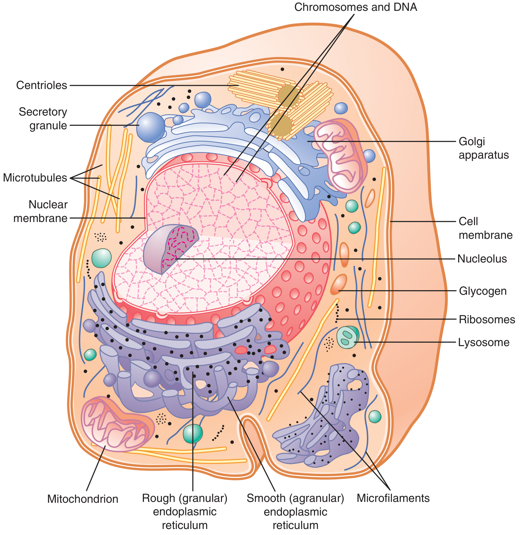
Figure 2.2 - Guyton & Hall: Reconstruction of a typical cell showing organelles
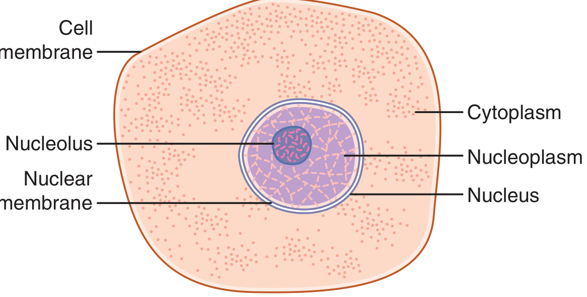
Figure 2.1 - Guyton & Hall: Cell structures visible with a light microscope
Chemical Composition of the Cell
| Substance | % of Cell Mass |
|---|---|
| Water | 70 - 85% |
| Proteins | 10 - 20% |
| Lipids | ~2% |
| Carbohydrates | ~1% (up to 6% in liver) |
| Other substances | Electrolytes, nucleic acids |
Proteins (10-20% of cell mass) fall into two main types:
- Structural proteins - Long filaments forming microtubules (cytoskeleton of cilia, nerve axons, mitotic spindles); fibrillar proteins form collagen and elastin in connective tissue, blood vessel walls, tendons, and ligaments
- Functional proteins - Mainly enzymes in tubular-globular form. Enzymes catalyze chemical reactions (e.g., those splitting glucose and combining it with oxygen to produce CO2, water, and energy)
Lipids: Phospholipids and cholesterol (~2% of cell mass) are insoluble in water and form the cell membrane and intracellular membrane barriers. Triglycerides stored in adipocytes can account for up to 95% of fat cell mass, representing the body's main energy store.
Carbohydrates: Play a major role in nutrition and (as glycoproteins) structural roles. About 1% of cell mass, up to 3% in muscle cells and 6% in liver cells. Stored as glycogen (insoluble polymer of glucose), rapidly depolymerized when energy is needed.
Cell Organelles
Cell Membrane (Plasma Membrane)
A thin, pliable, elastic structure about 7.5 to 10 nanometers thick. Composed mainly of a lipid bilayer with embedded proteins. The membrane:
- Controls the passage of substances into and out of the cell
- Contains receptor proteins for hormones and neurotransmitters
- Contains transport proteins (channels and carriers) for ion and molecular movement
- Has an outer coat (glycocalyx) of carbohydrate-protein complexes involved in cell recognition
Three types of proteins in the membrane:
- Integral proteins (transmembrane proteins) - span the entire membrane; act as channels, carriers, or receptors
- Peripheral proteins - attached to the cytoplasmic surface; often enzymatic or structural
- Lipid-linked proteins - attached to lipids of the bilayer
Endoplasmic Reticulum (ER)
A network of tubules and flat, vesicular structures running throughout the cytoplasm.
- Rough ER (granular ER): Has ribosomes on its outer surface. Synthesizes proteins that are to be secreted from the cell or used in membranes. Proteins pass into the lumen of the ER and are carried to the Golgi apparatus.
- Smooth ER (agranular ER): Lacks ribosomes. Functions include synthesis of lipid substances and steroid hormones, drug detoxification (especially in the liver), and calcium storage/release (in muscle cells, it is called the sarcoplasmic reticulum).
Golgi Apparatus
Located near the nucleus. Receives proteins from the ER and:
- Processes, modifies, and packages them (adds carbohydrate chains to form glycoproteins)
- Sorts proteins - directing them to lysosomes, the cell membrane (secretory vesicles), or extracellular secretion
The Golgi apparatus is especially well developed in secretory cells (e.g., pancreatic acinar cells, pituitary cells).
Mitochondria
The "powerhouses" of the cell. Variable in size (0.5 to 1 micrometer in diameter) and number (few to thousands per cell, depending on energy demand). Mitochondria:
- Generate ATP (adenosine triphosphate) via oxidative phosphorylation
- Contain their own circular DNA, RNA, and ribosomes (remnant of ancient symbiotic bacteria)
- Have an outer membrane and a highly folded inner membrane (cristae) that increases surface area for ATP synthesis
- Operate via the Krebs cycle (TCA cycle) and the electron transport chain
The complete oxidation of 1 molecule of glucose generates approximately 38 ATP molecules.
Key enzymes include:
- Citrate synthase
- Succinate dehydrogenase (Complex II)
- ATP synthase (Complex V)
Lysosomes
Tiny sac-like structures throughout the cytoplasm containing acid hydrolases (proteases, lipases, nucleases, carbohydrases) that can digest:
- Bacteria and foreign substances (from phagocytic vesicles)
- Damaged or excess intracellular organelles (autophagy)
- Cellular debris
After pinocytosis or phagocytosis, lysosomes attach to the vesicle and empty their enzymes into it, forming a digestive vesicle. Products (amino acids, glucose, phosphates) diffuse into the cytoplasm. Indigestible material remains as a residual body, eventually expelled by exocytosis.
When a cell is damaged, lysosomes may rupture and cause autolysis - digestion of the entire cell. This is important in tissue regression and development (e.g., involution of the uterus postpartum, tadpole tail resorption).
Peroxisomes
Similar to lysosomes but contain oxidases rather than hydrolases. Many of their reactions involve hydrogen peroxide (H2O2), which is broken down by catalase. Peroxisomes are especially numerous in liver cells, where they detoxify harmful substances (alcohol) and participate in fatty acid beta-oxidation.
Centrosome and Centrioles
The centrosome is a small region near the nucleus containing two centrioles oriented at right angles to each other. During cell division, the centrioles organize the formation of the mitotic spindle, which pulls chromosomes to opposite poles of the dividing cell.
Nucleus
The nucleus is the control center of the cell. It contains:
- Chromatin - DNA combined with histone proteins (forms chromosomes when condensed during cell division)
- Nucleolus - a dense structure within the nucleus that synthesizes ribosomal RNA (rRNA) and assembles early ribosomal subunits
- Nuclear envelope - double membrane punctuated by large nuclear pores through which mRNA, rRNA, and ribosomal subunits pass into the cytoplasm
The human nucleus contains approximately 6 feet (1.8 m) of DNA, distributed among 46 chromosomes (23 pairs), with 20,000 to 25,000 protein-coding genes.
Cellular Ingestion - Endocytosis
Cells ingest substances from the extracellular fluid by two processes:
Pinocytosis
- Ingestion of small fluid droplets and dissolved substances
- Initiated when protein molecules bind to specific receptors on coated pits (lined with clathrin)
- The pit invaginates inward, pinches off to form a pinocytotic vesicle inside the cytoplasm
- Requires energy (ATP) and calcium ions (Ca2+)
Phagocytosis
- Ingestion of large particles (bacteria, dead cells, tissue debris)
- Occurs mainly in macrophages and some white blood cells
- The particle binds to surface receptors; the membrane evaginates outward in a zipper-like manner to surround the particle, forming a phagocytic vesicle (phagosome)
- Actin and myosin filaments contract and pinch off the vesicle into the cytoplasm
- Opsonization (coating with antibodies) enhances phagocytosis of bacteria
Cell Movements
The most important movement mechanisms:
- Ameboid movement - The cell sends out pseudopods (false feet) by polymerizing actin filaments at the leading edge, creating attachment to a substrate, then contracting the rear portion. Characteristic of macrophages and neutrophils.
- Cilia and flagella - Hair-like protrusions driven by a 9+2 arrangement of microtubules (nine double peripheral tubules + two central single tubules), called the axoneme. Dynein proteins use ATP to generate sliding force between adjacent tubules, producing the whiplike beat. Cilia move mucus in the respiratory tract; flagella propel sperm.
Cell Metabolism and Energy
All cells extract chemical energy from nutrients and use it to:
- Maintain membrane transport systems
- Synthesize new molecules (proteins, lipids, nucleic acids)
- Perform mechanical work (muscle contraction, ciliary movement, cell division)
The ATP-ADP cycle is the universal energy currency:
- Catabolism (nutrient oxidation) produces ATP from ADP + Pi
- Anabolism (biosynthesis, transport) consumes ATP, regenerating ADP + Pi
Energy production from glucose:
- Glycolysis (cytoplasm): 1 glucose → 2 pyruvate + 2 ATP (net) + 2 NADH
- Krebs cycle (mitochondrial matrix): acetyl-CoA → CO2 + NADH + FADH2
- Oxidative phosphorylation (inner mitochondrial membrane): NADH/FADH2 → ATP (32-34 ATP per glucose)
CHAPTER 3: Genetic Control of Protein Synthesis, Cell Function, and Cell Reproduction
Genes Control Cell Function
Genes, located in the nuclei of all cells, control:
- Heredity - from parents to children
- Daily cell function - by determining which structures, enzymes, and chemicals are synthesized
Each gene is composed of deoxyribonucleic acid (DNA) and controls the formation of ribonucleic acid (RNA). This RNA then spreads through the cell to control the formation of a specific protein.
The entire process - from transcription of the genetic code in the nucleus to translation of the RNA code and protein formation in the cytoplasm - is called gene expression.
The human body has approximately 20,000 to 25,000 protein-coding genes per cell, capable of producing at least 100,000 different proteins (due to alternative splicing and post-translational modifications).
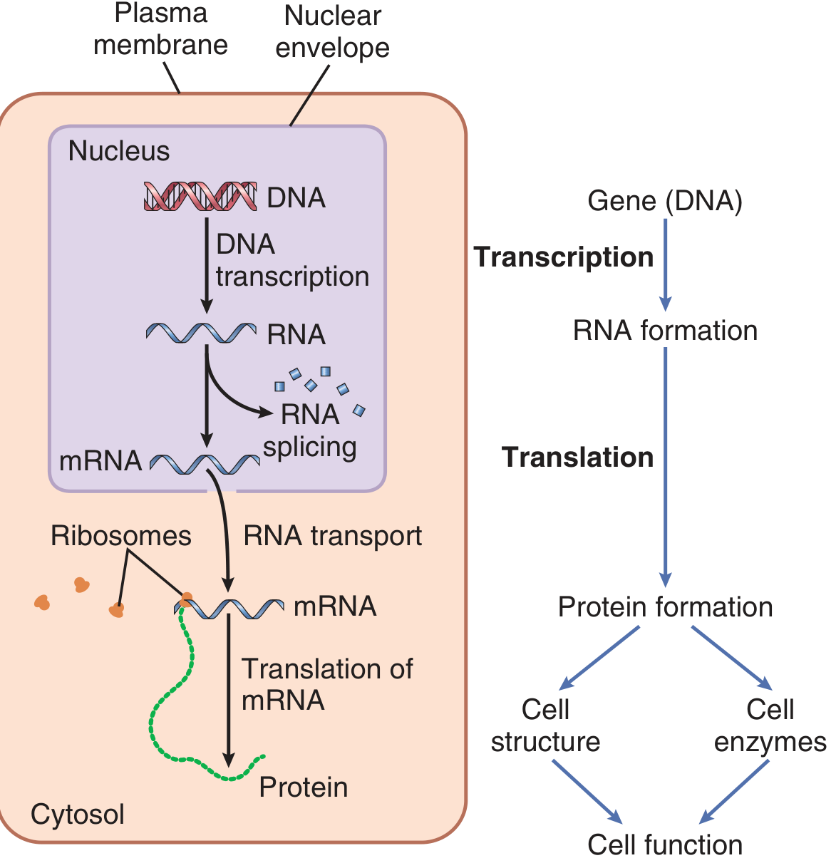
Figure 3.1 - Guyton & Hall: General schema of genetic control via mRNA
Structure of DNA
Building Blocks of DNA
DNA is composed of:
- Phosphoric acid
- Deoxyribose (a 5-carbon sugar)
- Four nitrogenous bases:
- Purines: Adenine (A) and Guanine (G)
- Pyrimidines: Thymine (T) and Cytosine (C)
The phosphoric acid and deoxyribose form the two helical backbone strands. The nitrogenous bases lie between the strands and connect them via hydrogen bonds (A=T, G≡C).
Nucleotides
Each nucleotide = 1 phosphoric acid + 1 deoxyribose + 1 nitrogenous base. Four nucleotides are assembled in specific sequences to form the DNA chain.
The DNA Double Helix:
- Two antiparallel strands wound around each other
- Base pairing rules: A pairs with T (2 H-bonds), G pairs with C (3 H-bonds)
- The sequence of base pairs constitutes the genetic code
The Genetic Code:
- Coded in triplets (codons) of three successive nucleotide bases
- Each triplet codes for one specific amino acid
- 64 possible codons encode 20 amino acids (the code is therefore degenerate - most amino acids are coded by more than one codon)
- Special codons: AUG = start (methionine); UAA, UAG, UGA = stop codons
The Genetic Code - From DNA to Protein
Step 1: Transcription (DNA → mRNA)
The process by which the DNA base sequence is copied into a complementary messenger RNA (mRNA) strand:
- The enzyme RNA polymerase attaches to a promoter region on DNA
- The double helix unwinds and one strand serves as the template strand
- RNA polymerase adds ribonucleotides complementary to the template (A→U, T→A, G→C, C→G)
- A pre-mRNA (primary transcript) is formed
- In the nucleus, RNA processing occurs:
- 5' capping (adds modified guanosine cap)
- 3' poly-A tailing (adds ~200 adenines)
- Splicing - introns (non-coding sequences) are removed; exons (coding sequences) are joined by a spliceosome
- The mature mRNA exits through nuclear pores into the cytoplasm
Step 2: Translation (mRNA → Protein)
Translation occurs on ribosomes in the cytoplasm:
Transfer RNA (tRNA):
- Small cloverleaf-shaped RNA molecules (~80 nucleotides)
- Each tRNA carries a specific amino acid attached to its 3' end
- Contains an anticodon (triplet of bases) that base-pairs with the complementary codon on mRNA
- There is at least one specific tRNA for each of the 20 amino acids
Ribosomal RNA (rRNA):
- Constitutes ~60% of the ribosome; the rest is ~75 types of protein
- Ribosomal subunits are assembled in the nucleolus and transported to the cytoplasm
- A ribosome has two subunits: small (40S) + large (60S) → functional 80S ribosome
Steps of translation:
- Initiation - small ribosomal subunit binds mRNA at the 5' cap and scans to the start codon (AUG); initiator tRNA (carrying methionine) binds; large subunit joins
- Elongation - tRNA with the correct anticodon enters the A-site of the ribosome; a peptide bond forms between the new amino acid and the growing chain; the ribosome advances 3 nucleotides along the mRNA (translocation)
- Termination - when a stop codon (UAA, UAG, or UGA) is reached, release factors cause the completed polypeptide to be released
Polyribosomes (Polysomes): Multiple ribosomes can translate a single mRNA simultaneously, dramatically increasing protein production efficiency.
Types of RNA Involved in Protein Synthesis
| RNA Type | Location | Function |
|---|---|---|
| mRNA (messenger RNA) | Nucleus → cytoplasm | Carries the genetic code from DNA to ribosomes |
| tRNA (transfer RNA) | Cytoplasm | Carries amino acids to the ribosome; anticodon recognizes mRNA codons |
| rRNA (ribosomal RNA) | Nucleolus → ribosome | Structural and catalytic component of the ribosome |
| miRNA (microRNA) | Nucleus → cytoplasm | 21-23 nucleotide fragments that regulate gene expression by binding to complementary mRNA and inhibiting translation or causing mRNA degradation |
| snRNA (small nuclear RNA) | Nucleus | Component of the spliceosome; participates in RNA splicing |
Regulation of Gene Expression
Not all genes are expressed at all times. Gene expression is controlled by:
- Transcription factors - proteins that bind to specific DNA sequences (promoters and enhancers) to activate or repress transcription
- Epigenetic mechanisms - chemical modifications to histones (acetylation, methylation) or DNA (methylation) that alter chromatin structure and gene accessibility without changing the DNA sequence
- miRNA and siRNA - post-transcriptional regulation through mRNA silencing
- Feedback inhibition - the protein product itself can inhibit its own synthesis
Cell Reproduction (Cell Division)
Cell Life Cycle
The cell cycle consists of:
| Phase | Duration | Events |
|---|---|---|
| G1 (Gap 1) | Variable (hours to days) | Cell grows; prepares for DNA synthesis |
| S (Synthesis) | ~10-12 hours | DNA replication occurs |
| G2 (Gap 2) | ~4-6 hours | Cell continues to grow; prepares for mitosis |
| M (Mitosis) | ~1 hour | Cell division |
Cells can also exit the cycle into G0 (quiescence) - a resting state where they neither divide nor prepare to divide. Neurons and cardiac muscle cells are largely in G0.
DNA Replication
DNA replication occurs during the S phase and ensures each daughter cell receives an identical copy of the genome. Key steps:
- Unwinding - DNA helicase breaks hydrogen bonds between base pairs, separating the double helix into a replication fork (Y-shaped)
- Primer binding - DNA primase synthesizes a short RNA primer at the 3' end of the leading strand, providing a starting point
- Elongation:
- Leading strand - replicated continuously in the 5' to 3' direction by DNA polymerase
- Lagging strand - replicated discontinuously as Okazaki fragments (short DNA segments, each preceded by its own RNA primer), also in the 5' to 3' direction
- Ligation - DNA ligase joins Okazaki fragments together and seals the final phosphodiester bonds
- Proofreading - DNA polymerase checks for and corrects errors (~1 error per 10 billion base pairs after correction)
Two identical double-stranded DNA molecules result (semiconservative replication - each new helix contains one original and one new strand).
Mitosis (Nuclear Division)
After DNA replication, the cell undergoes mitosis - division of the nucleus into two identical daughter nuclei with identical sets of 46 chromosomes.
Stages of Mitosis:
1. Prophase:
- Chromatin condenses into visible chromosomes (each consisting of two identical chromatids joined at the centromere)
- The nucleolus disappears
- Centrioles move to opposite poles; the mitotic spindle begins to form from microtubules
2. Prometaphase:
- The nuclear envelope breaks down
- Spindle microtubules attach to chromosomes at the kinetochore (a protein complex at the centromere)
3. Metaphase:
- Chromosomes align along the equatorial plane (metaphase plate) of the cell
- Each chromosome is attached to microtubules from both poles
4. Anaphase:
- Sister chromatids separate and are pulled to opposite poles by shortening of spindle microtubules
- The cell elongates
5. Telophase:
- Chromosomes arrive at opposite poles and begin to decondense
- Nuclear envelopes re-form around each set of chromosomes
- Nucleoli reappear
Cytokinesis (Cytoplasmic Division)
Following telophase, the cytoplasm divides via a cleavage furrow - a ring of actin and myosin filaments contracts around the cell equator, pinching the cell into two equal daughter cells, each with a complete set of chromosomes and organelles.
Mutations and DNA Repair
Mutations are heritable changes in the DNA sequence:
- Point mutations - substitution of one base pair
- Insertions/deletions (indels) - addition or removal of bases, often causing frameshifts
- Causes: Radiation (UV, X-rays), chemical mutagens, replication errors
DNA repair mechanisms:
- Base excision repair - removes damaged single bases
- Nucleotide excision repair - removes bulky lesions (e.g., UV-induced thymine dimers)
- Mismatch repair - corrects mismatches after replication
- Double-strand break repair - uses homologous recombination or non-homologous end joining
If DNA damage is too extensive, the cell either undergoes apoptosis (programmed cell death) or, if repair mechanisms fail, may become a cancer cell.
Summary Table
| Topic | Key Concept |
|---|---|
| Internal environment | ECF (~14 L) - the milieu in which all cells live |
| Homeostasis | Maintenance of nearly constant ECF conditions by negative feedback |
| Cell size | ~10 µm; 35-40 trillion cells in the human body |
| Cell membrane | 7.5-10 nm lipid bilayer with integral and peripheral proteins |
| Mitochondria | ATP production via Krebs cycle + oxidative phosphorylation |
| Lysosomes | Acid hydrolases for intracellular digestion |
| Gene number | ~20,000-25,000 protein-coding genes per cell |
| DNA structure | Double helix; A=T (2H), G≡C (3H); antiparallel strands |
| Transcription | DNA → mRNA via RNA polymerase in the nucleus |
| Translation | mRNA → protein via ribosomes in the cytoplasm |
| tRNA anticodon | Recognizes mRNA codon; delivers correct amino acid |
| Cell cycle | G1 → S (DNA replication) → G2 → M (mitosis) |
| Mitosis phases | Prophase → Prometaphase → Metaphase → Anaphase → Telophase |
| DNA replication | Semiconservative; leading strand continuous, lagging strand as Okazaki fragments |
All content sourced from: Guyton and Hall Textbook of Medical Physiology, 14th Edition, Chapters 1, 2, and 3.
This is a shared conversation. Sign in to Orris to start your own chat.