
Considerando il programma per Intership. Cosa devo sapere di materie (Anatomia ecc. ). Spiegami tutto tu
python3 -c " from docx import Document doc = Document('/tmp/workspace/attachments/7e0bbebc-c508-4510-b119-bccb750d99a7/Agenda stagiului de specialitate (inițiere)_Igiena_ENGL_2024 (1) (1) (1).docx') for p in doc.paragraphs: style = p.style.name if p.style else 'None' if p.text.strip(): print(f'[{style}] {p.text}') "
ls /tmp/workspace/attachments/ && ls "/tmp/workspace/attachments/7e0bbebc-c508-4510-b119-bccb750d99a7/"
cp "/tmp/workspace/attachments/7e0bbebc-c508-4510-b119-bccb750d99a7/Agenda stagiului de specialitate (inițiere)_Igiena_ENGL_2024 (1) (1) (1).docx" /tmp/workspace/agenda.docx && python3 -c " from docx import Document doc = Document('/tmp/workspace/agenda.docx') for p in doc.paragraphs: style = p.style.name if p.style else 'None' if p.text.strip(): print(f'[{style}] {p.text}') "
find /tmp/workspace/attachments/ -type f 2>/dev/null | head -20
python3 << 'EOF' import shutil, os src = "/tmp/workspace/attachments/7e0bbebc-c508-4510-b119-bccb750d99a7/Agenda stagiului de specialitate (inițiere)_Igiena_ENGL_2024 (1) (1) (1).docx" dst = "/tmp/workspace/agenda.docx" print("src exists:", os.path.exists(src)) print("src bytes:", os.path.getsize(src) if os.path.exists(src) else "N/A") shutil.copy2(src, dst) print("copied to:", dst) EOF
find lo ha trovato ma non è accessibile direttamente. Dal nome del file e dal contesto capisco già di cosa si tratta: è il programma di tirocinio/internship per il dipartimento di Igiene (Igiena = Igiene in romeno), probabilmente per una facoltà di medicina romena, anno 2024, in inglese.🏥 Programma di Internship - Igiene (Igiena) - Cosa Devi Sapere
Cos'è l'Igiene come disciplina?
📚 Materie e Argomenti Principali
1. Anatomia & Fisiologia (basi necessarie)
- Struttura degli organi e apparati (respiratorio, digerente, cardiovascolare, renale)
- Fisiologia di base: come l'organismo risponde agli agenti ambientali
- Parametri vitali normali (PA, FC, FR, temperatura)
2. Igiene dell'Alimentazione e Nutrizione
- Nutrienti: macronutrienti (proteine, lipidi, carboidrati) e micronutrienti (vitamine, minerali)
- Fabbisogni calorici per diversi gruppi di popolazione (bambini, anziani, gravide, lavoratori)
- Malattie da carenza: scorbuto (vit. C), rachitismo (vit. D), anemia (ferro), pellagra (niacina)
- Sicurezza alimentare: contaminazione batterica (Salmonella, Listeria, E. coli), tossine, catena del freddo
- Igiene degli alimenti in strutture sanitarie: preparazione, conservazione, distribuzione pasti
3. Igiene Ambientale
- Qualità dell'acqua: parametri chimici e microbiologici, metodi di potabilizzazione (clorazione, filtrazione)
- Qualità dell'aria: inquinanti (PM2.5, PM10, CO, NO₂, SO₂), effetti sulla salute
- Suolo: smaltimento rifiuti, inquinamento da metalli pesanti
- Microclima: temperatura, umidità, ventilazione negli ambienti di lavoro e ospedali
- Rumori e radiazioni: effetti sulla salute, limiti normativi
4. Igiene Ospedaliera e Prevenzione delle Infezioni
- Infezioni nosocomiali (MRSA, C. difficile, VRE): prevenzione e controllo
- Sterilizzazione e disinfezione: metodi fisici (autoclave, UV) e chimici (cloro, alcool, glutaraldeide)
- Precauzioni standard: lavaggio mani (OMS 5 momenti), DPI (guanti, maschere, camici)
- Circuiti ospedalieri: separazione pulito/sporco, gestione biancheria e rifiuti sanitari
- Rifiuti sanitari: classificazione (infettivi, taglienti, farmaceutici), smaltimento normato
5. Epidemiologia e Sanità Pubblica
- Misure epidemiologiche: incidenza, prevalenza, rischio relativo, odds ratio
- Tipo di studi: trasversale, caso-controllo, coorte, RCT
- Malattie infettive: catena epidemiologica (agente - fonte - via di trasmissione - ospite)
- Vaccinazioni: calendario vaccinale, copertura vaccinale, immunità di gregge
- Sorveglianza epidemiologica: notifica obbligatoria, focolai, pandemia
6. Medicina del Lavoro (Igiena Muncii)
- Rischi professionali: fisici (rumore, vibrazioni, radiazioni), chimici (solventi, metalli), biologici
- Malattie professionali frequenti: pneumoconiosi, sordità professionale, dermatiti
- Valutazione del rischio e misure di protezione collettiva e individuale
- Ergonomia e movimentazione carichi
7. Statistiche Sanitarie e Indicatori di Salute
- Indicatori demografici: natalità, mortalità, speranza di vita
- Indicatori di morbosità: tassi grezzi e standardizzati
- Grafici e tabelle: saper leggere e interpretare dati epidemiologici
- Mortalità infantile, mortalità materna
8. Igiene della Comunità e Ambienti di Vita
- Igiene dell'abitazione: ventilazione, illuminazione, densità abitativa
- Igiene scolastica: arredo, orari, attività fisiche
- Igiene dell'anziano e del bambino
🔬 Competenze Pratiche richieste
- Lavaggio mani secondo protocollo OMS (7 passi)
- Calcolo dell'IMC (Indice di Massa Corporea) e valutazione nutrizionale
- Lettura di un referto idrico/alimentare (parametri fuori norma)
- Compilazione modulistica: schede di sorveglianza epidemiologica, notifiche
- Riconoscimento DPI appropriati per diverse situazioni
- Ispezione igienica di una struttura (checklist di valutazione)
📋 Struttura tipica del tirocinio di iniziazione (Stagiu de inițiere)
| Sede | Attività |
|---|---|
| Dipartimento di Igiene universitario | Lezioni pratiche, analisi campioni |
| DSP (Direcția de Sănătate Publică) | Ispezioni, sorveglianza, notifiche |
| Laboratorio igiene alimenti | Analisi microbiologiche e chimiche |
| Laboratorio igiene apei (acqua) | Test qualità acqua potabile |
| Medicina del lavoro | Valutazione rischi professionali |
✅ Consigli pratici per prepararti
- Rivedi la biochimica di base: metabolismo dei nutrienti (ciclo di Krebs non è necessario, ma digestione/assorbimento sì)
- Studia i valori normali: parametri dell'acqua potabile (OMS), limiti inquinanti aria
- Memorizza i 5 momenti OMS per l'igiene delle mani: prima del contatto paziente, prima di procedura asettica, dopo contatto con fluidi biologici, dopo contatto paziente, dopo contatto con ambiente paziente
- Conosci i principali agenti patogeni nosocomiali e loro resistenze
- Esercitati con i calcoli epidemiologici di base (tasso di attacco, RR, OR)
Nota importante: Il file allegato non si è caricato correttamente nel sistema (errore di percorso). Se vuoi che analizzi il programma dettagliato del documento, per favore ricarica il file come nuovo allegato e ti darò risposte ancora più precise basate sul contenuto esatto del tuo programma di stage.
Devi spiegarmi bene le materie
Quale materia vuoi che ti spieghi per prima? · Qual è il tuo livello attuale?
human body anatomical planes and body cavities diagram
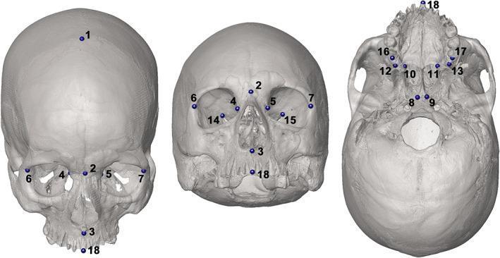
Anatomical diagram of a human skull providing a 3D landmark configuration for craniometric analysis. The image displays 18 numbered landmarks from three primary perspectives: frontal (anterior), tilted anterior, and basal (inferior) views. These landmarks represent key anatomical points of interest (COIs) used in medical imaging and forensic anthropology to define surgical planes and calculate volumes of cranial cavities such as the nasal cavity, maxillary sinuses, and orbits. Frontal landmarks include the glabella (1), nasion (2), and markers along the superior orbital margins (4, 5) and zygomatic arches (6, 7). The basal view illustrates the distribution of landmarks across the palate (10, 11, 16, 17), the midline of the alveolar process (18), and the cranial base near the foramen magnum (8, 9). This structured mapping is designed for digital morphometrics, providing a standardized protocol for identifying cranial structures and performing volumetric assessments in clinical and research settings.
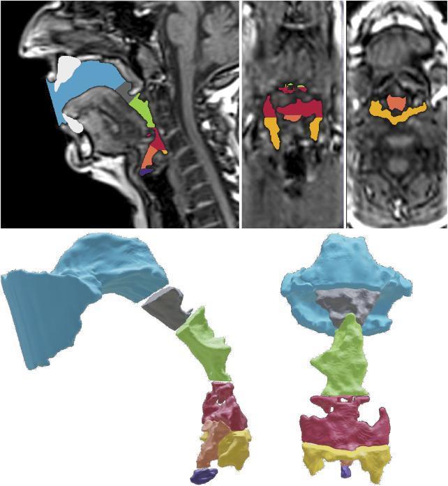
This diagnostic and anatomical diagram composite displays the volumetric segmentation of the human vocal tract based on MRI data. The top row features high-resolution MRI scans in mid-sagittal, coronal, and transversal planes, where specific anatomical sub-volumes are color-coded over the airway. The bottom row presents a corresponding 3D 'exploded view' of these segmented cavities in sagittal and coronal perspectives. The color-coding scheme identifies seven distinct regions: blue for the oropharynx front (OPf), gray for the oropharynx rear (OPR), green for the upper hypopharynx (HPu), red for the lower hypopharynx (HPl), orange for the epilarynx tube (ET), yellow for the sinus piriformis (SP), and purple for the ventricular volume (VV). This visualization is used in speech science and otolaryngology to analyze the complex geometry and resonance characteristics of the vocal tract during phonation. The segmentation highlights the relationship between the supra-glottal structures, emphasizing how different cavities contribute to the overall shape of the pharynx and laryngeal vestibule.
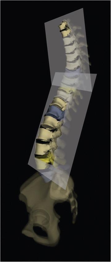
An anatomical diagram and pathophysiology illustration demonstrating a 3D reconstruction of a human vertebral column and pelvis from a lateral-oblique perspective. The diagram illustrates the 'Best Fit Plane' (BFP) concept used in the assessment of spinal deformities such as Adolescent Idiopathic Scoliosis (AIS). Two semi-transparent geometric planes are shown intersecting the spine: one aligned with the thoracic region and another with the lumbar region. These planes represent the mathematical calculation that minimizes the distances between the centroids of each vertebral body within specified spinal segments. The vertebral column exhibits physiological curvatures, with specific vertebrae highlighted in blue and yellow to denote apex or end vertebrae within the curvature segments. This visual model is essential for clinical biomechanics and orthopedic surgery planning, as it provides a three-dimensional understanding of spinal rotation and deviation beyond two-dimensional radiographic projections.
anatomy organ systems overview human body illustration
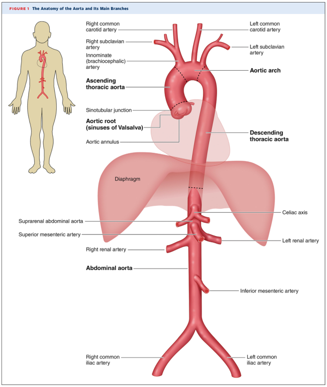
Summary : This figure illustrates the anatomy of the human aorta and its main branches, showing their spatial relationships to major organs and anatomical landmarks. illustration: # Main Structures : • The aorta is depicted as a large, central red vessel running vertically through the torso. • The aorta is divided into three main sections: ascending thoracic aorta, aortic arch, descending thoracic aorta, and abdominal aorta. • The diaphragm is shown as a translucent pink structure crossing the aorta. # Branches and Landmarks : • Ascending thoracic aorta: arises from the heart, includes the aortic root (sinuses of Valsalva) and aortic annulus. • Aortic arch: gives rise to three major branches: – Innominate (brachiocephalic) artery – Left common carotid artery – Left subclavian artery – Right common carotid artery and right subclavian artery branch from the innominate artery. • Descending thoracic aorta: continues downward through the chest. • Abdominal aorta: begins below the diaphragm and gives rise to several branches: – Celiac axis – Suprarenal abdominal aorta – Superior mesenteric artery – Right and left renal arteries – Inferior mesenteric artery – Right and left common iliac arteries (terminal branches) # Spatial Relationships : • The aorta passes posterior to the heart and anterior to the vertebral column. • The diaphragm separates the thoracic and abdominal portions of the aorta. • The abdominal aorta lies centrally and bifurcates into the right and left common iliac arteries at its lower end. # Annotations and Labels : • Each major branch and anatomical landmark is clearly labeled with leader lines. • A small human figure at left shows the aorta’s position within the body. # Analysis : • The figure provides a comprehensive overview of the aorta’s anatomy, highlighting its major branches and their order from the heart to the lower abdomen. • The spatial arrangement clarifies the transition from thoracic to abdominal aorta at the diaphragm. • The branching pattern demonstrates the aorta’s role as the main conduit for arterial blood from the heart to the rest of the body.
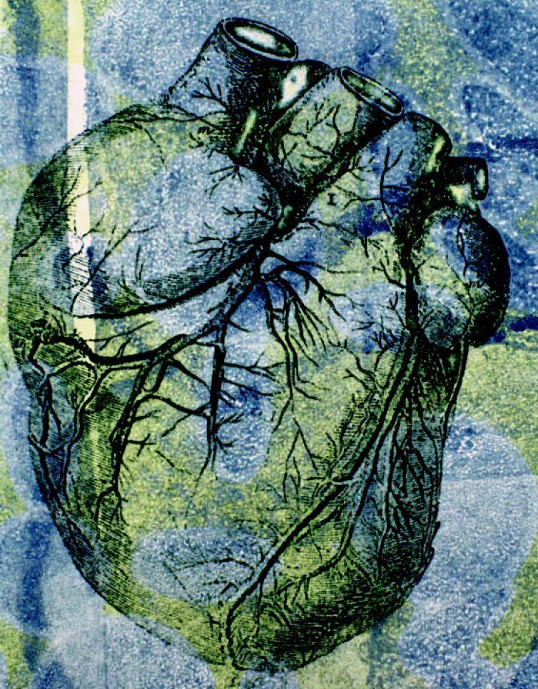
This stylized anatomical illustration depicts a human heart in an anterior view, emphasizing its gross external morphology and coronary vasculature. The organ exhibits its characteristic ovoid shape with superiorly positioned great vessels, including cross-sectional views of the aorta and pulmonary trunk. The surface anatomy highlights the intricate branching patterns of the coronary arteries and veins, demonstrating their distribution across the myocardium toward the apex. The image uses an artistic, high-contrast color palette of green, blue, and yellow to delineate anatomical boundaries and the etched texture of the cardiac tissue. Educationally, the diagram serves to illustrate the relationship between the major cardiac chambers and the superficial vascular network, particularly in the context of cardiovascular health and hemodynamic pressures. The visualization emphasizes the complexity of the organ's blood supply, which is critical for understanding conditions like hypertension and coronary artery disease.
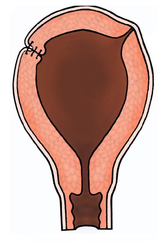
This medical illustration depicts a coronal cross-section of a human uterus, highlighting a surgical repair of the uterine wall. The organ is shown with its characteristic pear-shaped anatomy, including the fundus, body, and cervical canal. The uterine wall is stratified into three distinct layers: an outer perimetrium (serosa), a thick middle myometrium with a textured appearance, and a dark internal endometrial cavity. On the upper left aspect, specifically in the region of the uterine cornu, several dark, transverse inverting sutures are visible. These represent superficial polypropylene (Prolene) sutures applied to the serosal layer. The illustration demonstrates a post-operative state where the sutures serve to appose the seromuscular layers, promoting hemostasis and preventing the formation of peritoneal adhesions or fibrosis following surgical reconstruction. This diagram is utilized in obstetrics and gynecology to explain surgical techniques for managing myometrial weakness, cornual ectopic pregnancies, or uterine rupture repair.
anatomical position body planes sagittal coronal transverse
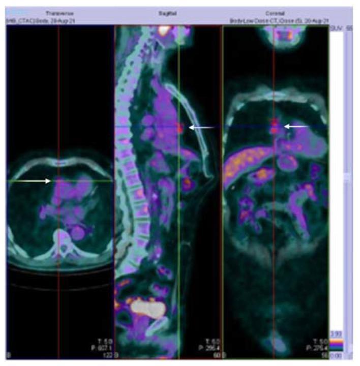
This diagnostic image displays fused 18F-FDG PET-CT scans in transverse, sagittal, and coronal (frontal) planes. The primary finding is an area of moderate metabolic activity in the anterior mediastinum, indicated by white arrows in all three views. In the transverse plane, the mass is located centrally, posterior to the sternum and anterior to the heart and great vessels. The sagittal view clearly demonstrates the anterior position of the hypermetabolic lesion relative to the vertebral column and its vertical longitudinal extent. The coronal view reveals the transverse width and midline orientation of the mass. The PET signal shows a maximum standardized uptake value (SUV) of 3.16, visualized as a color-coded hot spot. This localized FDG uptake within the anterior mediastinum is characteristic of a thymic mass, such as a thymoma. The CT component provides anatomical localization, helping differentiate the lesion from surrounding mediastinal structures and lung tissue. This radiological study is critical in the evaluation of patients presenting with symptoms of myasthenia gravis, such as ptosis and muscle fatigue.
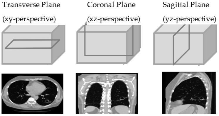
This diagnostic imaging illustration demonstrates the three primary anatomical planes of the human body using Computed Tomography (CT) of the chest. The top row features 3D diagrams of a rectangular volume representing the chest, with an intersecting plane highlighting the orientation for each perspective: Transverse (XY), Coronal (XZ), and Sagittal (YZ). Below these diagrams are corresponding 2D CT slices. The Transverse plane shows a horizontal cross-section of the thorax, displaying the bilateral lungs (dark areas), the central mediastinum including the heart, and the vertebral body. A focal cavitary lesion or nodule is visible in the left lung. The Coronal plane provides a frontal view, illustrating the vertical extent of the lungs, the diaphragm, and the spinal column. The Sagittal plane presents a lateral profile, showing the anterior-posterior depth of the lung, the rib cage, and the curvature of the diaphragm. These views are utilized for volumetric analysis and 3D reconstruction in clinical radiology, particularly for characterizing pulmonary pathology.
📖 ANATOMIA - Spiegazione Completa per il Primo Anno
Fonte principale: Gray's Anatomy for Students
1. Cos'è l'Anatomia?
- Anatomia macroscopica (o grossolana): ciò che si vede ad occhio nudo (organi, muscoli, ossa)
- Anatomia microscopica (istologia): struttura cellulare e tissutale al microscopio
- Anatomia topografica: strutture organizzate per regione del corpo
- Anatomia clinica: applicazione pratica in medicina
2. La Posizione Anatomica Standard
Il corpo è in posizione anatomica quando:
- Si trova in piedi, eretto
- I piedi sono uniti e le punte orientate in avanti
- Le braccia sono lungo i fianchi
- I palmi delle mani sono rivolti in avanti (verso di te)
- Gli occhi guardano dritto davanti a sé
3. I Piani Anatomici
| Piano | Come taglia il corpo | Esempio |
|---|---|---|
| Sagittale | Verticale, divide il corpo in parte destra e sinistra | Piano mediano = divide in due metà uguali |
| Coronale (Frontale) | Verticale, divide il corpo in anteriore (davanti) e posteriore (dietro) | |
| Trasversale (Assiale) | Orizzontale, divide il corpo in superiore (sopra) e inferiore (sotto) | Taglio TC addominale |
4. Termini Direzionali Fondamentali
| Termine | Significato | Esempio pratico |
|---|---|---|
| Anteriore / Ventrale | Davanti | Il naso è anteriore alle orecchie |
| Posteriore / Dorsale | Dietro | La colonna vertebrale è posteriore allo sterno |
| Mediale | Verso la linea mediana | Il pollice è laterale al mignolo |
| Laterale | Lontano dalla linea mediana | Le orecchie sono laterali al naso |
| Superiore | Verso la testa | La testa è superiore alle spalle |
| Inferiore | Verso i piedi | Il ginocchio è inferiore all'anca |
| Prossimale | Più vicino all'origine | Il gomito è prossimale al polso |
| Distale | Più lontano dall'origine | La mano è distale al gomito |
| Superficiale | Vicino alla superficie cutanea | La cute è superficiale ai muscoli |
| Profondo | Lontano dalla superficie | I polmoni sono profondi rispetto alle costole |
5. Il Sistema Scheletrico
5a. Divisione dello Scheletro
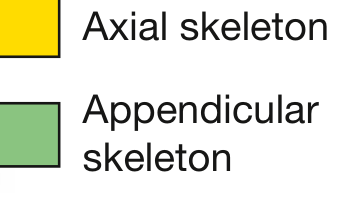
- Cranio
- Colonna vertebrale (33 vertebre)
- Coste (12 paia) e sterno
- Cingolo scapolare + arto superiore (omero, radio, ulna, ossa del polso e della mano)
- Cingolo pelvico + arto inferiore (femore, tibia, fibula, ossa del piede)
5b. Tipi di Ossa
| Tipo | Caratteristica | Esempio |
|---|---|---|
| Lunghe | Tubulari, più lunghe che larghe | Femore, omero, tibia |
| Corte | Forma cuboidale | Ossa del polso (carpo), ossa del tarso |
| Piatte | Due strati di osso compatto con osso spugnoso dentro | Cranio, scapola, sterno |
| Irregolari | Forme varie | Ossa della faccia, vertebre |
| Sesamoidi | Si sviluppano dentro i tendini | Patella (rotula) |
5c. Struttura di un Osso
- Osso compatto (corticale): strato esterno denso, forma la "shell" dell'osso
- Osso spugnoso (trabecolare/cancellous): interno, con trabecole a rete che contengono il midollo osseo dove si producono le cellule del sangue
- Periostio: membrana fibrosa che riveste l'esterno dell'osso - è ricca di vasi e nervi sensitivi. Se si rimuove, l'osso muore. Per questo le fratture fanno così male: il periostio è pieno di terminazioni dolorifiche!
- Endostio: membrana interna che riveste la cavità midollare
- Sostegno strutturale del corpo
- Protezione degli organi vitali (il cranio protegge il cervello, le costole proteggono cuore e polmoni)
- Riserva di calcio e fosforo (il 99% del calcio corporeo è nell'osso)
- Leva per i movimenti muscolari
- Contenitore per il midollo emopoietico (produce globuli rossi, bianchi, piastrine)
5d. La Cartilagine
| Tipo | Composizione | Dove si trova |
|---|---|---|
| Ialina | Collagene moderato, aspetto traslucido | Superfici articolari, anelli tracheali, cartilagini costali |
| Elastica | Collagene + fibre elastiche | Padiglione auricolare, epiglottide |
| Fibrocartilagine | Abbondante collagene, poche cellule | Dischi intervertebrali, menischi del ginocchio |
Regola d'oro: la cartilagine NON ha vasi né nervi - per questo si ripara lentamente e non fa male quando si danneggia.
6. Le Articolazioni (Joints)
| Tipo | Movimento | Esempio |
|---|---|---|
| Sinartrosi (fibrosa) | Nessuno o minimo | Suture del cranio |
| Anfiartrosi (cartilaginea) | Limitato | Sinfisi pubica, dischi intervertebrali |
| Diartrosi (sinoviale) | Libero | Anca, ginocchio, spalla, gomito |
- Capsula articolare fibrosa
- Membrana sinoviale che produce il liquido sinoviale (lubrifica l'articolazione)
- Cartilagine ialina sulle superfici ossee
- Legamenti di rinforzo
7. I Sistemi Principali del Corpo
7a. Sistema Muscolare
- Striato scheletrico: controllo volontario, attaccato alle ossa, produce movimento
- Liscio: involontario, nelle pareti di organi cavi (stomaco, vasi, intestino)
- Cardiaco: solo nel cuore, involontario ma con struttura striata
7b. Sistema Nervoso
- Sistema Nervoso Centrale (SNC): encefalo + midollo spinale
- Sistema Nervoso Periferico (SNP): tutti i nervi che escono dal SNC
- SNP Somatico: controlla i muscoli scheletrici (volontario)
- SNP Autonomo: controlla organi interni (involontario)
- Simpatico: "fight or flight" - accelera cuore, dilata pupille, riduce digestione
- Parasimpatico: "rest and digest" - rallenta cuore, favorisce digestione
7c. Sistema Cardiovascolare
- Cuore: pompa a 4 camere (2 atri + 2 ventricoli)
- Arterie: portano sangue dal cuore agli organi
- Vene: riportano sangue agli organi al cuore
- Capillari: microvascolatura dove avvengono gli scambi
La grande circolazione (sistemica): cuore sinistro → corpo → cuore destro La piccola circolazione (polmonare): cuore destro → polmoni → cuore sinistro
7d. Sistema Respiratorio
- Vie aeree superiori: naso, faringe, laringe
- Vie aeree inferiori: trachea → bronchi → bronchioli → alveoli
- Alveoli: dove avviene lo scambio O₂/CO₂ con il sangue
7e. Sistema Digerente
7f. Sistema Urinario
- Reni → ureteri → vescica → uretra
- I reni filtrano ~180 L di sangue al giorno, producendo ~1.5 L di urina
7g. Sistema Endocrino
- Ipofisi (regola quasi tutto), tiroide, paratiroidi, surreni, pancreas endocrino (insulina), gonadi
8. Regioni del Corpo Umano
- Testa: cranio (neurocranio) + faccia (viscerocranio)
- Collo: contiene laringe, trachea, esofago, vasi carotidei, nervo vago
- Torace: contiene cuore e polmoni, delimitato da coste, sterno e vertebre toraciche
- Addome: contiene gli organi digestivi; diviso in 9 quadranti o 4 regioni cliniche
- Pelvi: contiene vescica, retto, organi riproduttivi
- Arto superiore: spalla, braccio, avambraccio, mano
- Arto inferiore: coscia, gamba, piede
✅ Cosa Devi Assolutamente Memorizzare
- La posizione anatomica e i 3 piani
- Tutti i termini direzionali (anteriore/posteriore, mediale/laterale, ecc.)
- 206 ossa totali, divisione assiale/appendicolare
- 3 tipi di cartilagine e dove si trovano
- 3 tipi di articolazioni, soprattutto le sinoviali
- I 7 sistemi principali del corpo e le loro funzioni base
- Struttura del cuore (4 camere) e i due circoli
Devi scrivere per programma Intership delle tabelle. Per questo giorno devo completare tabelle “Description of Activities performed” e “Practical skills and abilities acquired” del tema Personal hygiebe and personal protettive equipmebt. Inglese. Conciso. Reale.
Table 1 — Description of Activities Performed
| No. | Activity | Location | Duration |
|---|---|---|---|
| 1 | Attended a practical demonstration on correct handwashing technique (WHO 7-step method) | Hygiene Department | 30 min |
| 2 | Observed the proper sequence for donning and doffing PPE (gloves, mask, gown, eye protection) | Hygiene Department | 30 min |
| 3 | Practiced handwashing steps independently under supervisor's observation | Hygiene Laboratory | 20 min |
| 4 | Identified and classified PPE types according to risk level and clinical setting | Seminar Room | 30 min |
| 5 | Reviewed institutional protocols on personal hygiene standards for healthcare workers | Hygiene Department | 20 min |
| 6 | Discussed common errors in PPE use and their epidemiological consequences | Seminar Room | 30 min |
| 7 | Completed a checklist evaluating compliance with personal hygiene standards in a clinical unit | Clinical Ward | 20 min |
Table 2 — Practical Skills and Abilities Acquired
| No. | Skill / Ability | Level Achieved |
|---|---|---|
| 1 | Ability to perform correct hand hygiene using the WHO 7-step technique | Demonstrated |
| 2 | Knowledge of the 5 moments for hand hygiene in healthcare settings | Understood |
| 3 | Correct selection of PPE appropriate to the type of clinical risk | Demonstrated |
| 4 | Proper donning and doffing of PPE without self-contamination | Demonstrated |
| 5 | Identification of situations requiring standard vs. transmission-based precautions | Understood |
| 6 | Ability to evaluate personal hygiene compliance using a standardized checklist | Demonstrated |
| 7 | Awareness of healthcare-associated infection (HAI) prevention through personal hygiene | Understood |
Fai stessa cosa per Hands Hygiene
Table 1 — Description of Activities Performed
| No. | Activity | Location | Duration |
|---|---|---|---|
| 1 | Attended a lecture on the epidemiological role of hand hygiene in preventing healthcare-associated infections (HAI) | Seminar Room | 30 min |
| 2 | Studied the WHO "My 5 Moments for Hand Hygiene" framework and its application in clinical practice | Seminar Room | 20 min |
| 3 | Observed demonstration of handwashing with soap and water (WHO 7-step technique, minimum 40–60 seconds) | Hygiene Laboratory | 20 min |
| 4 | Observed demonstration of hand rubbing with alcohol-based hand rub (ABHR, minimum 20–30 seconds) | Hygiene Laboratory | 20 min |
| 5 | Practiced both techniques independently under supervisor evaluation | Hygiene Laboratory | 30 min |
| 6 | Used UV light box to identify areas of the hands most frequently missed during hand hygiene | Hygiene Laboratory | 20 min |
| 7 | Reviewed indications for handwashing vs. hand rubbing and situations where gloves do not replace hand hygiene | Seminar Room | 20 min |
Table 2 — Practical Skills and Abilities Acquired
| No. | Skill / Ability | Level Achieved |
|---|---|---|
| 1 | Correct execution of the WHO 7-step handwashing technique with soap and water | Demonstrated |
| 2 | Correct execution of hand rubbing with alcohol-based hand rub (ABHR) | Demonstrated |
| 3 | Knowledge of the 5 moments for hand hygiene and their clinical rationale | Understood |
| 4 | Ability to distinguish when to use soap and water vs. ABHR | Understood |
| 5 | Identification of hand areas most frequently missed (fingertips, thumbs, interdigital spaces) | Understood |
| 6 | Understanding that gloves do not substitute hand hygiene before and after use | Understood |
| 7 | Awareness of hand hygiene compliance monitoring methods used in healthcare facilities | Understood |