
Femoral triangle
Reading File
Finding Sources
Searching Images
labeled anatomical diagram of femoral triangle boundaries contents femoral nerve artery vein lymphatics
Searching PubMed
"femoral triangle" anatomy
Searching the Web
femoral triangle anatomy boundaries contents clinical relevance
Reading File
Reading File
Reading File
Femoral triangle
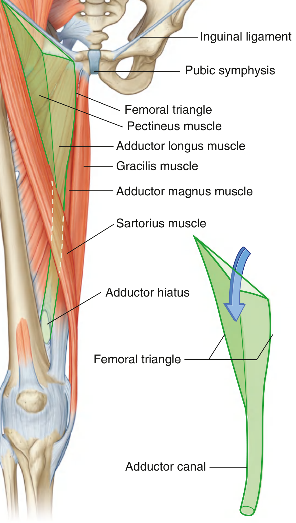
A triangular, subfascial depression in the upper anterior thigh. It is a passageway for major nerves, vessels, and lymphatics between the abdomen and lower limb.
Boundaries
| Part | Formed by |
|---|---|
| Base (superior) | Inguinal ligament |
| Lateral boundary | Medial border of sartorius |
| Medial boundary | Medial border of adductor longus |
| Apex | Where sartorius crosses adductor longus, continuing into the adductor canal |
| Floor | Iliopsoas laterally, pectineus and adductor longus medially |
| Roof | Skin, superficial fascia, fascia lata, including cribriform fascia at the saphenous opening |
Contents
From lateral to medial, remember NAVEL:
- N: Femoral nerve and its branches
- A: Femoral artery and branches
- V: Femoral vein and tributaries, notably the great saphenous vein
- E: Empty space, the femoral canal
- L: Deep inguinal lymphatics/nodes
The femoral nerve lies outside the femoral sheath. The femoral sheath encloses the femoral artery, femoral vein, and femoral canal. The canal is its medial compartment and contains lymphatics. Gray's Anatomy for Students, p. 675.
Clinical importance
- Femoral pulse: felt just inferior to the inguinal ligament at the mid-inguinal point, halfway between the ASIS and pubic symphysis.
- Vascular access: femoral artery or vein can be accessed here for catheterization.
- Femoral nerve block: the nerve is lateral to the artery.
- Femoral hernia: protrudes through the femoral ring into the femoral canal, typically presenting inferolateral to the pubic tubercle. The narrow, rigid canal predisposes to obstruction or strangulation.
- Lymph nodes: superficial/deep inguinal nodes may enlarge with infections or malignancy affecting the lower limb, external genitalia, or lower anterior abdominal wall.
The femoral artery and vein continue through the adductor canal and become the popliteal vessels posterior to the knee. Gray's Anatomy for Students, p. 675. A useful external anatomy reference also summarizes the NAVEL arrangement.
Knee joint
Reading File
Finding Sources
Finding Sources
Searching Images
labeled anatomy diagram knee joint ligaments menisci cruciate collateral
Searching PubMed
"knee joint" anatomy
Searching the Web
knee joint anatomy ligaments menisci movements reputable medical reference
Reading File
Reading File
Knee joint
The knee is the largest synovial joint in the body. It is a modified hinge joint that mainly permits flexion and extension, with a small degree of rotation when flexed.
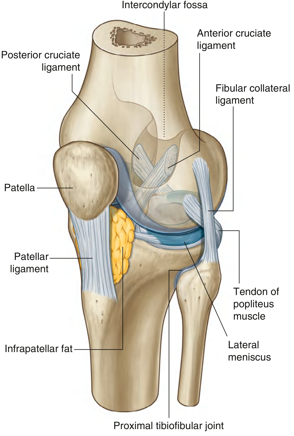
It has two articulations within one joint cavity:
- Tibiofemoral joint: between femoral condyles and tibial condyles. This bears body weight.
- Patellofemoral joint: between posterior patella and the patellar surface of femur. It improves the line of pull and mechanical advantage of quadriceps.
Gray's Anatomy for Students, p. 709.
Articular surfaces
- Medial and lateral femoral condyles
- Superior articular surfaces of medial and lateral tibial condyles
- Posterior articular surface of patella
- All articular surfaces are covered by hyaline cartilage.
Menisci
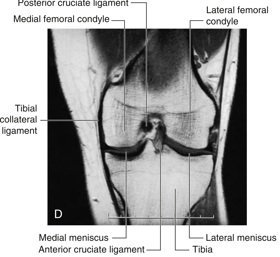
Two C-shaped fibrocartilaginous structures lie between femur and tibia:
| Medial meniscus | Lateral meniscus |
|---|---|
| More firmly attached to capsule and medial collateral ligament | More mobile |
| More commonly injured | Less commonly injured |
| Less circular, C-shaped | Nearly circular |
Functions: deepen tibial articular surfaces, improve congruence, absorb shock, distribute load, and assist joint stability.
Ligaments
Extracapsular ligaments
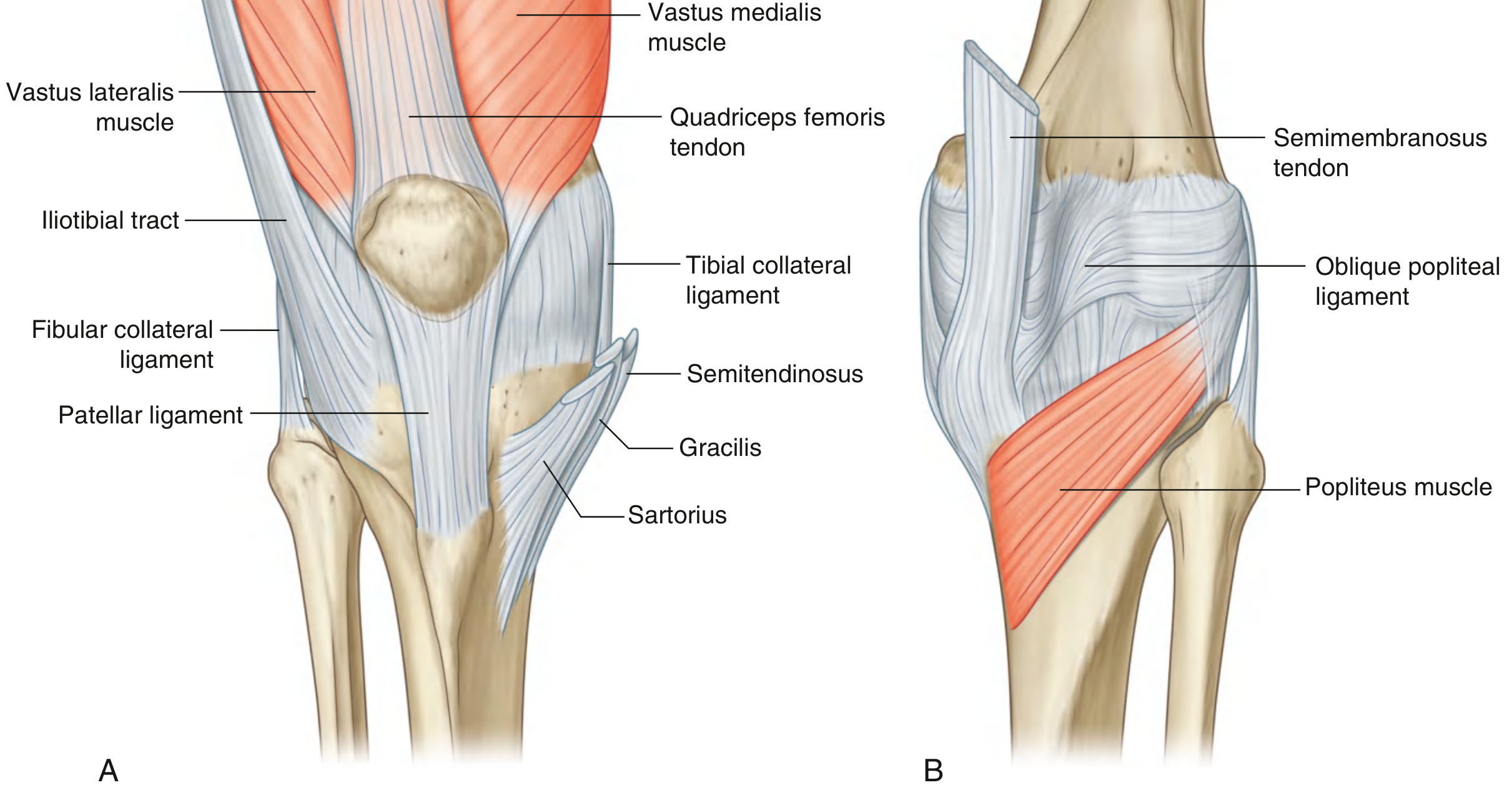
- Patellar ligament: continuation of quadriceps tendon from apex of patella to tibial tuberosity.
- Tibial collateral ligament (MCL): medial femoral epicondyle to medial tibia. Resists valgus stress.
- Fibular collateral ligament (LCL): lateral femoral epicondyle to head of fibula. Resists varus stress.
- Oblique popliteal ligament and arcuate popliteal ligament: reinforce posterior capsule.
Intracapsular, extrasynovial ligaments
- Anterior cruciate ligament (ACL): resists anterior displacement of tibia relative to femur and limits hyperextension.
- Posterior cruciate ligament (PCL): resists posterior displacement of tibia relative to femur and limits hyperflexion.
The principal ligaments are the patellar, medial and lateral collateral, and anterior and posterior cruciate ligaments. Gray's Anatomy for Students, p. 714.
Movements and muscles
- Flexion: hamstrings, sartorius, gracilis, gastrocnemius, popliteus
- Extension: quadriceps femoris
- Medial rotation in flexion: semitendinosus, semimembranosus, sartorius, gracilis, popliteus
- Lateral rotation in flexion: biceps femoris
Locking and unlocking
In terminal extension, medial rotation of the femur on tibia in weight-bearing, or lateral rotation of tibia on femur in non-weight-bearing, produces the locking mechanism. This makes standing more energy-efficient.
The popliteus unlocks the knee by laterally rotating the femur on a fixed tibia, or medially rotating the tibia when the limb is free.
Blood supply
Mainly from the genicular anastomosis, formed by branches of popliteal, femoral, and anterior tibial arteries.
Nerve supply
Articular branches from:
- Femoral nerve
- Tibial nerve
- Common fibular nerve
- Obturator nerve
Applied anatomy
- ACL tear: often due to twisting or sudden deceleration; anterior drawer/Lachman test may be positive.
- PCL tear: classically after a posterior force on flexed knee, such as dashboard injury.
- MCL injury: valgus force; may be associated with medial meniscus and ACL injury.
- Meniscal tear: locking, joint-line tenderness, and clicking may occur.
- Patellar dislocation: commonly lateral, especially with valgus stress.
- Knee effusion: can distend the suprapatellar bursa.
Long
Reading File
Finding Sources
Finding Sources
Searching PubMed
"knee joint" AND anatomy
Searching the Web
knee joint anatomy capsule ligaments menisci movements blood supply NCBI
Knee Joint
The knee joint is the largest and one of the most complex synovial joints in the body. It connects the thigh to the leg, bears body weight, permits locomotion, and provides stability in standing.
Although it functions mainly as a modified hinge joint, it also permits a small degree of rotation, especially when flexed.
Gray's Anatomy for Students, p. 709.
1. Type and articulations
The knee is a compound synovial joint with a common synovial cavity. It includes:
-
Tibiofemoral articulation
- Between the medial and lateral femoral condyles and corresponding tibial condyles.
- It is the principal weight-bearing component.
-
Patellofemoral articulation
- Between the posterior surface of the patella and patellar surface of the femur.
- The patella improves the leverage of quadriceps and protects the anterior joint surface.
The articular surfaces are covered by hyaline cartilage.
2. Articular surfaces
| Bone | Articular part |
|---|---|
| Femur | Medial and lateral condyles; patellar surface |
| Tibia | Superior surfaces of medial and lateral condyles, also called tibial plateau |
| Patella | Posterior articular surface, divided into medial and lateral facets |
The femoral condyles are convex and unequal. The tibial condyles are relatively flat, so the menisci increase congruence between them.
3. Capsule of the knee joint
The fibrous capsule surrounds the joint but is deficient anteriorly, where it is replaced by the quadriceps tendon, patella, and patellar ligament.
Attachments
- Superiorly: around the femoral condyles
- Inferiorly: around the margins of the tibial condyles
- Anteriorly: reinforced by quadriceps tendon, patella, patellar ligament, medial and lateral patellar retinacula
- Posteriorly: strengthened by oblique and arcuate popliteal ligaments
Synovial membrane
The synovial membrane:
- Lines the inner surface of the fibrous capsule.
- Covers the infrapatellar fat pad.
- Does not cover the cruciate ligaments, which are intracapsular but extrasynovial.
- Extends superiorly deep to the quadriceps tendon as the suprapatellar bursa.
The suprapatellar bursa commonly communicates with the knee joint cavity. Therefore, a large knee effusion may cause swelling above the patella.
4. Menisci
The knee has two wedge-shaped fibrocartilaginous menisci:
- Medial meniscus
- Lateral meniscus
They lie between the femoral condyles and the tibial plateau.
Functions of menisci
- Increase congruence between femur and tibia.
- Deepen the relatively shallow tibial articular surfaces.
- Distribute weight and load across the knee.
- Absorb shock.
- Improve stability.
- Assist lubrication and nutrition of articular cartilage.
- Contribute to proprioception.
Comparison of medial and lateral menisci
| Feature | Medial meniscus | Lateral meniscus |
|---|---|---|
| Shape | C-shaped | Nearly circular |
| Mobility | Less mobile | More mobile |
| Attachment | Firmly attached to capsule and MCL | Not attached to LCL |
| Injury | More commonly injured | Less commonly injured |
The medial meniscus is more vulnerable because it is relatively fixed by attachment to the fibrous capsule and tibial collateral ligament. Sudden rotation of a partially flexed, weight-bearing knee can tear it.
5. Ligaments of the knee
The primary ligaments are:
- Patellar ligament
- Tibial collateral ligament
- Fibular collateral ligament
- Anterior cruciate ligament
- Posterior cruciate ligament
Gray's Anatomy for Students, p. 714.
A. Patellar ligament
It is the continuation of the quadriceps tendon below the patella.
- Superior attachment: apex and lower border of patella
- Inferior attachment: tibial tuberosity
- Function: transmits force from quadriceps to tibia to produce extension of the knee.
The deep infrapatellar bursa lies between the patellar ligament and upper tibia.
B. Tibial collateral ligament
Also called the medial collateral ligament, or MCL.
- Broad, flat ligament on the medial side.
- Extends from the medial femoral epicondyle to the medial surface of the tibia.
- Deep fibers attach to the medial meniscus.
Function
- Resists valgus stress, meaning force that drives the knee medially.
- Helps prevent excessive abduction and medial rotation of the tibia.
Clinical relevance
A valgus injury may damage the MCL. Since it is attached to the medial meniscus, the meniscus may also be injured.
C. Fibular collateral ligament
Also called the lateral collateral ligament, or LCL.
- A cord-like ligament on the lateral side.
- Extends from lateral femoral epicondyle to the head of fibula.
- Separated from the lateral meniscus by the tendon of popliteus.
- It is not attached to the lateral meniscus.
Function
- Resists varus stress, meaning force that pushes the knee laterally.
- Stabilizes the lateral aspect of the joint.
D. Anterior cruciate ligament
The ACL runs from the anterior intercondylar area of tibia upward, backward, and laterally to attach to the posteromedial surface of the lateral femoral condyle.
Functions
- Prevents anterior displacement of tibia on femur.
- Prevents posterior displacement of femur on tibia in weight-bearing.
- Limits hyperextension.
- Helps control rotational stability of the knee.
Clinical relevance
ACL injury commonly occurs with:
- Sudden stopping or deceleration
- Pivoting or twisting
- Hyperextension
- Landing awkwardly after a jump
Common features include a popping sensation, rapid swelling from hemarthrosis, instability, and a positive Lachman test or anterior drawer test.
E. Posterior cruciate ligament
The PCL runs from the posterior intercondylar area of tibia upward, forward, and medially to attach to the lateral surface of medial femoral condyle.
Functions
- Prevents posterior displacement of tibia on femur.
- Prevents anterior displacement of femur on tibia in weight-bearing.
- Limits excessive flexion.
The PCL is stronger than the ACL.
Clinical relevance
PCL injury can occur from a blow to the anterior tibia while the knee is flexed, classically a dashboard injury in a motor vehicle collision. It can produce a positive posterior drawer test or posterior sag sign.
F. Oblique popliteal ligament
- Expansion of the tendon of semimembranosus.
- Runs superolaterally across the posterior aspect of knee capsule.
- Reinforces the posterior capsule.
G. Arcuate popliteal ligament
- Arises from posterior aspect of head of fibula.
- Curves superomedially over popliteus tendon.
- Strengthens posterolateral aspect of joint capsule.
6. Movements at the knee
Main movements
- Flexion
- Extension
Accessory movements
When the knee is flexed, slight medial and lateral rotation can occur.
| Movement | Main muscles |
|---|---|
| Extension | Quadriceps femoris |
| Flexion | Hamstrings, gastrocnemius, sartorius, gracilis, plantaris, popliteus |
| Medial rotation of tibia in flexion | Semitendinosus, semimembranosus, sartorius, gracilis, popliteus |
| Lateral rotation of tibia in flexion | Biceps femoris |
7. Locking and unlocking mechanism
The knee has a locking mechanism that stabilizes it in full extension and reduces muscular effort in standing.
Locking
During the last phase of extension:
- In an open chain movement, the tibia rotates laterally on the femur.
- In weight-bearing, the femur rotates medially on the tibia.
This tightens the cruciate and collateral ligaments, stabilizing the joint in extension.
Unlocking
The popliteus muscle unlocks the knee:
- It medially rotates the tibia on the femur when the foot is free.
- It laterally rotates the femur on the fixed tibia during weight-bearing.
After unlocking, flexion can begin.
8. Bursae around the knee
Important bursae include:
| Bursa | Location | Clinical condition |
|---|---|---|
| Suprapatellar bursa | Between quadriceps tendon and distal femur | Distends in knee effusion |
| Prepatellar bursa | Between skin and patella | Prepatellar bursitis, "housemaid's knee" |
| Superficial infrapatellar bursa | Between skin and patellar ligament | Inflammation due to kneeling |
| Deep infrapatellar bursa | Between patellar ligament and tibia | May become inflamed with overuse |
| Pes anserine bursa | Between pes anserinus tendons and medial tibia | Pes anserine bursitis |
| Semimembranosus bursa | Posteromedial knee | Can communicate with joint and contribute to Baker cyst |
A Baker cyst, or popliteal cyst, is fluid distension of a bursa in the popliteal region, commonly associated with intra-articular knee disease.
9. Blood supply
The knee receives a rich arterial network called the genicular anastomosis.
Main contributors include:
- Superior medial genicular artery
- Superior lateral genicular artery
- Inferior medial genicular artery
- Inferior lateral genicular artery
- Middle genicular artery
- Descending genicular branch of femoral artery
- Descending branch of lateral circumflex femoral artery
- Anterior tibial recurrent artery
- Circumflex fibular branch of posterior tibial artery
This extensive network maintains blood flow during knee flexion, when the popliteal artery may be partly compressed.
10. Nerve supply
The knee follows Hilton's law and receives articular branches from nerves supplying muscles that move the joint.
- Femoral nerve
- Tibial nerve
- Common fibular nerve
- Obturator nerve
The posterior articular branch of the tibial nerve and branches of common fibular nerve contribute substantially to innervation of the posterior and lateral knee.
11. Important clinical anatomy
1. Unhappy triad
A severe valgus and twisting injury may damage:
- Tibial collateral ligament
- Medial meniscus
- Anterior cruciate ligament
This combination is commonly termed the unhappy triad.
2. Meniscal injury
Typical symptoms:
- Joint-line pain
- Swelling
- Locking or catching
- Clicking
- Reduced range of motion
A torn meniscus may mechanically block full extension and cause a locked knee.
3. ACL injury
Features include:
- Audible or felt pop
- Immediate swelling
- Instability or giving way
- Positive Lachman test
4. PCL injury
Features include:
- Posterior knee pain
- Posterior tibial sag
- Positive posterior drawer test
5. Patellar dislocation
The patella usually dislocates laterally. Risk factors include increased Q-angle, weak vastus medialis, and a shallow trochlear groove.
6. Prepatellar bursitis
Inflammation of prepatellar bursa, often from prolonged kneeling. It is called housemaid's knee.
7. Osteoarthritis
Because the knee is a weight-bearing joint, degeneration of articular cartilage is common. It may cause pain, stiffness, crepitus, reduced function, and deformity.
Quick revision summary
- Type: Largest synovial, compound, modified hinge joint.
- Articulations: Tibiofemoral and patellofemoral.
- Menisci: Medial is less mobile and more commonly injured.
- MCL: Resists valgus force.
- LCL: Resists varus force.
- ACL: Prevents anterior tibial translation.
- PCL: Prevents posterior tibial translation.
- Main extensor: Quadriceps femoris.
- Main flexors: Hamstrings.
- Unlocking muscle: Popliteus.
- Common triad injury: ACL + MCL + medial meniscus.
This is a shared conversation. Sign in to Orris to start your own chat.