
FIGO staging of cervical cancer 2026
FIGO staging cervical cancer 2025 2026 revised
FIGO 2026 cervical cancer staging update revised classification
FIGO 2024 cervical cancer staging revised
https://pmc.ncbi.nlm.nih.gov/articles/PMC11399491
PMID: 39280203
FIGO staging cervical cancer diagram 2018
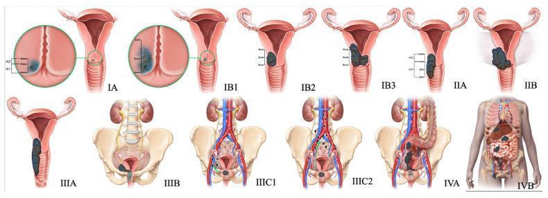
Anatomical diagrams illustrating the 2018 FIGO staging system for cervical cancer, progressing from Stage IA to IVB. The top row depicts cervical and vaginal spread: Stage I is confined to the cervix with subdivisions based on depth (IA) and diameter (IB1-IB3); Stage II shows extension beyond the uterus into the upper two-thirds of the vagina (IIA) and parametrial invasion (IIB). The bottom row illustrates advanced regional and distant spread: Stage IIIA involves the lower one-third of the vagina, while IIIB shows extension to the pelvic sidewall and associated hydroureter/hydronephrosis. Stage IIIC indicates lymphatic involvement, further categorized into pelvic (IIIC1) and para-aortic (IIIC2) lymph nodes. Stage IV demonstrates local invasion of the bladder or rectum (IVA) and distant metastasis beyond the pelvis (IVB). Key anatomical landmarks included are the uterus, cervix, vagina, ureters, kidneys, pelvic bones, and major blood vessels. The diagrams use color-coded masses to represent tumor growth and invasion depth, serving as a comprehensive educational guide for gynecologic oncology staging.
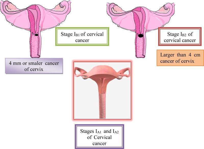
A multi-panel medical illustration and anatomical diagram depicting the FIGO staging of cervical cancer (Stages IA and IB). The central bottom panel displays a 3D-style anatomical diagram representing Stages IA1 and IA2, where the cervix appears macroscopically normal, indicating microinvasive disease that is not visible to the naked eye. The top left panel illustrates Stage IB1, showing a cross-section of the uterus and cervix with a small, localized dark lesion on the cervical canal; it is annotated as a cancer of 4 mm or smaller. The top right panel illustrates Stage IB2, showing a significantly larger, darker, and more prominent exophytic/ulcerative lesion on the cervix, annotated as being larger than 4 cm. All panels show relevant reproductive anatomy including the uterus, fallopian tubes, and ovaries. This educational graphic serves to distinguish clinical stages of cervical carcinoma based on tumor size and macroscopic visibility, relevant for gynecologic oncology and diagnostic pathology training.
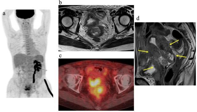
This composite diagnostic image illustrates a case of FIGO stage IVA cervical cancer through multiple modalities. (a) Maximum-intensity-projection (MIP) PET image showing high metabolic activity in the pelvic region and identifying secondary left hydronephrosis due to ureteral obstruction. (b) Axial T2-weighted MRI and (c) corresponding axial PET/CT fusion image provide detailed anatomical localization of the primary tumor, correlating high FDG uptake with the cervical mass. (d) Sagittal T2-weighted MRI demonstrates an intermediate signal intensity tumor originating in the uterine cervix. Yellow arrows highlight the tumor's extensive local invasion into the uterine body, the vagina, and anteriorly into the posterior wall of the bladder. This series serves as an educational example of advanced gynecological malignancy staging, emphasizing the importance of multimodality imaging (MRI for local soft tissue invasion and PET/CT for metabolic staging) in assessing tumor margins and visceral involvement.
FIGO Staging of Cervical Cancer (Current System as of 2026)
Key Principles of FIGO 2018 (+ 2024 corrigendum)
- Imaging and pathology may supplement clinical findings at all stages. Pathologic findings supersede imaging and clinical findings when available.
- Horizontal (lateral) extent is no longer used for Stage IA (microinvasive disease) - only depth of invasion matters.
- Vascular/lymphatic space involvement does not alter staging.
- When doubt exists, the earlier (lower) stage is assigned.
- A new Stage IIIC was introduced for lymph node involvement regardless of tumor size, with notations: (r) for radiologic diagnosis, (p) for pathologic diagnosis.
- Micrometastases in lymph nodes also qualify for Stage IIIC (clarified in 2024 corrigendum).
Full Staging Table
| Stage | Description |
|---|---|
| I | Carcinoma confined strictly to the cervix (extension to uterine corpus is disregarded) |
| IA | Invasive carcinoma diagnosed only by microscopy; deepest invasion ≤5 mm |
| IA1 | Stromal invasion ≤3 mm in depth |
| IA2 | Stromal invasion >3 mm and ≤5 mm in depth |
| IB | Invasive carcinoma with deepest invasion >5 mm; lesion limited to the cervix |
| IB1 | Invasion >5 mm depth, and <2 cm in greatest dimension |
| IB2 | ≥2 cm and <4 cm in greatest dimension |
| IB3 | ≥4 cm in greatest dimension |
| II | Carcinoma invades beyond uterus but NOT to pelvic wall or lower third of vagina |
| IIA | Upper two-thirds of vagina involved, no parametrial invasion |
| IIA1 | Tumor ≤4 cm in greatest dimension |
| IIA2 | Tumor >4 cm in greatest dimension |
| IIB | Parametrial invasion present, but NOT to the pelvic wall |
| III | Extension to pelvic wall and/or lower third of vagina; and/or hydronephrosis/non-functioning kidney; and/or lymph node involvement |
| IIIA | Extension to lower third of vagina, but NOT to pelvic wall |
| IIIB | Extension to pelvic wall and/or hydronephrosis or non-functioning kidney (unless due to another cause) |
| IIIC | Pelvic and/or para-aortic lymph node involvement (regardless of tumor size/extent) |
| IIIC1 | Pelvic lymph node metastasis only |
| IIIC2 | Para-aortic lymph node metastasis |
| IV | Extension beyond the true pelvis or biopsy-proven involvement of bladder/rectal mucosa |
| IVA | Spread to adjacent pelvic organs (bladder or rectal mucosa) |
| IVB | Spread to distant organs |
What Changed from FIGO 2009 → FIGO 2018
| Feature | FIGO 2009 | FIGO 2018 |
|---|---|---|
| Stage IA | Depth ≤5 mm AND horizontal ≤7 mm | Depth ≤5 mm only (horizontal dropped) |
| Stage IB | IB1 (≤4 cm), IB2 (>4 cm) | IB1 (<2 cm), IB2 (2-<4 cm), IB3 (≥4 cm) |
| Lymph nodes | Not formally staged | Stage IIIC added |
| Imaging | Not used | Permitted to supplement |
| Pathology | Not used | Can supersede clinical findings |
2024 Corrigendum (Minor Updates)
- Confirmed that micrometastases (not just macrometastases) in lymph nodes assign Stage IIIC.
- Clarified that pathological staging takes precedence over radiological findings, which in turn take precedence over clinical findings.
- Aligned with TNM classification regarding lymph node classification.
Visual Summary
Clinical Implications
- Stage IA1: Cone biopsy or simple hysterectomy (fertility-sparing possible)
- Stage IA2 - IIA: Radical hysterectomy with pelvic lymph node dissection OR concurrent chemoradiation
- Stage IIB - IVA: Primary concurrent chemoradiation (cisplatin-based) + brachytherapy
- Stage IVB: Palliative systemic therapy; pembrolizumab is now incorporated in first-line regimens (KEYNOTE-826 data)
- Stage IIIC: Upstaged due to lymph nodes - chemoradiation field extended to cover positive nodes
Bottom line: As of August 2026, no new FIGO 2026 revision for cervical cancer staging has been published. The FIGO 2018 system (with the minor 2024 corrigendum) remains current. The most recent major updates in gynecologic FIGO staging in 2023-2024 were for endometrial cancer (FIGO 2023), not cervical cancer.
Post menopausal bleeding ACOG 2026
"postmenopausal bleeding" AND "ACOG" AND management
ACOG 2025 2026 postmenopausal bleeding guidelines evaluation management update
https://www.guidelinecentral.com/guideline/5213608
"postmenopausal bleeding" AND "endometrial biopsy" AND "transvaginal ultrasonography"
https://consultqd.clevelandclinic.org/new-guidance-supports-…
PMID: 41990335
ACOG 2026 Guidelines: Postmenopausal Bleeding
Definition
Why the 2026 Update Was Needed
- Rising incidence: Uterine cancer incidence increased from 23.5 to 28.8 per 100,000 women (2000-2022). Over 69,000 new cases and ~14,000 deaths in 2025 alone. Deaths from endometrial cancer now exceed those from ovarian cancer.
- Falling sensitivity of TVUS: As disease prevalence rises, the NPV of the 4-mm cutoff declines. Recent data show unacceptably low sensitivity.
- Racial disparities: In non-Hispanic Black women, the 4-mm cutoff had a sensitivity of only 47.5% and an NPV of only 91.7%. Using ultrasound alone misses ~25% of serous endometrial cancer (an aggressive subtype) in this population.
- High-grade histological subtypes (serous, clear cell, carcinosarcoma) - not estrogen-driven - are increasing and are not reliably detected by endometrial thickness measurement.
- Technical limitations: No standardized definition of "recurrent bleeding"; measurement variability in TVUS.
- Health equity concerns: Barriers to follow-up care mean relying on "come back if bleeding recurs" is inadequate for many patients.
Core Recommendation (ACOG April 2026)
For most patients with postmenopausal bleeding, the initial evaluation should include BOTH transvaginal ultrasonography AND endometrial tissue sampling.
The Exception: When TVUS Alone May Be Acceptable
| Criterion | Requirement |
|---|---|
| Episode count | Single episode of postmenopausal bleeding only |
| Endometrial thickness | Sonographically fully visualized endometrium ≤4 mm |
| Risk factors | No factors strongly associated with endometrial cancer (see list below) |
| Counseling | Patient counseled that continued or recurrent bleeding requires immediate re-evaluation |
| Access to care | No significant barriers to prompt gynecologic evaluation |
Risk Factors That Favor Immediate Biopsy
- History of hormone therapy (with or without progestin)
- Tamoxifen use
- BMI >30 (obesity)
- Nulliparity
- Genetic predisposition to endometrial cancer (Lynch syndrome, PTEN mutations)
- Diabetes mellitus
- Race (non-Hispanic Black women - higher risk of high-grade subtypes)
- Family history of endometrial cancer
- Late menopause
Technical Notes on TVUS
- Endometrial thickness is measured as the thickest portion of the endometrial stripe on a long-axis uterine view, measured anterior to posterior.
- The endometrium must be fully visualized - if not, biopsy is required regardless of any measured thickness.
- TVUS remains valuable for characterizing uterine anatomy (polyps, fibroids, submucosal lesions) even when biopsy is also performed.
Endometrial Tissue Sampling Methods
| Method | Notes |
|---|---|
| Office endometrial biopsy (Pipelle) | First-line, minimally invasive |
| Sonohysterography + biopsy | Better characterization of focal lesions |
| Hysteroscopy + directed biopsy | For failed office biopsy, focal lesions, or persistent bleeding despite negative initial workup |
| D&C | For cases where office sampling fails or is inadequate |
Summary: 2018 vs 2026
| Feature | ACOG 2018 | ACOG 2026 |
|---|---|---|
| Initial evaluation | TVUS alone if ET ≤4 mm | TVUS + endometrial biopsy for most patients |
| Biopsy threshold | Biopsy only if ET >4 mm or risk factors present | Biopsy for most; TVUS alone only in very select low-risk cases |
| Risk factor consideration | Biopsy regardless of thickness if risk factors present | Same, but risk factors now broaden the "biopsy for most" approach |
| Health equity | Not explicitly addressed | Explicitly cited as a driver for the change |
Causes of Postmenopausal Bleeding (Differential - for context)
- Atrophic vaginitis / endometrial atrophy - most common (~60-80%)
- Endometrial polyps
- Endometrial hyperplasia (with or without atypia / EIN)
- Endometrial carcinoma (~10% of PMB cases)
- Cervical pathology (polyps, carcinoma)
- Exogenous hormones (HRT, tamoxifen)
- Submucosal fibroids
- Ovarian tumors (estrogen-secreting)
- Uterine sarcoma (rare)
Key takeaway: ACOG 2026 marks a significant paradigm shift - ultrasound alone is no longer sufficient for initial workup in most patients with PMB. Dual assessment (TVUS + endometrial biopsy) is now the standard, driven by rising endometrial cancer rates, racial disparities in diagnostic accuracy, and the increased prevalence of high-grade subtypes that evade ultrasound detection.