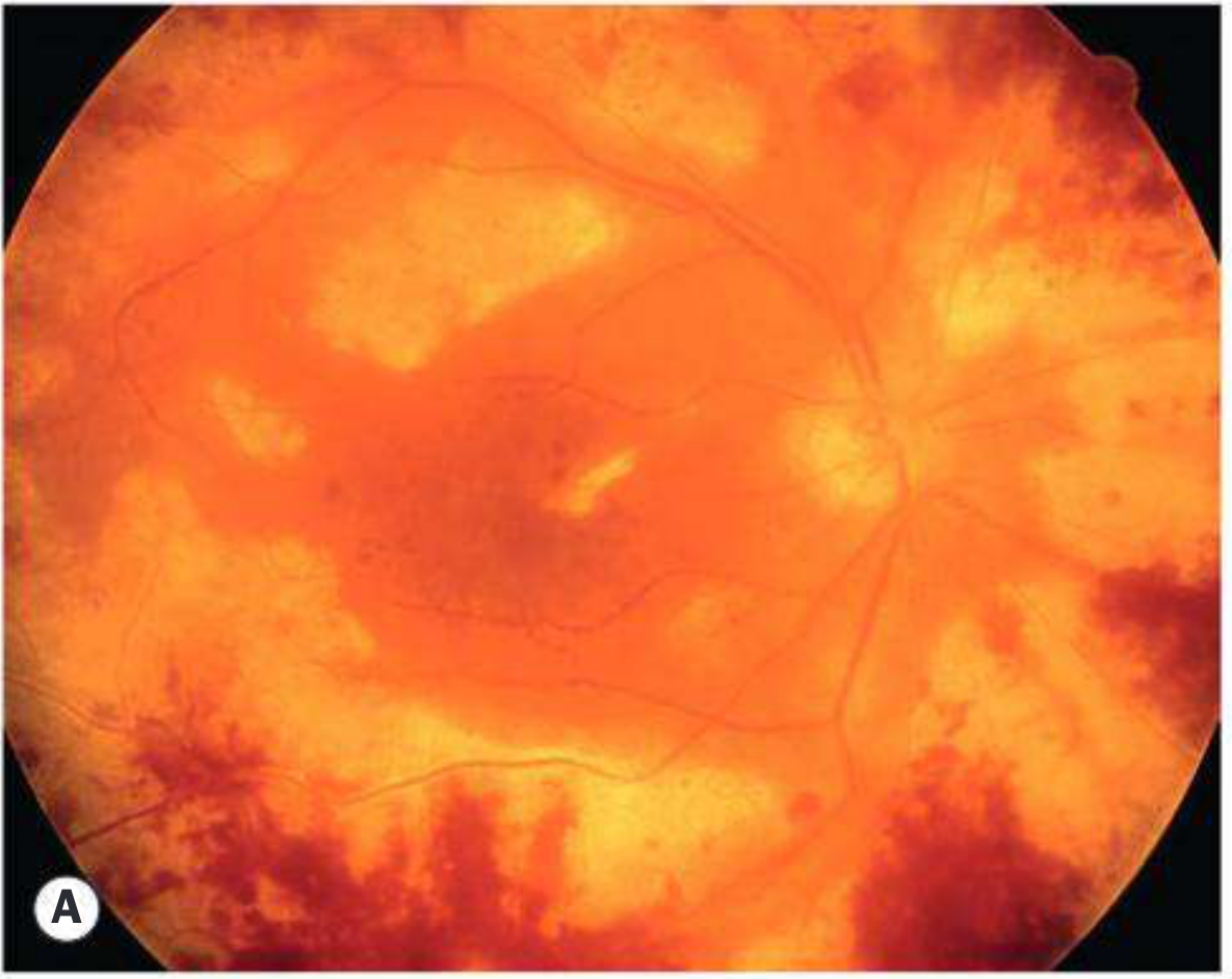
does lsd ude cause any retinopathies

Reading File
Finding Sources
Finding Sources
Searching PubMed

LSD retinopathy lysergic acid diethylamide

Finding Sources
Reading Article

PMID: 32064962

Searching the Web

LSD retinopathy ocular effects lysergic acid diethylamide macular damage

Finding Sources
Reading Web Page

https://en.wikipedia.org/wiki/Drug_abuse_retinopathy

Finding Sources
Finding Sources
Reading File
Reading File
Reading File
Here is a thorough answer synthesizing textbook and research evidence:

LSD and Retinopathy

Does LSD directly cause retinopathy?

LSD (lysergic acid diethylamide) does not cause a classic, well-defined retinopathy through its pharmacological action alone. However, there are two distinct ways it is linked to retinal damage:

1. Solar / Phototoxic Maculopathy (most well-established)

The most clinically documented ocular injury from LSD use is solar maculopathy - not from LSD itself, but from sun-gazing behavior induced while under its influence. A 23-year-old man who sustained severe macular damage from sun gazing during an LSD-induced hallucinogenic state is a documented case in the literature (PMID 1266919). Sequential fundus photography and fluorescein angiography confirmed prominent focal injury to the retinal pigment epithelium (RPE).
The mechanism is photochemical: solar radiation causes oxidative damage to photoreceptors and the RPE at the fovea. LSD removes the protective inhibition that normally prevents people from staring at the sun.
Signs of solar retinopathy (from Kanski's Clinical Ophthalmology, 10th ed.):
  • Small yellow or red foveolar spot shortly after exposure
  • Evolves to a sharply defined foveolar defect with irregular borders or a lamellar hole
  • OCT: foveal thinning with focal hyporeflective area involving photoreceptor inner and outer segments
  • Variable reduction in visual acuity, with a small central scotoma
  • No specific treatment; prognosis is generally good, with improvement over 6 months, though some patients retain permanent visual loss

2. Direct Retinal Toxicity (animal/experimental evidence)

A 2020 mouse study (PMID 32064962, Chen et al., Cutaneous and Ocular Toxicology) demonstrated that:
  • LSD administered intraperitoneally to C57 mice at escalating doses (0.2-1.2 mg/kg twice daily for 5 days) caused measurable retinal damage
  • Electroretinography (ERG) showed decreased response
  • Histology showed loss of photoreceptor cells
  • The mechanism involved upregulation of p-JAK1/p-STAT1, promoting photoreceptor apoptosis via the JAK/STAT pathway
This is animal data only - direct extrapolation to human clinical retinopathy requires caution. The doses used are supra-therapeutic compared to typical human recreational use.

3. Hallucinogen-Persisting Perception Disorder (HPPD)

While not a structural retinopathy, LSD users can develop HPPD - a condition where visual symptoms persist long after drug cessation. These include:
  • Enhanced color sensitivity, halos around lights
  • Trailing and stroboscopic perception of moving objects
  • Afterimages, palinopsia, macropsia/micropsia
Per Bradley and Daroff's Neurology in Clinical Practice, these perceptual changes "can persist for years." HPPD is thought to reflect cortical disinhibition (loss of serotonergic filtering in the visual cortex) rather than structural retinal damage, but the visual complaints can mimic maculopathy.
Per Kaplan & Sadock's Comprehensive Textbook of Psychiatry, HPPD is more likely in those who chronically use hallucinogens or combine them with cannabis, MDMA, or alcohol.

Summary Table

MechanismType of DamageEvidence Level
Sun-gazing while intoxicatedSolar maculopathy (RPE + foveal photoreceptor damage)Case reports in humans
Direct retinal toxicity via JAK/STAT apoptosisPhotoreceptor cell lossAnimal study (mouse)
HPPD (cortical, not retinal)Persistent visual disturbances, no structural lesionClinical/observational

Bottom line: LSD itself does not have a well-documented direct retinopathy in clinical human studies, but it is associated with retinal injury through sun-gazing behavior, and experimental animal data suggest a potential for direct photoreceptor toxicity via the JAK1/STAT1 pathway. Clinicians evaluating visual symptoms in LSD users should also consider HPPD as a diagnosis of exclusion after ruling out structural causes.

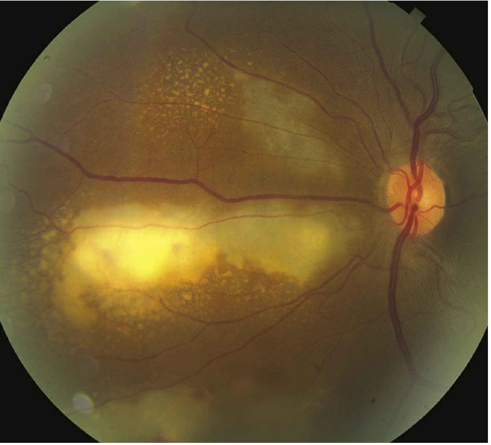
Post viral retinopathies

Finding Sources
Finding Sources
Searching PubMed

post-viral retinopathy retinal

Reading File
Reading File
Finding Sources
Reading File
Finding Sources
Finding Sources
Reading File
Finding Sources
Finding Sources
Reading File
Reading File
Searching PubMed

viral retinitis retina herpes CMV

Reading Article

PMID: 39770716

Finding Sources
Searching the Web

post viral retinopathy types causes mechanisms 2024

Reading File
Finding Sources
Loading Image
Loading Image
Loading Image
Here is a detailed, textbook-grounded overview of post-viral retinopathies, with clinical images:

Post-Viral Retinopathies

Post-viral retinal disease can occur through two broad mechanisms:
  1. Direct viral invasion of retinal cells (necrotizing, cytolytic)
  2. Immune-mediated / inflammatory damage triggered by viral antigens (vasculitis, pigmentary changes, uveitis)

1. Herpetic Necrotizing Retinopathies

This is the most clinically important group. A 2024 review (PMID 39770716, Nguyen & Conrady, Microorganisms) describes the molecular mechanisms by which herpesviruses (HSV, VZV, CMV) invade and destroy retinal tissue, noting the retina's particular vulnerability due to its immune-privileged status.

A. Acute Retinal Necrosis (ARN)

Causative viruses: VZV (older patients), HSV-1/HSV-2 (younger patients), rarely CMV or EBV.
Population: Typically immunocompetent individuals.
American Uveitis Society diagnostic criteria:
  • One or more discrete foci of peripheral retinal necrosis with well-defined borders
  • Rapid circumferential progression without antiviral therapy
  • Occlusive vasculopathy with arterial involvement (retinal arteritis)
  • Prominent anterior chamber and vitreous inflammation (panuveitis)
Clinical features:
  • Blurred vision, floaters, ocular pain, photophobia
  • Deep yellow-white peripheral infiltrates
  • Retinal haemorrhages (less prominent than CMV)
  • Optic neuritis in some cases
  • Secondary rhegmatogenous retinal detachment (RRD) in ~70% of cases - major cause of visual morbidity
  • May involve the second eye in one-third of cases within weeks to months
ARN - Advanced disease reaching the posterior pole with confluent necrosis, haemorrhages, and vasculitis
Acute Retinal Necrosis - advanced disease with peripheral necrosis, haemorrhage, and pigmentary changes (Kanski's Clinical Ophthalmology, 10th ed.)
Diagnosis: Vitreous/aqueous PCR for viral DNA (preferred over serology).
Treatment:
  • Oral valacyclovir 1-2 g TID or famciclovir 500 mg TID + supplemental intravitreal foscarnet or ganciclovir injections 1-2x/week
  • Alternative: IV acyclovir 10 mg/kg TID for 5-14 days, then oral maintenance for minimum 2 months
  • Topical cycloplegic + steroids for anterior inflammation
  • Prophylactic barrier laser photocoagulation (efficacy unclear)
  • Pars plana vitrectomy for associated complex RRD
  • Systemic steroids (delayed at least 24 hours after antiviral initiation)

B. Progressive Outer Retinal Necrosis (PORN)

A variant of herpetic retinitis seen in immunocompromised patients (HIV/AIDS, transplant).
  • Rapidly progressive, characterized by sheet-like opacification deep to normal-looking retinal vessels
  • Vitreous is clear (minimal vitritis) - key distinguishing feature from ARN
  • Macula involved early (unlike ARN where it is spared early)
  • Pain and anterior uveitis are minimal
  • Frequently leads to rapid bilateral blindness due to retinal detachment
  • Prompt diagnosis and treatment is critical

C. CMV Retinitis

Population: Severe immunocompromise - HIV/AIDS (CD4 <50 cells/µL), leukemia, post-transplant, or local ocular immunosuppression (steroid injections). CMV is the leading cause of retinal blindness in AIDS patients.
Symptoms: Scotoma, decreased vision, floaters, photopsias. Pain and photophobia are uncommon. Often initially asymptomatic.
Two clinical forms:
  • Indolent form: Peripheral granular opacities ± hemorrhage
  • Fulminant form: Confluent areas of necrosis with prominent hemorrhage, starting along the major vascular arcades ("brushfire" or "pizza pie" pattern)
CMV Retinitis - wedge-shaped area of retinal whitening and haemorrhage centred along vessels, with granular peripheral lesions
CMV Retinitis - confluent necrosis with haemorrhage following the vascular arcades (Wills Eye Manual)
Other features: Mild anterior uveitis, minimal vitritis, RPE atrophy and pigment clumping after resolution, RRD in ~1/3 of cases (risk rises when >25% retina involved).
Treatment (from Wills Eye Manual):
DrugInductionMaintenance
Ganciclovir (IV)5 mg/kg IV BID x 14 days5 mg/kg IV daily
Valganciclovir (oral)900 mg PO BID x 21 days900 mg PO daily
Foscarnet90 mg/kg IV BID90-120 mg/kg IV daily
Cidofovir5 mg/kg IV weekly x 25 mg/kg IV every 2 weeks
Intravitreal ganciclovir implant is also an option for local disease control.

2. Congenital Viral Retinopathies (Pigmentary)

A. Rubella Retinopathy

Virus: Rubella (German measles) - transplacental transmission to fetus.
Ocular manifestations of congenital rubella:
  • "Salt and pepper" pigmentary retinopathy - the hallmark finding, caused by irregular RPE pigmentation across the posterior pole
  • Cataract, anterior uveitis, glaucoma, microphthalmos
  • Latent rubella can cause chronic anterior uveitis (Fuchs uveitis syndrome association)
Rubella retinopathy - characteristic 'salt and pepper' pigmentary disturbance of the RPE throughout the posterior pole
Rubella retinopathy - 'salt and pepper' RPE appearance (Kanski's Clinical Ophthalmology, 10th ed.)

B. Congenital CMV Retinopathy

CMV is the leading cause of congenital sensorineural hearing loss and intellectual disability (TORCH syndrome). Retinitis in neonates follows the same pattern as in immunocompromised adults.

C. Congenital Zika Virus

Virus: Zika (Aedes mosquito-borne flavivirus).
  • Microcephaly and CNS malformations in neonates
  • Ocular effects: chorioretinal and macular atrophy, focal pigmentary macular changes, optic nerve abnormality, glaucoma
  • In adults with acute infection: conjunctivitis (most common), then uveitis
  • Virus persists in the lacrimal gland and retinal pigment epithelium

3. Post-Viral Immune-Mediated Retinopathy

A. Subacute Sclerosing Panencephalitis (SSPE) - Measles

Virus: Measles (delayed CNS reactivation 6-15 years after infection).
  • Progressive neurological degeneration (personality change → choreoathetosis → myoclonus → death)
  • Ocular: posterior uveitis, papilloedema, optic neuritis, nystagmus
  • Ischaemic retinal vein occlusion secondary to vasculitis (rare)
  • Classical macular lesion: white/grey-white patch resolving to macular scarring and RPE atrophy
Measles (acute): Can also cause retinitis with macular and disc oedema in active infection.

B. Ebola Virus Disease

Post-Ebola Virus Disease Syndrome (PEVDS):
  • Uveitis in up to 1/3 of survivors, occurring within 2 months of discharge
  • Anterior, posterior, or panuveitis
  • Up to 15% develop cataract
  • Optic nerve swelling in 10%
  • Ebola virus detected in aqueous and can infect and persist in RPE cells
  • Treatment: topical + oral corticosteroids; must start early to prevent permanent visual loss

Summary Comparison

VirusPopulationRetinal PatternKey Feature
HSV/VZV (ARN)ImmunocompetentPeripheral necrotizing retinitisOcclusive arteritis, RRD in 70%
VZV/CMV (PORN)ImmunocompromisedOuter retinal necrosis, starts posteriorlyClear vitreous, rapid blindness
CMVAIDS/transplantVascular-arcuate necrosis + haemorrhage"Pizza pie" pattern, minimal inflammation
Rubella (congenital)NeonatesDiffuse RPE pigmentary changes"Salt and pepper" fundus
Measles/SSPEChildren/teensMacular scarring, vasculitisDelayed CNS + retinal disease
Zika (congenital)NeonatesMacular/chorioretinal atrophyOptic nerve anomalies
Ebola (post-infectious)SurvivorsUveitis, RPE involvementVirus persists in RPE

Sources: Kanski's Clinical Ophthalmology 10th ed.; Wills Eye Manual; Nguyen & Conrady, Microorganisms 2024, PMID 39770716

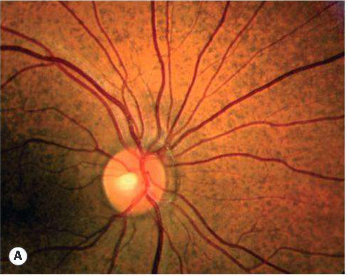
Does cannabis usage cause retinopathies

Finding Sources
Searching PubMed

cannabis marijuana retina retinopathy

Searching the Web

cannabis cannabinoids retinopathy retinal damage ocular effects 2024

Reading Article

PMID: 30773228

Searching PubMed

cannabis THC CBD ocular retinal function electroretinogram

Reading Web Page

https://pmc.ncbi.nlm.nih.gov/articles/PMC11613704

Searching PubMed

cannabis marijuana retinal vascular occlusion macular

Finding Sources
Finding Sources
Reading Article

PMID: 32947368

Searching PubMed

cannabis electroretinogram retinal function neurotoxicity

Reading Web Page

https://www.mdpi.com/2813-2564/4/4/22

Searching PubMed

cannabis retinal artery occlusion vasospasm young

Finding Sources
Here is a thorough, evidence-based answer on cannabis and retinopathy:

Cannabis Use and Retinopathy

The short answer is: cannabis does not cause a classic, defined retinopathy in the way that, say, chloroquine or radiation does. However, mounting evidence - from electrophysiology, vascular case reports, and molecular studies - shows that cannabis use can cause measurable retinal dysfunction and, in heavy use scenarios, structural vascular damage.

1. The Endocannabinoid System in the Retina

The retina expresses both CB1 and CB2 cannabinoid receptors, as well as endocannabinoid-synthesizing and degrading enzymes throughout all its neural layers. This means exogenous cannabinoids (THC, CBD) directly interact with retinal tissue - the retina is not a passive bystander.
Because the retina is embryologically an outgrowth of the brain (part of the CNS), retinal electrophysiology (ERG) serves as a measurable window into cannabinoid-induced neural dysfunction - a concept highlighted in a key 2019 review (PMID 30773228, Schwitzer et al., Neuroscience & Biobehavioral Reviews).

2. Neuroretinal Dysfunction (Functional Damage)

Electroretinographic (ERG) Changes

Studies using ERG in regular cannabis users have demonstrated:
  • Delayed implicit times in photopic (cone-driven) responses
  • Increased retinal background noise - a 2019 study found elevated ERG noise in co-users of cannabis and alcohol (PMID 30292729, Lucas et al., Progress in Neuropsychopharmacology)
  • Abnormal electrooculography (EOG) and electrically evoked phosphene thresholds in a heavy cannabis user later diagnosed with HPPD, suggesting direct cannabinoid effects on the retinal pigment epithelium (RPE) function (Zobor et al., 2015, cited in Bondok et al., Clin Ophthalmol 2024)
These findings indicate that chronic heavy use can disrupt the synaptic transmission machinery in the inner and outer retina, particularly affecting dopaminergic and glutamatergic pathways that are under endocannabinoid modulation.

3. Vascular Retinopathy - Retinal Artery Occlusion

The most dramatic structural retinal injury linked to cannabis is ischaemic vascular occlusion.
A well-documented 2022 case report (PMID 32947368, Ramtohul, Freund & Sarraf, Retinal Cases) described:
  • A 21-year-old healthy male with no systemic risk factors
  • Smoking ~15 g/day of cannabis during COVID-19 lockdown
  • Acute branch retinal artery occlusion (BRAO) with right inferotemporal retinal whitening
  • OCT showed paracentral acute middle maculopathy (PAMM) - ischaemic hyperreflectivity of inner and middle retinal layers
  • OCT-angiography: flow signal loss in the deep capillary plexus
  • Fluorescein angiography and full systemic workup: unremarkable (no other cause found)
  • Proposed mechanism: transient arterial vasospasm induced by cannabis
This mirrors the pattern seen with cocaine and other vasospastic drugs in young patients with retinal artery occlusion at an unexpected age.
A separate case (Aktaş et al., 2016, cited in Bondok et al. 2024) reported a 55-year-old male with sudden painless visual loss due to haemorrhagic macular infarction following inhaled cannabis use.
Mechanism of vascular injury: Cannabis causes a biphasic cardiovascular response - initial tachycardia and hypertension followed by vasodilatation. This haemodynamic instability, combined with potential platelet aggregation changes and vasospasm from high-THC strains, can precipitate retinal vascular events, particularly in susceptible individuals.

4. Retinal Vascular Changes with Chronic Use

An observational study of 106 young adults found that mean arteriolar diameter was significantly wider in frequent cannabis smokers vs. controls (Bondok et al. 2024), likely reflecting residual vasodilatory effects. A clinical trial with oral dronabinol (synthetic THC) showed increased optic nerve head blood flow and decreased retinal arteriovenous passage time - effects that may be transiently beneficial but could contribute to vascular instability with chronic, heavy use.

5. The THC:CBD Ratio - Neuroprotective vs. Neurotoxic

A 2024 review (MDPI Cannabinoids) highlighted a critically important concept:
THC:CBD RatioRetinal Effect
Low THC + adequate CBD (older strains, ~10:1)Neuroprotective - reduces oxidative stress, protects photoreceptors
High THC + low CBD (modern strains, ~100:1)Neurotoxic - promotes photoreceptor apoptosis, oxidative damage
Modern commercial cannabis strains have been selectively bred for very high THC content with dramatically reduced CBD. This shift means the neuroprotective advantage seen in older research may no longer apply to contemporary street or dispensary cannabis. High-THC cannabis at recreational doses is increasingly recognized as potentially neurotoxic to retinal cells.
CBD alone has antioxidant properties (scavenges reactive oxygen species, independent of CB1 receptors) that are mildly neuroprotective. THC alone at high concentrations promotes apoptosis and oxidative damage.

6. Potential Therapeutic Effects (Context)

Cannabis and cannabinoids have also been studied for retinal protection:
  • IOP reduction - THC lowers IOP by 25-30% (transient, lasting 3-4 hours; not a substitute for sustained glaucoma therapy)
  • Diabetic retinopathy - preclinical evidence that CBD reduces retinal inflammation, neurotoxicity, and blood-retinal barrier breakdown
  • Ischaemia - cannabinoid receptor agonists reduce retinal ischaemia-reperfusion injury in animal models
These therapeutic properties do not offset the risks of chronic high-THC recreational use.

Summary

EffectEvidenceMechanism
ERG dysfunction (delayed responses, increased noise)Human studiesDisrupted dopaminergic/glutamatergic synaptic transmission
RPE dysfunction (EOG changes)Case reportsDirect cannabinoid effect on RPE
Retinal artery occlusion / PAMMCase reports (young adults)Vasospasm, haemodynamic instability
Haemorrhagic macular infarctionCase reportsVascular occlusion
Wider arteriolar diameterObservational studyResidual vasodilation
Neurotoxicity (high-THC strains)Animal/lab dataOxidative stress, apoptosis - dose-dependent
Bottom line: Cannabis does not cause a stereotyped retinopathy like chloroquine or radiation. However, heavy or chronic use - especially with modern high-THC strains - is associated with measurable neuroretinal dysfunction on ERG, RPE disturbance, and rare but serious retinal vascular occlusions in young individuals via vasospasm. The retinal endocannabinoid system makes the eye genuinely susceptible to cannabinoid effects, and the growing THC:CBD ratio in modern cannabis shifts the balance from neuroprotection toward potential neurotoxicity.
Sources: Schwitzer et al. PMID 30773228; Ramtohul et al. PMID 32947368; Lucas et al. PMID 30292729; Bondok et al., Clin Ophthalmol 2024 PMC11613704
This is a shared conversation. Sign in to Orris to start your own chat.