
Upper and motor neuron lesions
Reading File
Now let me search the library for UMN and LMN lesion content in parallel.
Finding Sources
Finding Sources
Excellent. Let me now read the primary sections in parallel - the detailed UMN/LMN table and the main discussion.
Reading File
Reading File
Reading File
Let me read the full Adams & Victor Table 3-1 and the LMN section from Neuroanatomy, plus the Ganong physiology section.
Reading File
Reading File
Reading File
Now let me also fetch the LMN section from Adams & Victor and look for a figure showing the UMN/LMN anatomy diagram.
Finding Sources
Finding Sources
Reading File
Reading File
I now have comprehensive content from multiple authoritative textbooks. Let me fetch a key diagram image to include in the answer.
Loading Image
I now have all the content needed to write a thorough, well-organized response. Here is the comprehensive answer:
Upper Motor Neuron (UMN) vs. Lower Motor Neuron (LMN) Lesions
Anatomy: Defining the Two Systems
Upper motor neurons (UMN) are neurons whose cell bodies lie in the cerebral cortex (primarily motor cortex) or brainstem and whose axons descend via the corticospinal tract (for limbs/trunk) or corticobulbar tract (for cranial nerve motor nuclei). They do not directly innervate muscle - they synapse onto lower motor neurons.
Lower motor neurons (LMN) are the "final common pathway." Their cell bodies sit in the anterior horn of the spinal cord (or motor nuclei of cranial nerves in the brainstem), and their axons travel via peripheral nerves directly to skeletal muscle. Each motor neuron + its axon + the muscle fibers it innervates = one motor unit.
- Neuroanatomy through Clinical Cases, 3rd Ed.: "Upper motor neurons of the corticospinal tract project from the cerebral cortex to lower motor neurons located in the anterior horn of the spinal cord. Lower motor neurons, in turn, project via peripheral nerves to skeletal muscle."
Core Signs: Side-by-Side Comparison
The classic comparison table (from Neuroanatomy through Clinical Cases and Adams & Victor's Principles of Neurology):
| Sign | UMN Lesion | LMN Lesion |
|---|---|---|
| Weakness | Yes | Yes |
| Atrophy | Mild (disuse only) | Pronounced - up to 70% of bulk |
| Fasciculations | No | Yes |
| Muscle tone | Increased (spasticity)^b | Decreased (flaccidity/hypotonia) |
| Deep tendon reflexes | Increased (hyperreflexia)^b | Decreased/absent |
| Plantar reflex | Extensor - Babinski sign | Flexor (normal) or absent |
| Muscles affected | Groups; never individual muscles | Individual muscles can be isolated |
| EMG/NCS | Normal nerve conduction; no denervation potentials | Abnormal NCS; fibrillations, positive sharp waves, fasciculations on EMG |
^b With acute UMN lesions, tone and reflexes may initially be decreased (spinal shock), gradually evolving to spasticity over hours to months.
UMN Lesions in Detail
Spasticity
Increased tone with a velocity-dependent quality - resistance increases as passive movement speeds up, sometimes with a "clasp-knife" release. The mechanism is loss of descending inhibitory pathways traveling alongside the corticospinal tract, leading to increased excitability of anterior horn cells. Importantly, spasticity is NOT produced by selective corticospinal tract lesions alone; the lateral reticulospinal and other pathways contribute.
Biochemically (Adams & Victor): glycine (inhibitory transmitter of interneurons) is reduced; loss of descending noradrenergic, dopaminergic, and serotonergic fibers also contributes.
Treatment of spasticity:
- Baclofen (GABA-B agonist) - reduces excitatory transmitter release from primary afferent terminals. Intrathecal baclofen via implanted pump is used for severe cases.
- Diazepam/benzodiazepines - potentiate postsynaptic GABA receptors
- Tizanidine - alpha-2 adrenergic agonist; increases presynaptic inhibition
- Botulinum toxin - locally blocks ACh release at neuromuscular junction
Pathological Reflexes
- Babinski sign: extension (dorsiflexion) of the great toe + fanning of other toes when the lateral sole is stroked from heel upward. Normal in infants (immature corticospinal tracts); pathological in adults. May be absent if toe extensor muscles are severely atrophied.
- Hoffmann sign: in the upper limbs - flicking the distal phalanx of the middle finger produces reflex flexion of thumb and index finger.
- Jaw jerk: exaggerated in bilateral corticobulbar lesions.
- Clonus: sustained rhythmic contractions at ankle or wrist with maintained stretch.
- Loss of abdominal reflexes
Pattern of Weakness
UMN weakness affects groups of muscles, never individual muscles in isolation. Typically:
- Upper limb: flexors relatively spared, extensors weaker (arm held in flexion)
- Lower limb: extensors relatively spared, flexors weaker (leg held in extension - classic spastic gait)
Pseudobulbar Palsy
When bilateral corticobulbar tracts are damaged, the motor nuclei of cranial nerves V, VII, IX, X, XII lose their supranuclear input. Features:
- Dysarthria (spastic), dysphagia, dysphonia
- Brisk jaw jerk, hyperactive gag reflex
- Emotional lability (involuntary crying/laughing, also called emotional incontinence) - pathognomonic of pseudobulbar palsy, distinguishing it from true (LMN) bulbar palsy.
LMN Lesions in Detail
Core Features
From Ganong's Review of Medical Physiology: "Damage to lower motor neurons is associated with flaccid paralysis, muscular atrophy, fasciculations, hypotonia, and hyporeflexia or areflexia."
- Flaccid paralysis with hypotonia - loss of all muscle tone since the connection to the muscle is severed
- Atrophy - rapid and severe (up to 70% of bulk), due to denervation atrophy (loss of trophic support), far greater than the mild disuse atrophy of UMN lesions
- Fasciculations - spontaneous, visible muscle twitches from random firing of denervated/diseased motor units
- Fibrillations on EMG - spontaneous firing of individual muscle fibers (not visible to the eye)
- Individual muscles can be affected in isolation, unlike UMN lesions
Topographic Patterns of LMN Damage
The pattern depends on where the LMN is lesioned (Adams & Victor):
- Anterior horn cell lesion: affects a myotome pattern (all muscles from one spinal segment), regardless of peripheral nerve distribution
- Ventral root lesion: similar to anterior horn, but sensory root may also be involved
- Peripheral nerve lesion: affects all muscles supplied by that nerve. E.g., L5 root lesion -> weakness of foot dorsiflexion AND inversion; peroneal nerve lesion -> foot drop but foot inversion spared (supplied via tibial nerve)
True Bulbar Palsy
LMN lesion of brainstem motor nuclei (CN IX, X, XII). Features include dysarthria (flaccid, nasal, breathy), dysphagia, tongue fasciculations and atrophy, depressed/absent gag. Distinguished from pseudobulbar by absent emotional lability and flaccid rather than spastic quality.
Key Disease Examples
| Disease | Neurons Involved |
|---|---|
| ALS (Amyotrophic Lateral Sclerosis) | Both UMN + LMN - simultaneous upper and lower motor neuron degeneration; pathognomonic combination |
| Stroke / MS | UMN only (corticospinal tract) |
| Polio / Spinal muscular atrophy | LMN (anterior horn cells) |
| Guillain-Barre syndrome | LMN (peripheral nerves) |
| Cervical myelopathy | UMN signs below the level; LMN signs at the level |
ALS is the classic combined UMN+LMN disease: patients have simultaneous fasciculations/atrophy (LMN) and hyperreflexia/Babinski/spasticity (UMN). The "amyotrophic" refers to LMN muscle wasting; "lateral sclerosis" refers to glial scarring of the corticospinal tracts. Riluzole (a glutamate antagonist/SK channel opener) modestly slows progression.
Spinal Shock (Acute UMN)
After acute spinal cord injury, there is an initial period of spinal shock in which all reflexes below the level of the lesion are absent and tone is flaccid - mimicking LMN. Over hours to months, classic UMN signs (spasticity, hyperreflexia, Babinski) gradually emerge as spinal cord interneuron circuits develop hypersensitivity to the loss of supraspinal input.
Clinical Exam Image
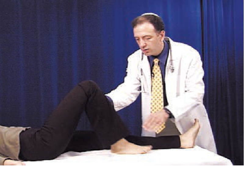
Summary Mnemonic
UMN = "WHIRRS": Weakness (grouped), Hyperreflexia, Increased tone, Reflex spread/clonus, Release signs (Babinski, Hoffmann), Spasticity
LMN = "WAFD": Weakness (individual), Atrophy (severe), Fasciculations/Fibrillations, Decreased/absent reflexes
Sources:
- Neuroanatomy through Clinical Cases, 3rd Ed. - Ch. 6.1 (Table 6.4)
- Adams and Victor's Principles of Neurology, 12th Ed. - Ch. 3 (Table 3-1), Ch. on The Lower Motor Neuron
- Ganong's Review of Medical Physiology, 26th Ed. - Lower vs. Upper Motor Neuron Damage
- Bradley and Daroff's Neurology in Clinical Practice - Box 97.2
Can u show me the exact postion of thalamus cordate nucleus and lantiform nucleus
Reading File
This is a visual anatomy question - image retrieval is critical. Let me search for diagrams of the thalamus, caudate nucleus, and lentiform nucleus simultaneously.
Finding Sources
Finding Sources
Excellent - found a Netter's Atlas diagram and a Neuroanatomy textbook figure. Let me read those sections and fetch the images in parallel.
Reading File
Reading File
Reading File
Now let me fetch the key images in parallel - the Netter's diagram with caudate/thalamus/lentiform, the horizontal section from Gray's, and the thalamus schematic.
Loading Image
Loading Image
Loading Image
I have the key diagrams. Let me also fetch the Gray's horizontal section diagram and the thalamus schematic.
Loading Image
Loading Image
I have excellent diagrams from Netter's Atlas. Here is the full visual and anatomical answer:
Thalamus, Caudate Nucleus, and Lentiform Nucleus - Position & Relationships
1. Netter's Atlas - Interrelationship Diagram (Left Lateral View)
This is the most important diagram showing how all three structures relate to each other:
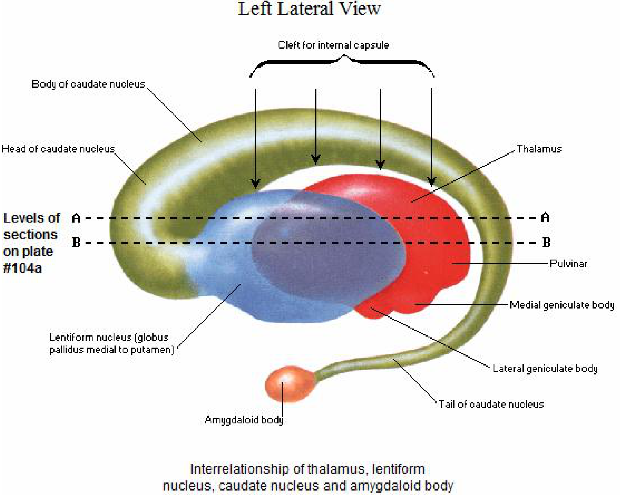
What this diagram shows:
- The caudate nucleus (olive/gold, C-shaped) wraps around the outside in a large arc - you can see its head (front), body (top), and tail (curling downward to the amygdala)
- The lentiform nucleus (blue/purple, lens-shaped) sits in the center - it consists of the putamen (outer) and globus pallidus (inner, medial)
- The thalamus (red) sits medially and posteriorly, nestled inside the arc of the caudate
- The internal capsule separates them - it sits in the cleft between the caudate/thalamus (medially) and the lentiform nucleus (laterally)
- The caudate's tail terminates at the amygdaloid body (small orange structure at the bottom)
2. Netter's Atlas - Horizontal Section through Cerebrum (Top-Down View)
This shows exactly where these structures sit when you cut across the brain:
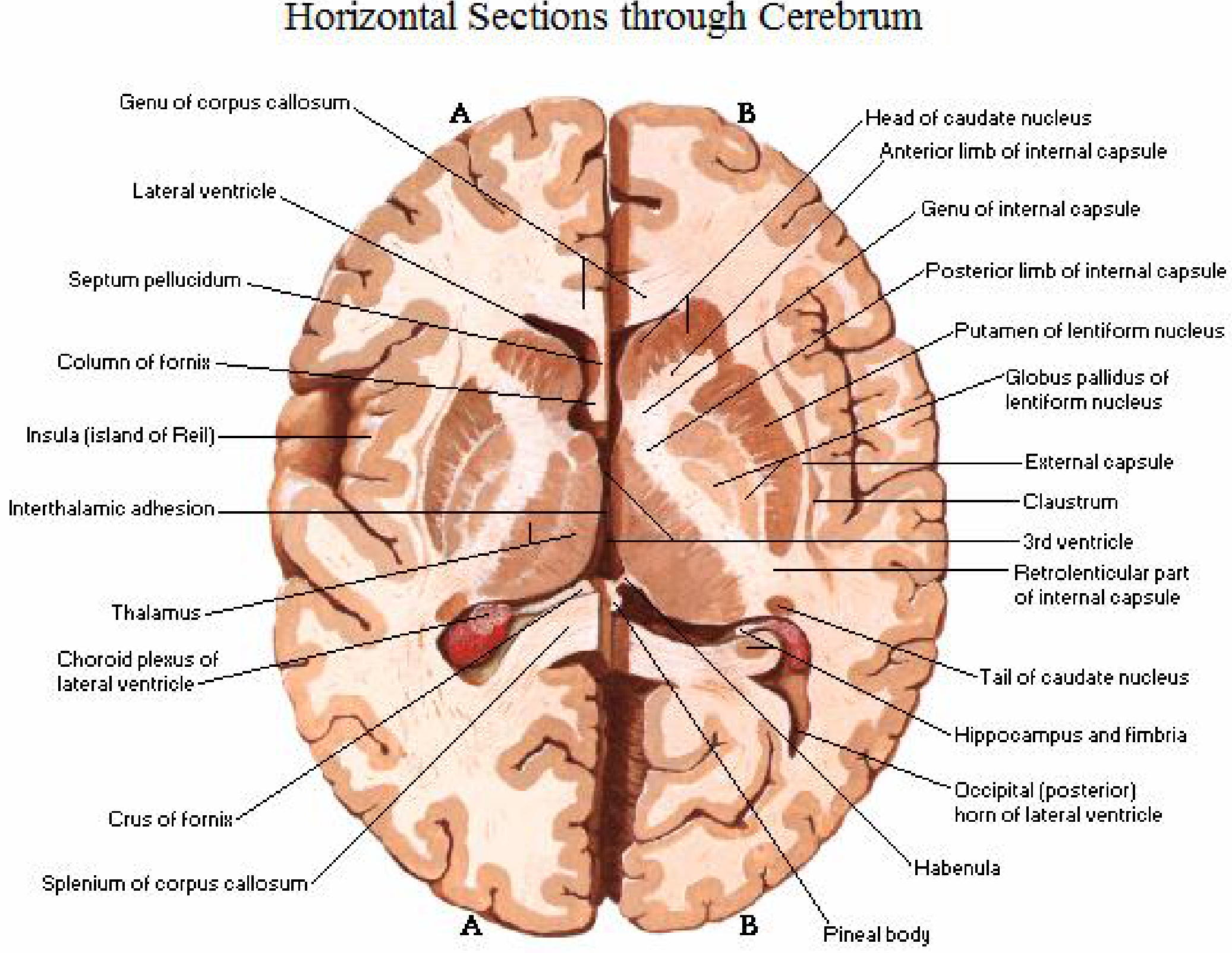
Key relationships visible in cross-section:
- Head of caudate nucleus - bulges into the lateral wall of the lateral ventricle (anteriorly)
- Tail of caudate nucleus - curves posteriorly and inferiorly along the floor of the lateral ventricle
- Lentiform nucleus (putamen + globus pallidus) - sits lateral to the internal capsule, lateral to the caudate/thalamus
- Thalamus - sits medially, forming the lateral walls of the 3rd ventricle, posterior to the caudate head
- Internal capsule - the V-shaped white matter cleft separating them: anterior limb (between caudate head and lentiform), genu (the bend), posterior limb (between thalamus and lentiform)
3. Netter's Thalamic Nuclei Diagram (3D Schematic)
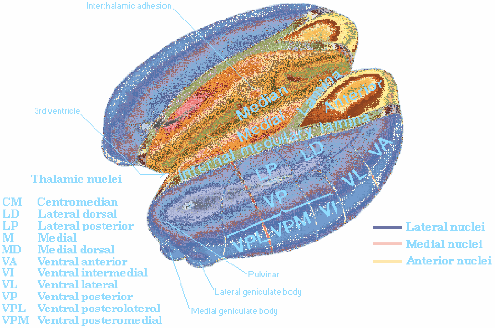
The thalamus itself is a paired egg-shaped diencephalic structure sitting on either side of the 3rd ventricle, with distinct nuclear groups (VA, VL, VPL, VPM, pulvinar, etc.) and the interthalamic adhesion connecting the two sides.
Summary of Positions
| Structure | Location | Shape | Key Landmark |
|---|---|---|---|
| Caudate Nucleus | Medial to internal capsule; wraps around lateral ventricle | Large C-shape (head → body → tail) | Bulges into floor of lateral ventricle |
| Lentiform Nucleus (putamen + globus pallidus) | Lateral to internal capsule; between insular cortex and internal capsule | Lens/wedge shape pointing medially | Lies lateral to internal capsule |
| Thalamus | Medial to posterior limb of internal capsule; forms lateral wall of 3rd ventricle | Paired oval masses | Bounded by 3rd ventricle medially, internal capsule laterally |
The internal capsule is the key white matter structure that separates them:
- Anterior limb: between caudate (medial) and lentiform (lateral)
- Posterior limb: between thalamus (medial) and lentiform (lateral)
- Genu: the bend between anterior and posterior limbs
Note: You used "caudate nucleus" and "lantiform nucleus" in your question - I've covered the caudate nucleus and lentiform nucleus (the correct term) above. The lentiform nucleus = putamen + globus pallidus combined.
Sources: Netter's Atlas of Human Anatomy; Gray's Anatomy for Students; Neuroanatomy through Clinical Cases, 3rd Ed.
This is a shared conversation. Sign in to Orris to start your own chat.