
Gunnie worm
Guinea worm (Dracunculus medinensis) is a parasitic nematode that causes dracunculiasis - a debilitating disease where a long worm slowly emerges through the skin, typically from the lower leg. It has no drug treatment; the worm is extracted by slowly winding it around a stick over days to weeks.
Searching sources...
Reading File
Searching the Web
Guinea worm Dracunculus medinensis dracunculiasis 2025 2026
Finding Sources
Reading File
Finding Sources
Reading File
Reading File
Guinea Worm (Dracunculus medinensis)
Overview
Guinea worm disease (dracunculiasis) is caused by the nematode Dracunculus medinensis - the world's longest tissue-dwelling parasite of humans (female worms reach 30 cm to 1 m in length; males are tiny, only ~4 cm, and die after copulation). It is on track to become the second human infectious disease ever eradicated, after smallpox.
Transmission and Life Cycle
The infection is acquired by drinking stagnant water containing infected Cyclops copepods (tiny freshwater crustaceans that serve as the intermediate host). The sequence:
- Ingested larvae penetrate the stomach or intestinal wall
- Worms mate and mature over approximately 12 months
- The impregnated female migrates to subcutaneous tissues, usually the lower extremity
- She induces a blister on the skin that ruptures and forms an ulcer
- On contact with fresh water, the female releases thousands of motile larvae - which infect new Cyclops and complete the cycle
Clinical Features
- Pre-emergence (~1 year after infection): No symptoms until just before the blister forms
- Prodrome: Sudden onset of fever, periorbital edema, wheezing, and urticaria (allergic reaction)
- Emergence: Local pain and swelling at the blister site; typically the lower leg but can occur on the genitalia, buttocks, or arms
- Complications: Secondary bacterial infection causing cellulitis, abscess, or tetanus; rarely, the worm becomes encapsulated and calcified in the tissues
Diagnosis
Purely clinical - based on the visible emergence of the adult worm through the skin. No specific lab test is needed.
Treatment
There is no effective drug therapy for dracunculiasis. The only treatment is the ancient technique of:
Gradual mechanical extraction - slowly winding a few centimeters of the worm onto a stick each day (~1 inch/day), taking great care not to rupture it (rupture causes severe inflammation). Full extraction may take days to weeks.
Surgical excision is possible but can exacerbate allergic symptoms.
Prevention
Since there is no vaccine or drug, eradication has relied entirely on behavioral and environmental measures:
- Filtering drinking water through finely woven cloth (removes Cyclops)
- Boiling or treating water with temephos (Abate), a larvicide safe for drinking water
- Educating infected individuals not to enter fresh water while the worm is emerging
- Provision of safe wells and clean water sources
Epidemiology and Eradication Progress
In 1986, there were an estimated 3.5 million cases annually across 21 countries. Through the Carter Center-led global campaign:
- As of end-2022: only 13 human cases in four African countries (Chad: 6, South Sudan: 5, Ethiopia: 1, Central African Republic: 1)
- Remaining endemic countries as of 2023: Chad, Mali, South Sudan, Angola, and Ethiopia
- A major obstacle to final eradication is animal reservoirs - dogs and cats in Chad serve as significant reservoirs, which was not appreciated until the 2010s
Historical Note
D. medinensis is one of medicine's oldest parasites - calcified worms have been found in 3,000-year-old Egyptian mummies. It is widely believed to be the "fiery serpent" referenced in the Bible (Numbers 21:6-9), and the symbol of a worm being wound around a stick is thought to be the origin of the Rod of Asclepius, the universal symbol of medicine.
Sources: Goldman-Cecil Medicine International Edition; Harrison's Principles of Internal Medicine 22E (2025); Quick Compendium of Clinical Pathology 5th ed.
Scabies
Scabies is a highly contagious skin infestation caused by the mite Sarcoptes scabiei var. hominis. It causes intense pruritus (worse at night) from the mite burrowing into the skin. Treatment is topical permethrin 5% or oral ivermectin.
Searching sources...
Finding Sources
Searching the Web
scabies treatment guidelines 2025
Reading File
Reading File
Reading File
Finding Sources
Reading File
Reading File
Now let me fetch the dermoscopy image from Fitzpatrick's since it was referenced:
Reading File
Scabies
Overview
Scabies is a highly contagious skin infestation caused by Sarcoptes scabiei var. hominis, a parasitic mite in the class Arachnida. It affects over 100 million people worldwide annually across all ages, races, and socioeconomic levels. Prevalence in some developing countries ranges from 4% to 100% of the general population.
The Mite
- Appearance: Pearl-like, translucent, white, oval, eyeless, with 4 pairs of short stubby legs
- Size: Adult female is 0.4 × 0.3 mm (just barely invisible to the naked eye); male is slightly smaller
- Survival off the host: 3 days in a sterile environment; up to 7 days in mineral oil
- Cannot fly or jump
- A normal host harbors 3 to 50 female mites; infants and the elderly may harbor 50-250; patients with crusted scabies can harbor millions
Life Cycle
The entire life cycle occurs on human skin. The female mite burrows into the stratum corneum (to the boundary of the stratum granulosum), excavating 0.5-5 mm per day, laying 0-4 eggs/day (up to 50 eggs in her lifetime). Larvae hatch and complete development to adults on the skin surface.
Transmission
- Primary route: Close, prolonged skin-to-skin contact (sexual and non-sexual)
- Fomites: Much less likely in normal scabies but highly relevant in crusted scabies (mites shed in huge numbers into bedding, furniture, floors)
- Incubation period: 4-6 weeks in first exposure; only 1-4 days on re-exposure (due to sensitization)
Clinical Features
Symptoms
- Intense pruritus, classically worse at night (sensitization to mite saliva, eggs, feces)
- On re-infestation, symptoms begin within 1-4 days
Distribution (classic sites)
- Interdigital web spaces of fingers
- Volar wrists and lateral palms
- Elbows, axillae
- Scrotum, penis, labia, areolae in women
- Head and neck spared in healthy adults; involved in infants, the elderly, and immunocompromised
Lesions
- Small (<5 mm) papules, pustules, and vesicles with excoriations
- Pathognomonic sign: The burrow - a thin, thread-like, linear or J-shaped tunnel 1-10 mm long in the stratum corneum
Here is what the burrow and mite look like under dermoscopy:
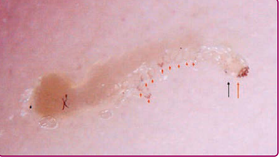
Dermoscopy showing the "delta/triangle sign" - dense mite head (long red arrow), translucent body (long black arrow), eggs (short red arrows), and classic S-shaped burrow. (Fitzpatrick's Dermatology)
And burrows in the web spaces of fingers:
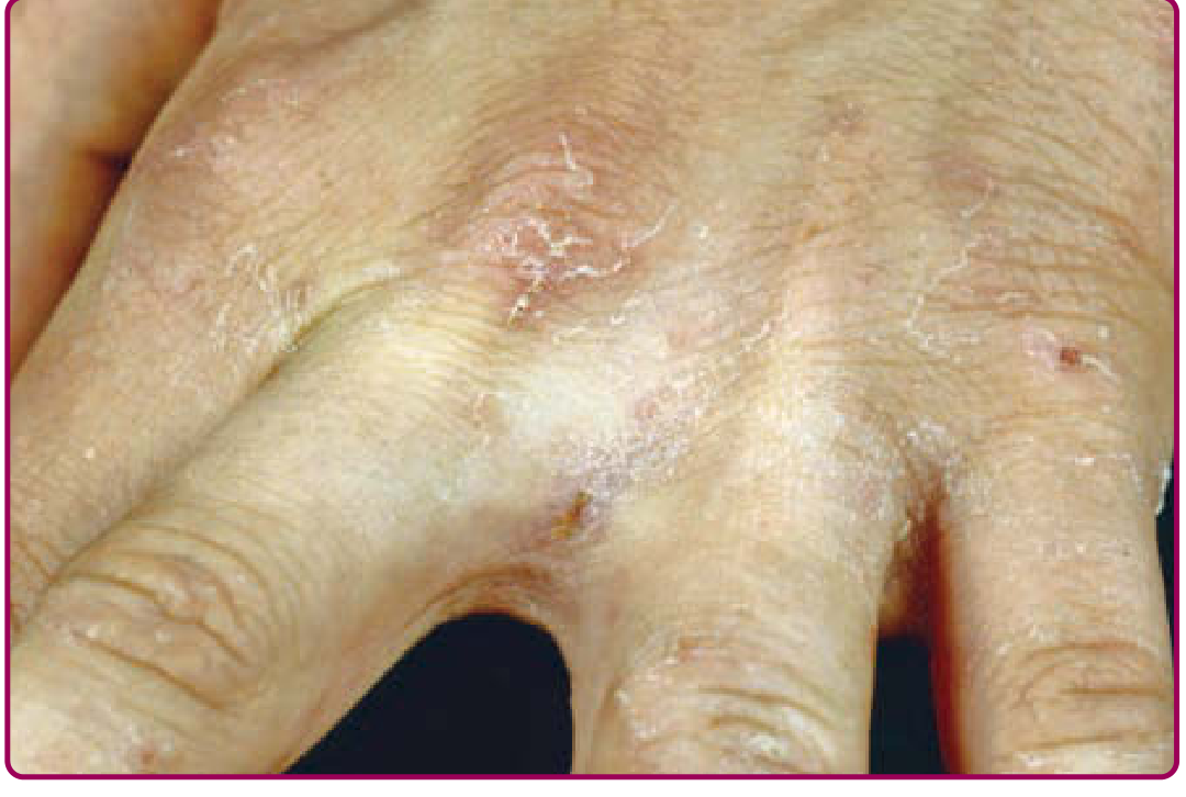
Thread-like burrows in the web spaces of the fingers - a classic location. (Fitzpatrick's Dermatology)
Crusted (Norwegian) Scabies
A severe variant in immunocompromised patients (HIV, elderly, dementia, neuropathy, leprosy):
- Hyperkeratotic plaques diffusely on palmar/plantar regions
- Thickening and dystrophy of toenails and fingernails
- Minimal or no pruritus (defective immune/sensory response)
- Millions of mites - extremely contagious; can spread to anyone in the vicinity
- Environmental mite burden is enormous (6,000 mites/g of shed debris from sheets, furniture)
Diagnosis
Clinical in most cases (history + characteristic distribution + pruritus worse at night).
Confirmatory tests:
- Skin scraping with a broad-blade scalpel from the terminal end of a burrow, placed in mineral oil on a glass slide - look for mites (330-450 μm), eggs, or scybala (fecal pellets). Low sensitivity.
- Dermoscopy (epiluminescence microscopy): Non-invasive; identifies the "delta-wing jet" or "triangle sign" of mite parts within the stratum corneum in vivo. Higher sensitivity.
- Burrow ink test: Apply black felt-tip marker to affected area, wipe off with alcohol - the burrow retains ink and becomes visible
- Reflectance confocal microscopy and PCR on swabbed skin are emerging high-sensitivity/specificity tools
Differential diagnosis: Atopic dermatitis, contact dermatitis, pityriasis rosea, papular urticaria, secondary syphilis, folliculitis, lichen planus, psoriasis, dermatitis herpetiformis
Treatment
First-line
| Agent | Regimen | Notes |
|---|---|---|
| Permethrin 5% cream | Apply neck-to-toe, leave on 8-14 hours, wash off; repeat in 1-2 weeks | Drug of choice; ovicidal; safe in pregnancy and infants >2 months |
| Oral ivermectin | 200 mcg/kg × 2 doses, 7-14 days apart | Not ovicidal - second dose essential; not FDA-approved for scabies; not established in <15 kg or pregnancy |
A Cochrane review found oral ivermectin is as effective as topical permethrin.
Crusted scabies
- Combination of oral ivermectin + topical permethrin is recommended (mite burden is too high for topical alone to be reliable)
Alternative agents
- Crotamiton 10% cream/lotion
- Precipitated sulfur 5-10% in petrolatum (safe in pregnancy and infants <2 months - youngest age group)
- Lindane 1% - avoid due to neurotoxicity; reserve only if all others fail or cannot be tolerated; contraindicated in children <10 years
Adjunct therapy
- Oral antihistamines and topical corticosteroids for pruritus relief (itching may persist for weeks after successful treatment due to ongoing hypersensitivity reaction)
- Topical/systemic antibiotics for secondary bacterial infection of excoriated lesions
Key Management Points
- Treat all contacts simultaneously - family members and close contacts, even if asymptomatic (many are asymptomatic mite carriers)
- Environment decontamination: Wash and dry all clothing, bedding, towels at high temperature; vacuum floors and furniture
- Pruritus persisting after treatment does not mean treatment failure - the hypersensitivity reaction can last weeks
- Hospitalized patients: Contact precautions until successfully treated
Sources: Fitzpatrick's Dermatology Vol 1-2; Rosen's Emergency Medicine; Red Book 2021 (Committee on Infectious Diseases); Goldman-Cecil Medicine International Edition
Characteristics findings fof kala azar
Kala-azar (visceral leishmaniasis) is caused by Leishmania donovani and is characterized by prolonged fever, massive splenomegaly, hepatomegaly, progressive wasting, and pancytopenia. The name "kala-azar" means "black fever" in Hindi, referring to the skin darkening seen in some patients.
Searching sources...
Finding Sources
Searching the Web
kala azar visceral leishmaniasis characteristic findings clinical features
Reading File
Finding Sources
Reading File
Reading File
Finding Sources
Reading File
I now have comprehensive information from multiple authoritative sources. Here is the full answer:
Kala-Azar (Visceral Leishmaniasis) - Characteristic Findings
What is it?
Kala-azar ("black fever/sickness" in Hindi) is the most severe form of leishmaniasis, caused by Leishmania donovani complex (L. donovani, L. infantum, L. chagasi). It is transmitted by the bite of an infected female phlebotomine sandfly. The parasite spreads throughout the reticuloendothelial system - invading macrophages in the spleen, liver, bone marrow, and lymph nodes as intracellular amastigotes (Leishman-Donovan bodies).
Characteristic Clinical Findings
1. Fever
- Prolonged, intermittent fever - temperatures of 39-40°C (102-104°F)
- A classic pattern is double daily temperature spikes (two fever peaks per day)
- Fever may persist for weeks to months
2. Massive Splenomegaly
- The most striking finding - the spleen can become enormously enlarged, sometimes reaching the pelvis
- Due to reticuloendothelial hyperplasia packed with parasitized macrophages
- The spleen is the most parasite-rich organ (>95% positivity on splenic aspirate)
3. Hepatomegaly
- Present alongside splenomegaly (hepatosplenomegaly is the hallmark)
- Hepatic fibrin-ring granulomas may be seen on biopsy
4. Progressive Wasting / Cachexia
- Severe weight loss, emaciation, weakness
- Patients develop a progressive wasting syndrome if untreated
- Advanced disease leads to GI symptoms and a wasting syndrome
5. Pancytopenia
All three cell lines are affected:
| Cell line | Finding | Consequence |
|---|---|---|
| Red cells | Anemia | Pallor, fatigue |
| White cells | Leukopenia / agranulocytosis | Susceptibility to secondary infections |
| Platelets | Thrombocytopenia | Epistaxis, purpura, bleeding |
6. Skin Darkening (the "Kala" of Kala-Azar)
- Patchy macular darkening of the skin due to melanin deposits
- Most marked on the forehead, temples, perioral area, and midabdomen
- Occurs in Indian patients; grayish/dark discoloration gave rise to the name
- NOT seen in all geographic variants
7. Hypergammaglobulinemia
- Marked polyclonal elevation of immunoglobulins (especially IgG)
- Basis of the Aldehyde (Napier's formol-gel) test
8. Hypoalbuminemia
- Nutritional depletion + protein loss
- Combined with hypergammaglobulinemia = reversal of albumin:globulin ratio
9. Lymphadenopathy
- Peripheral lymphadenopathy is particularly prominent in East Africa (Sudan, South Sudan)
- Less common in Asian variants
10. Secondary Complications
- Secondary bacterial infections (pulmonary, GI), noma/cancrum oris (oral ulceration)
- Hemophagocytic lymphohistiocytosis (HLH) - a recognized complication
- Tetanus and hemorrhage in late disease
- Death in ~2 years if untreated (advanced, untreated cases are almost universally fatal)
Post-Kala-Azar Dermal Leishmaniasis (PKDL)
A distinct sequela that develops after apparent successful treatment of visceral leishmaniasis:
- Indian subcontinent variant: Polymorphic lesions - hypopigmented macules/patches coexisting with papulonodules on the face and trunk
- Sudanese variant: Papular or nodular lesions
- The skin acts as a reservoir maintaining transmission even after the visceral disease is "cured"
Diagnostic Tests
1. rK39 Rapid Diagnostic Test (Dipstick) - First Line
- Immunochromatographic assay detecting antibodies to L. donovani using recombinant k39 antigen
- Results in 5 minutes; simple to perform in the field
- Mainstay of diagnosis in India
- Limitations: Do NOT use in relapse, reinfection, or HIV co-infection cases
2. Parasitological Diagnosis - Gold Standard (Confirmatory)
Direct demonstration of Leishman-Donovan (LD) bodies (amastigotes) in tissue aspirates:
| Site | Sensitivity | Risk |
|---|---|---|
| Spleen pulp aspirate | >95% | ~1% mortality risk - used cautiously |
| Bone marrow (sternal) | >85% | Safer, preferred |
| Liver tissue | Moderate | |
| Lymph node aspirate | Lower | |
| Peripheral blood | Only in Indian kala-azar | Low |
Organisms can also be cultured on NNN (Novy-MacNeal-Nicolle) medium.
3. Aldehyde Test (Napier's Formol-Gel Test) - Historical
- Add 1-2 drops of 40% formalin to 1-2 mL of patient serum
- Positive: Jellification to milk-white opacity (like white of hard-boiled egg) - newsprint cannot be read through it
- Strongly positive if occurs within 2-20 minutes
- Reflects extreme hypergammaglobulinemia (nonspecific - also positive in other conditions causing hyperglobulinemia)
Epidemiology
| Region | Species | Vector | Reservoir |
|---|---|---|---|
| India, Bangladesh | L. donovani donovani | Phlebotomus argentipes | Humans (anthroponotic) |
| Mediterranean, China, Near East | L. donovani infantum | P. perniciosus, P. ariasi | Dogs |
| South America | L. donovani chagasi | Lutzomyia longipalpis | Dogs |
| East Africa | L. donovani | Phlebotomus spp. | Humans/animals |
Treatment
| Drug | Notes |
|---|---|
| Liposomal Amphotericin B | Treatment of choice in India (due to antimony resistance); single-dose regimen available |
| Miltefosine | First oral drug for VL; as effective as amphotericin B; used widely in India and Ethiopia |
| Pentavalent antimony (sodium stibogluconate, meglumine antimonate) | Traditional first-line; widespread resistance in India |
| Amphotericin B deoxycholate | Effective but toxic; requires IV administration |
| Pentamidine | Second-line; significant toxicity |
Sources: Red Book 2021 (AAP Committee on Infectious Diseases); Andrews' Diseases of the Skin; Fishman's Pulmonary Diseases; Park's Textbook of Preventive and Social Medicine
This is a shared conversation. Sign in to Orris to start your own chat.