
APH OBSTATRIC IN DETAIL FOR FULL QUE
placenta previa types diagram antepartum hemorrhage
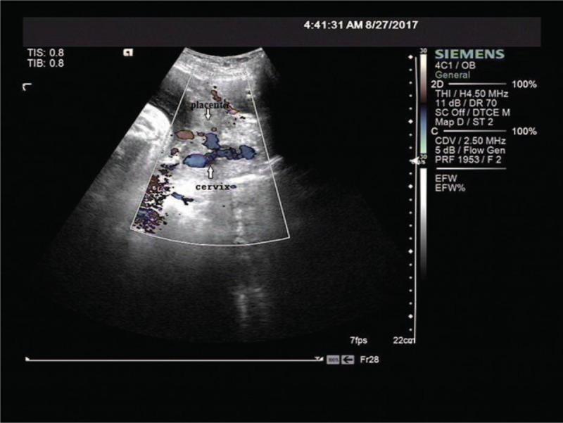
This diagnostic image is a transabdominal obstetric ultrasound with color Doppler, illustrating a longitudinal view of the uterus. The primary focus is on the relationship between the placenta and the cervix. The placenta is shown implanted on the anterior uterine wall; however, its lower edge extends inferiorly to completely cover the internal cervical os, a hallmark sign of placenta previa. The placental parenchyma displays mixed echogenicity with areas of low echoes (lacunae), and the color Doppler overlay reveals significant vascularity in the lower uterine segment near the cervical junction. The surrounding uterine tissue near the cervix appears more heterogeneous and less echogenic than the superior portions of the myometrium. This visual is clinically significant for diagnosing placenta previa, a condition requiring careful management due to the high risk of antepartum and intrapartum hemorrhage. This content is suitable for intermediate to advanced medical learners studying obstetrics and fetal imaging.
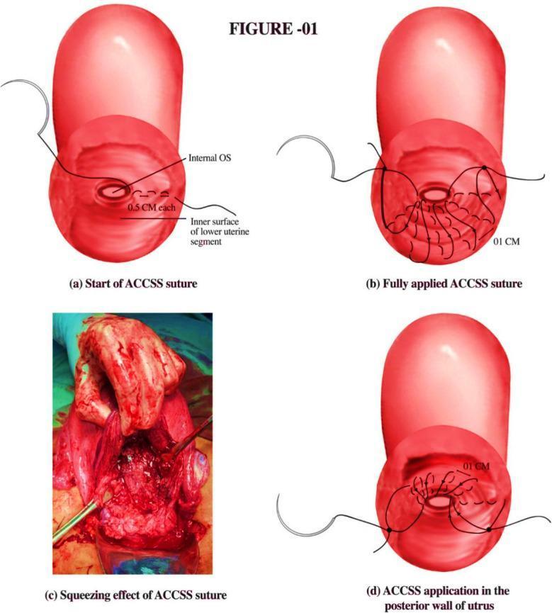
This medical illustration and clinical photograph composite describes the application of the A. Chohan Continuous Squeezing Suture (ACCSS) technique, a surgical intervention for managing hemorrhage during cesarean sections for placenta previa or accreta. The figure consists of four panels: (a) an anatomical diagram showing the initiation of the suture with 0.5 cm bites at the inner surface of the lower uterine segment near the internal os; (b) the progression to a fully applied continuous suture pattern with 1 cm intervals; (c) a clinical photograph demonstrating the intraoperative squeezing effect on the uterine tissue, showing significant tissue compression and congestion intended to achieve hemostasis; and (d) an illustration of the suture application on the posterior uterine wall. The technique utilizes a half-thickness suturing approach to compress placental site sinuses while maintaining the patency of the internal os. This educational material is designed for obstetric surgeons and trainees focusing on advanced surgical techniques for postpartum hemorrhage management.
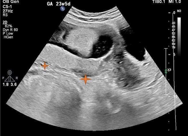
This diagnostic image is an abdominal obstetric ultrasound of a pregnant uterus at 23 weeks and 5 days of gestation. The scan demonstrates the placenta as a homogenous, relatively hyperechoic structure located in the lower uterine segment. Orange star markers indicate the placental tissue extending across and covering the region of the internal cervical os, confirming a diagnosis of placenta previa. Above the placenta, the gestational sac is visible, containing hypoechoic amniotic fluid and fetal parts. The myometrial-placental interface appears clear, with no sonographic evidence of retroplacental hematoma or abnormal placental lacunae. This visual is used to teach placental localization, the sonographic features of placenta previa in the second trimester, and the evaluation of the retroplacental space to rule out abruption. The clinical relevance focuses on managing high-risk pregnancies where placental positioning may lead to antepartum hemorrhage or complicate delivery planning.
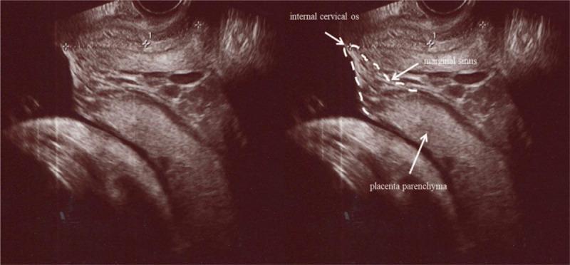
This diagnostic image consists of a side-by-side comparison of a sagittal grayscale transvaginal ultrasonography (TVUS) of the lower uterine segment during pregnancy. The right panel is annotated to identify key anatomical landmarks relevant to obstetric complications. The placenta parenchyma is shown as a homogeneous, iso-echogenic structure occupying the lower uterine segment. At the placental edge, a heterogeneous area with hypoechoic spaces is labeled as the marginal sinus. The internal cervical os is delineated by a dashed white line, demonstrating the close proximity of the placental margin and the marginal sinus to the cervical opening. This spatial relationship confirms the diagnosis of placenta previa. The imaging illustrates the use of high-resolution TVUS in the second or third trimester to evaluate placental position and identify vascular structures like the marginal sinus, which is clinically significant for assessing the risk of antepartum hemorrhage and determining delivery management in cases of low-lying or previa placentation.
abruptio placentae placental abruption concealed revealed hemorrhage uterus diagram
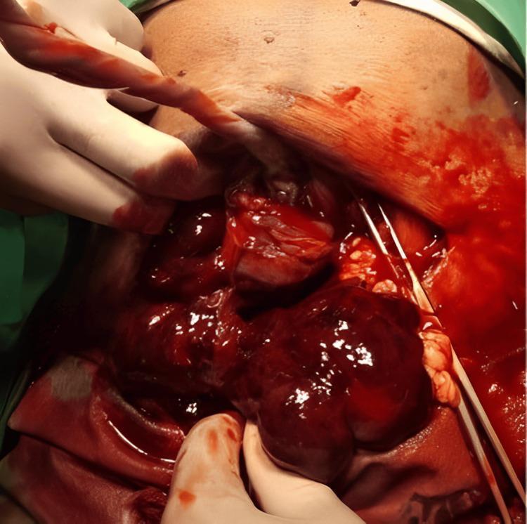
This clinical photograph captures an intraoperative view of a cesarean section complicated by placental abruption. The primary visual focus is on the uterus, which demonstrates the characteristic features of a Couvelaire uterus, also known as uteroplacental apoplexy. The myometrium appears dark, bluish-purple, and severely congested due to the widespread extravasation of blood into the uterine musculature. An incision in the uterine wall reveals a significant accumulation of dark red blood and organized clots within the hysterotomy site. The surgical field is heavily blood-stained, indicating active hemorrhage or hemoperitoneum. Gloved hands are seen manipulating the tissues, and surgical instruments, including forceps, are visible within the abdominal cavity. The umbilical cord is also seen emerging from the surgical site. This image serves as a clear educational example of the gross pathological appearance of uterine ecchymosis following severe abruptio placentae, highlighting the critical nature of obstetric hemorrhage and the visual markers of impaired uterine contractility in this clinical scenario.
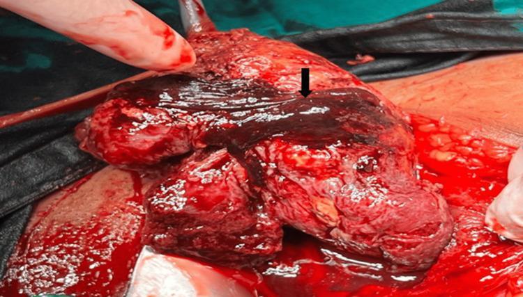
This clinical photograph captures an intraoperative view during an obstetric surgical procedure, illustrating a concealed placental abruption. The primary focus is the exposed uterus, which appears highly vascularized and congested with significant hemoperitoneum. A prominent, dark-red to black retroplacental hematoma is indicated by a black arrow, representing a large blood clot typical of placental abruption. The blood varies in color from bright red (fresh hemorrhage) to deep purple/black (older, organized clot), covering a substantial portion of the uterine tissue. The surgical context is reinforced by the presence of a gloved hand and a metal retractor holding the surgical margins. This image serves as an educational example of placental pathology and obstetric complications, specifically demonstrating the gross appearance of retroplacental bleeding and localized tissue necrosis following placental separation from the uterine wall.
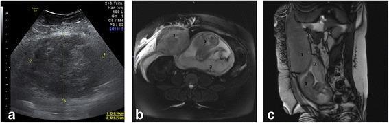
This composite of diagnostic images illustrates a Type III rectus sheath hematoma (RSH) in a 27-week pregnant patient. (a) Transverse transabdominal ultrasound shows a large, well-demarcated, heterogeneous mass measuring approximately 9.16 x 9.73 cm. The mass is partly hypoechoic and isoechoic, located adjacent to the uterine wall. (b) Axial MRI scan confirms a massive, hyperintense right-sided rectus sheath hematoma (1) that displaces the gravid uterus toward the contralateral side. The fetus (3) is visible in cephalic presentation, and edema is present in the overlying subcutaneous tissues. (c) Coronal MRI scan demonstrates the longitudinal extent of the hematoma (1), which measures 11 x 12 x 20 cm. It is positioned anterior and lateral to the uterus. The placenta (2) is clearly visualized on the posterior uterine wall, appearing normal with no signs of abruptio placentae or retroplacental hemorrhage. This case highlights the role of multimodality imaging in differentiating RSH from obstetric complications such as uterine myoma infarction or placental abruption during pregnancy.
ANTEPARTUM HEMORRHAGE (APH) - COMPLETE DETAILED NOTES
DEFINITION
- Barash Clinical Anesthesia, 9e - p. 3515
CAUSES / CLASSIFICATION
| Category | Examples |
|---|---|
| Placental causes | Placenta previa, Abruptio placentae (major causes) |
| Local/Cervical causes | Cervical erosion, cervical polyp, cervical carcinoma, vaginitis |
| Fetal vessel causes | Vasa previa |
| Unclassified | Marginal sinus rupture, idiopathic |
- Placenta Previa (~30%)
- Abruptio Placentae/Placental Abruption (~30%)
- "Unclassified" accounts for the remaining ~40%
PART 1: PLACENTA PREVIA
Definition
Classification / Types (4 types)
| Type | Description |
|---|---|
| Total / Complete Previa | The cervical os is completely covered by the placenta (~20% of cases - most severe, highest risk of exsanguination) |
| Partial Previa | Placenta covers a portion of the cervical os |
| Marginal Previa | Placenta extends just to the edge of the cervix |
| Low-lying Placenta | Placental implantation in the lower uterine segment but not covering the os |
"Central or total previa... can cause severe hemorrhage, with the risk of exsanguination for the fetus and mother." - Rosen's Emergency Medicine, p. 3355
Incidence
- 1 in 200-250 pregnancies
- Second trimester: ~5% of pregnancies have complete previa on USS; 90% of these resolve by term (placental migration)
Risk Factors (Mnemonic: STAMP-MC)
- S - Scarred uterus / previous Caesarean section (most important - dose-dependent relationship)
- T - Tobacco use (smoking)
- A - Advanced maternal age (AMA)
- M - Multiparity / multiple gestation
- P - Prior placenta previa
- M - Multiple uterine surgeries / miscarriages / induced abortions
- C - Cocaine use; assisted reproductive technology (IVF)
The risk for placenta previa and placenta accreta spectrum increases in a "dose-dependent" manner with the number of previous cesarean deliveries and greater parity. - Barash, p. 3515
Pathophysiology
- The lower uterine wall elongates during the 3rd trimester, OR
- With cervical dilation near the time of delivery
Clinical Features
| Feature | Description |
|---|---|
| Bleeding | Painless, bright red vaginal bleeding - the classic hallmark |
| Onset | Usually after 7th month; average first bleed at 27-32 weeks |
| Pattern | Initial bleed is usually not fatal; spontaneously stops and recurs |
| Uterine tone | Soft, non-tender uterus |
| Fetal lie | Abnormal lie common (transverse/breech) - because placenta occupies lower segment |
| Maternal vitals | Usually stable unless severe hemorrhage |
| Fetal status | Usually good initially |
"All patients with painless, second-trimester vaginal bleeding should be assumed to have placenta previa until proven otherwise." - Rosen's Emergency Medicine
Key Clinical Rule: NEVER do a vaginal examination in suspected placenta previa until ultrasound excludes it - digital/instrumental probing can precipitate catastrophic hemorrhage.
Diagnosis
- Transvaginal Ultrasonography (TVUS): Gold standard - safe, more accurate than transabdominal
- Transabdominal USS: Accuracy 93-98%; false positives with bladder distension or uterine contractions
- Important: Empty bladder before scan to avoid over-diagnosis; full bladder pushes lower uterine segment against posterior wall creating false appearance of previa
Management
- Two large-bore IV lines
- IV fluid resuscitation
- Continuous fetal monitoring (CTG)
- Baseline Hb, blood type and crossmatch
- Coagulation studies: platelet count, PT, PTT, fibrinogen, fibrin split products
- Normal fibrinogen in pregnancy: 400-450 mg/dL; values <300 mg/dL indicate significant coagulation factor consumption
- If Rh-negative and not yet received prophylaxis at 28 weeks: 300 μg Rh immune globulin within 72 hours
| Situation | Management |
|---|---|
| Bleeding not profuse + fetus immature | Expectant management - prolong pregnancy; admit to high-risk unit if contractions/acute bleeding |
| Severe bleeding OR mature fetus at symptom onset | Prompt Cesarean birth |
| Vaginal birth | Contraindicated in placenta previa |
- Neuraxial anesthesia is preferred for cesarean delivery (hemodynamically stable, no contraindications)
- General anesthesia is associated with greater blood loss and greater transfusion need
- Uterine atony - leading to PPH
- Placenta accreta spectrum (accreta, increta, percreta) - risk increases from 3% in primary cesarean with previa to 61% with 3 prior cesareans
- Postpartum hemorrhage
PART 2: ABRUPTIO PLACENTAE (Placental Abruption)
Definition
Incidence
- Complicates approximately 1% of deliveries
- Highest incidence between 24-32 weeks gestation
- Perinatal mortality: 9-12% in developed countries
- Maternal mortality: <1% (usually from coagulopathy or exsanguination)
Types Based on Bleeding Pattern
| Type | Description |
|---|---|
| Revealed (External) | Blood tracks down between membranes and uterine wall, escapes through cervix - visible bleeding |
| Concealed | Placental margins remain attached; blood collects behind placenta forming a retroplacental hematoma - no external bleeding despite significant internal hemorrhage |
| Mixed | Combination of both |
"Bleeding may be concealed if the placental margins have remained attached to the uterine wall." - Barash, p. 3516
Pathophysiology
- Spontaneous hemorrhage into the decidua basalis occurs
- Hematoma forms, causing further placental separation
- Hematoma compresses adjacent placenta, compromising fetal blood supply
- Leads to: increased intrauterine pressure, uterine tenderness, hypertonus, frequent contractions, fetal distress/demise
- In extensive abruption: retroplacental blood may penetrate the entire thickness of the uterine wall into the peritoneal cavity - called "Couvelaire Uterus" (uteroplacental apoplexy)
Couvelaire Uterus
- Retroplacental blood penetrates the myometrium due to increased intrauterine pressure
- Myometrium becomes weakened (blue-black, ecchymotic appearance)
- May rarely rupture, leading to life-threatening emergency
- Visible intraoperatively as a dark, bluish-purple congested uterus
Risk Factors (Mnemonic: CHAMP-T)
- C - Cocaine use (major risk)
- H - Hypertension (chronic) / eclampsia / preeclampsia (40-50% of cases have underlying hypertension)
- A - Advanced maternal age (>35) or young age (<20)
- M - Multiparity (parity ≥3)
- P - Prior abruption / Preterm PROM
- T - Trauma (motor vehicle accidents, intimate partner violence - 4-8% of pregnancies)
- Also: smoking, thrombophilia, oligohydramnios, chorioamnionitis, cocaine use, multiple gestation, sudden uterine decompression (delivery of first twin)
Clinical Features (Grades/Severity)
| Feature | Mild Abruption | Severe Abruption |
|---|---|---|
| Vaginal bleeding | Slight or absent | No or heavy (often dark, clotted) |
| Uterine tenderness | Mild | Severe - board-like rigidity |
| Uterine tone | Normal or slightly increased | Hypertonus/tetanic contractions |
| Fetal status | Normal | Distress or demise |
| Maternal vitals | Normal | Hypotension, tachycardia, shock |
| Fibrinogen | Normal | <150 mg/dL (DIC) |
| Coagulopathy | Absent | Present (DIC) |
"In severe abruptio placentae (15% of cases), the uterus is tetanically contracted and very painful, maternal hypotension results from visible or concealed uterine blood loss, fibrinogen levels are less than 150 mg/dL, and fetal death can occur." - Rosen's Emergency Medicine
- Painful vaginal bleeding (contrast with previa which is painless)
- Dark, clotted blood (contrast with previa - bright red)
- Uterine tenderness and hypertonus
- Fetal distress disproportionate to visible bleeding (due to concealed hemorrhage)
Diagnosis
- Clinical diagnosis (based on symptoms + signs)
- Ultrasound: NOT sensitive for fresh abruption (fresh blood is isoechoic with placenta); has 100% negative predictive value on cardiotocography (CTG) when monitoring is reassuring
- Electronic fetal monitoring (CTG): Very sensitive for identifying fetal distress; most important monitoring tool
- MRI: Diagnostic but impractical for unstable patients
Management
- CBC with platelets
- PT, PTT, fibrinogen, fibrin degradation products (FDP), D-dimer
- Blood type and crossmatch
- Kleihauer-Betke acid elution test (detect fetomaternal transfusion)
- Urine output monitoring
| Situation | Management |
|---|---|
| Mild abruption, stable mother, term fetus | Artificial rupture of membranes (ARM) + oxytocin augmentation; prepare for urgent delivery |
| Mild abruption, stable, preterm (distant from term) | Expectant management with close observation |
| Non-reassuring fetal status | Emergency Cesarean birth |
| Fetal death + severe abruption + stable mother | Attempted vaginal birth is reasonable |
| DIC | Replace coagulation factors (FFP, cryoprecipitate, platelets) |
| Severe/massive hemorrhage | Massive transfusion protocol; emergency delivery |
- Fresh-frozen plasma (FFP) or fresh whole blood
- Monitor fibrinogen closely
- Fetoplacental transfusion: administer RhoGAM if Rh-negative
- Neuraxial analgesia/anesthesia is appropriate only if mother is hemodynamically stable AND coagulation parameters are normal
Complications
- DIC - most feared coagulopathy complication
- Hemorrhagic shock
- Couvelaire uterus
- Acute renal failure / multi-organ failure
- Amniotic fluid embolism
- Uterine rupture (rare)
- Fetal distress / stillbirth (when separation >50% - stillbirth is the likeliest outcome)
PART 3: VASA PREVIA
Definition
Associations
- Velamentous insertion of the umbilical cord (almost always coexists)
- Placenta previa, bilobed/succenturiate lobe placenta
- In vitro fertilization
Clinical Danger
- When membranes rupture (spontaneously or artificially), fetal vessels tear - leads to rapid fetal exsanguination
- Fetal mortality: 33-100% if undiagnosed
- Pulsatile vessels may be palpated preceding the fetal vertex on digital examination - if this occurs, STOP examination
Diagnosis
- Transvaginal color Doppler ultrasound - shows vessels crossing the os
- Amniocentesis: APT test or Kleihauer-Betke to differentiate fetal from maternal blood
Management
- Elective Cesarean delivery before membrane rupture (usually at 35-37 weeks)
- If membranes rupture before delivery - immediate emergency Cesarean
PART 4: PLACENTA ACCRETA SPECTRUM (PAS)
| Type | Depth of Invasion | Frequency |
|---|---|---|
| Accreta | Placenta invades into myometrium but NOT full thickness | 82% |
| Increta | Invasion INTO full thickness myometrium | 12% |
| Percreta | Invasion THROUGH uterine wall into surrounding structures (bladder, bowel) | 6% |
- Risk escalates dramatically with number of prior cesarean deliveries
- Ultrasound features: loss of retroplacental clear zone, placental lacunae, abnormal vascularity on Doppler
COMPARISON TABLE: Placenta Previa vs. Abruptio Placentae
| Feature | Placenta Previa | Abruptio Placentae |
|---|---|---|
| Definition | Abnormally implanted placenta (lower segment) | Premature separation of normally implanted placenta |
| Incidence | 1 in 200-250 | 1 in 100 |
| Bleeding character | Painless, bright red, fresh | Painful, dark, clotted |
| Onset | Spontaneous, unprovoked | Often sudden, after precipitating event |
| Uterus | Soft, non-tender | Tender, rigid, board-like |
| Shock | Proportionate to visible bleeding | May be disproportionate (concealed) |
| Fetal presentation | Abnormal lie common | Usually normal |
| Coagulopathy (DIC) | Uncommon | Common (major complication) |
| USG utility | Highly useful (diagnostic) | Limited (fresh blood isoechoic) |
| Vaginal exam | ABSOLUTELY CONTRAINDICATED | Cautious but permissible |
| Delivery mode | Always Cesarean | Vaginal possible if fetal death + stable mother |
| Recurrence | Low | Higher (~10%) |
| Fetal risk | Due to prematurity | Due to placental insufficiency + DIC |
GENERAL MANAGEMENT OF APH (All Causes)
Initial Emergency Steps (ABCDE + Obstetric)
- Assess - vitals, conscious level, bleeding amount
- Position - left lateral tilt (relieve aortocaval compression)
- Airway + Breathing - oxygen 10-15 L/min by face mask
- IV access - two large-bore (14-16G) IV lines
- Fluids - crystalloid resuscitation; blood products as needed
- Labs: CBC, coagulation studies, LFTs, renal function, blood group, crossmatch, Kleihauer-Betke
- CTG - continuous fetal monitoring
- Urinary catheter - monitor urine output (target >30 mL/hr)
- Call obstetric team + neonatology, anesthesia
- Rh prophylaxis if Rh-negative: 300 μg anti-D immunoglobulin within 72 hours
Massive Hemorrhage Protocol
- Packed RBCs + FFP + Platelets in a 1:1:1 ratio
- Cryoprecipitate if fibrinogen <200 mg/dL
- Tranexamic acid 1g IV (evidence-based for PPH; used in APH too)
- Avoid hypothermia, acidosis, coagulopathy (the "lethal triad")
EXAM HIGH-YIELD POINTS (Mnemonics & Memory Aids)
Placenta Previa Key Points:
- Painless + Bright red + Soft uterus = Previa
- "P" for Painless = Previa
- Never do per vaginum exam until previa excluded by USS
- Always Cesarean delivery
- Risk with previous C-section is dose-dependent
Abruptio Placentae Key Points:
- Painful + Dark blood + Rigid uterus = Abruption
- "A" for Agonizing pain = Abruption
- Couvelaire uterus = blood infiltrates myometrium
- DIC is the most dangerous complication
- Shock may be disproportionate to visible bleeding (concealed hemorrhage)
- Kleihauer-Betke test for fetomaternal hemorrhage
Vasa Previa Key Points:
- Velamentous cord insertion = high association
- Rupture of membranes = immediate fetal exsanguination
- Elective Cesarean before term = definitive management
- Fetal blood = APT test / Kleihauer-Betke