

Write in firm of a flowvhart and give it in form of a image
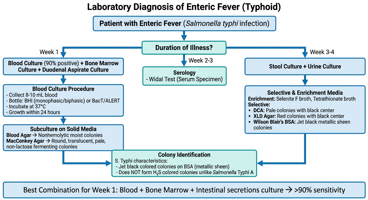
A detailed medical flowchart titled "Laboratory Diagnosis of Enteric Fever (Typhoid)" with the following structure: TOP: Start box "Patient with Enteric Fever (Salmonella typhi infection)" Arrow down to: "Duration of Illness?" Three branches: 1. LEFT: "Week 1" → Box: "Blood Culture (90% positive) + Bone Marrow Culture + Duodenal Aspirate Culture" 2. CENTER: "Week 2-3" → Box: "Serology - Widal Test (Serum Specimen)" 3. RIGHT: "Week 3-4" → Box: "Stool Culture + Urine Culture" From "Blood Culture" box, arrow down to: "Blood Culture Procedure" box: "- Collect 8-10 mL blood - Bottle: BHI (monophasic/biphasic) or BacT/ALERT - Incubate at 37°C - Growth within 24 hours" Arrow down to: "Subculture on Solid Media" box: "Blood Agar → Nonhemolytic moist colonies MacConkey Agar → Round, translucent, pale, non-lactose fermenting colonies" From "Stool/Urine Culture" box, arrow to: "Selective & Enrichment Media" box: "Enrichment: Selenite F broth, Tetrathionate broth Selective: • DCA: Pale colonies with black center • XLD Agar: Red colonies with black center • Wilson Blair's BSA: Jet black metallic sheen colonies" From both culture results, arrows point to: "Colony Identification" box: "S. Typhi characteristics: - Jet black colored colonies on BSA (metallic sheen) - Does NOT form H₂S colored colonies unlike Salmonella Typhi A" Bottom combined box: "Best Combination for Week 1: Blood + Bone Marrow + Intestinal secretions culture → >90% sensitivity" Clean white background, professional medical style, blue and teal color scheme for boxes, clear arrows, readable fonts, well-spaced layout.
-
Specimen selection by week of illness
- Week 1: Blood culture, bone marrow, duodenal aspirate
- Week 2-3: Serology (Widal test)
- Week 3-4: Stool and urine culture
-
Blood culture procedure - bottle types, incubation at 37°C, growth within 24 hrs, subculture on blood agar and MacConkey agar
-
Colony appearances - nonhemolytic on blood agar; round, pale, non-lactose fermenting on MacConkey
-
Stool/urine selective media - DCA, XLD agar, Wilson Blair's BSA with S. Typhi's characteristic jet black metallic sheen colonies
-
Best diagnostic combination - Blood + bone marrow + intestinal secretions in Week 1 gives >90% sensitivity
Also add a small note on wilson blair's in the image from the image provided
A detailed medical flowchart titled "Laboratory Diagnosis of Enteric Fever (Typhoid)" with the following structure: TOP: Start box "Patient with Enteric Fever (Salmonella typhi infection)" Arrow down to: "Duration of Illness?" Three branches: 1. LEFT: "Week 1" → Box: "Blood Culture (90% positive) + Bone Marrow Culture + Duodenal Aspirate Culture" 2. CENTER: "Week 2-3" → Box: "Serology - Widal Test (Serum Specimen)" 3. RIGHT: "Week 3-4" → Box: "Stool Culture + Urine Culture" From "Blood Culture" box, arrow down to: "Blood Culture Procedure" box: "- Collect 8-10 mL blood - Bottle: BHI (monophasic/biphasic) or BacT/ALERT - Incubate at 37°C - Growth within 24 hours" Arrow down to: "Subculture on Solid Media" box: "Blood Agar → Nonhemolytic moist colonies MacConkey Agar → Round, translucent, pale, non-lactose fermenting" From "Stool/Urine Culture" box, arrow to: "Selective and Enrichment Media" box: "Enrichment: Selenite F broth, Tetrathionate broth Selective: - DCA: Pale colonies with black center - XLD Agar: Red colonies with black center - Wilson Blair BSA: Jet black metallic sheen colonies" Below the Selective Media box, a highlighted side note box with a light yellow background and border, labeled "NOTE - Wilson Blair's Brilliant Green Bismuth Sulfite Agar (BSA)": "- Particularly useful for isolation of S. Typhi from HEAVILY CONTAMINATED specimens - S. Typhi: produces JET BLACK colored colonies with METALLIC SHEEN - Mechanism: due to H2S production - S. Paratyphi A and others that do NOT form H2S produce GREEN colored colonies instead - Key differentiator: Black = S. Typhi | Green = S. Paratyphi A" Bottom combined box in green: "Best Combination (Week 1): Blood + Bone Marrow + Intestinal secretions culture → >90% sensitivity" Clean white background, professional medical style, blue and teal color scheme for main boxes, yellow highlighted note box for Wilson Blair, clear arrows, readable fonts, well-spaced layout.
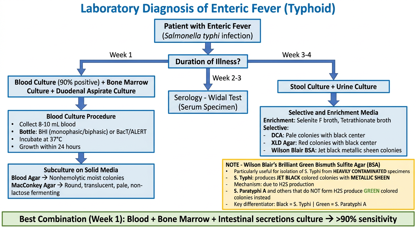
- Purpose - particularly useful for isolating S. Typhi from heavily contaminated specimens
- S. Typhi colonies - jet black with metallic sheen (due to H₂S production)
- S. Paratyphi A - produces green colored colonies (does NOT form H₂S)
- Key differentiator - Black = S. Typhi | Green = S. Paratyphi A
Add note about when to use stoo land urine culture from the given image and gerenrate new image
A detailed and well-spaced medical flowchart titled "Laboratory Diagnosis of Enteric Fever (Typhoid)" on a clean white background, professional medical style, blue and teal color scheme. TOP: Start rounded box "Patient with Enteric Fever (Salmonella typhi infection)" Arrow down to diamond decision box: "Duration of Illness?" Three branches going down: LEFT BRANCH - "Week 1": Box: "Blood Culture (90% positive) + Bone Marrow Culture + Duodenal Aspirate Culture" Arrow down to box: "Blood Culture Procedure: - Collect 8-10 mL blood - Bottle: BHI (monophasic/biphasic) or BacT/ALERT - Incubate at 37 degrees C - Growth within 24 hours" Arrow down to box: "Colony Appearance: Blood Agar: Nonhemolytic moist colonies MacConkey Agar: Round, translucent, pale, non-lactose fermenting" CENTER BRANCH - "Week 2-3": Box: "Serology Widal Test (Serum Specimen)" RIGHT BRANCH - "Week 3-4": Box: "Stool Culture + Urine Culture" Below the "Stool Culture + Urine Culture" box, a LIGHT GREEN highlighted note box with a dark green border titled "NOTE - Stool and Urine Culture": "When to use: - 3rd and 4th week of illness - Remain POSITIVE even after antibiotic treatment - Also used for detection of CARRIERS Urine Culture: - Seldom positive (salmonellae shed infrequently) - Urine centrifuged, deposit inoculated on MacConkey agar Stool Culture: - Done similar to Shigella method - Use appropriate media to inhibit commensals" Arrow from stool/urine box down to: "Selective and Enrichment Media for Stool" box: "Enrichment: Selenite F broth, Tetrathionate broth, GN broth Low selective: MacConkey agar Highly selective: - DCA: Pale colonies with black center - XLD Agar: Red colonies with black center - Wilson Blair BSA: Jet black metallic sheen colonies" Below that, a YELLOW highlighted side note box titled "NOTE - Wilson Blair's Brilliant Green Bismuth Sulfite Agar (BSA)": "- Particularly useful for isolation of S. Typhi from heavily contaminated specimens - S. Typhi: JET BLACK colonies with METALLIC SHEEN (due to H2S production) - S. Paratyphi A: GREEN colonies (does NOT form H2S) - Key differentiator: Black = S. Typhi | Green = S. Paratyphi A" BOTTOM wide box in green: "Best Combination (Week 1): Blood + Bone Marrow + Intestinal secretions culture = more than 90% sensitivity" Clear arrows between all boxes, readable font, well spaced, no overlapping text.
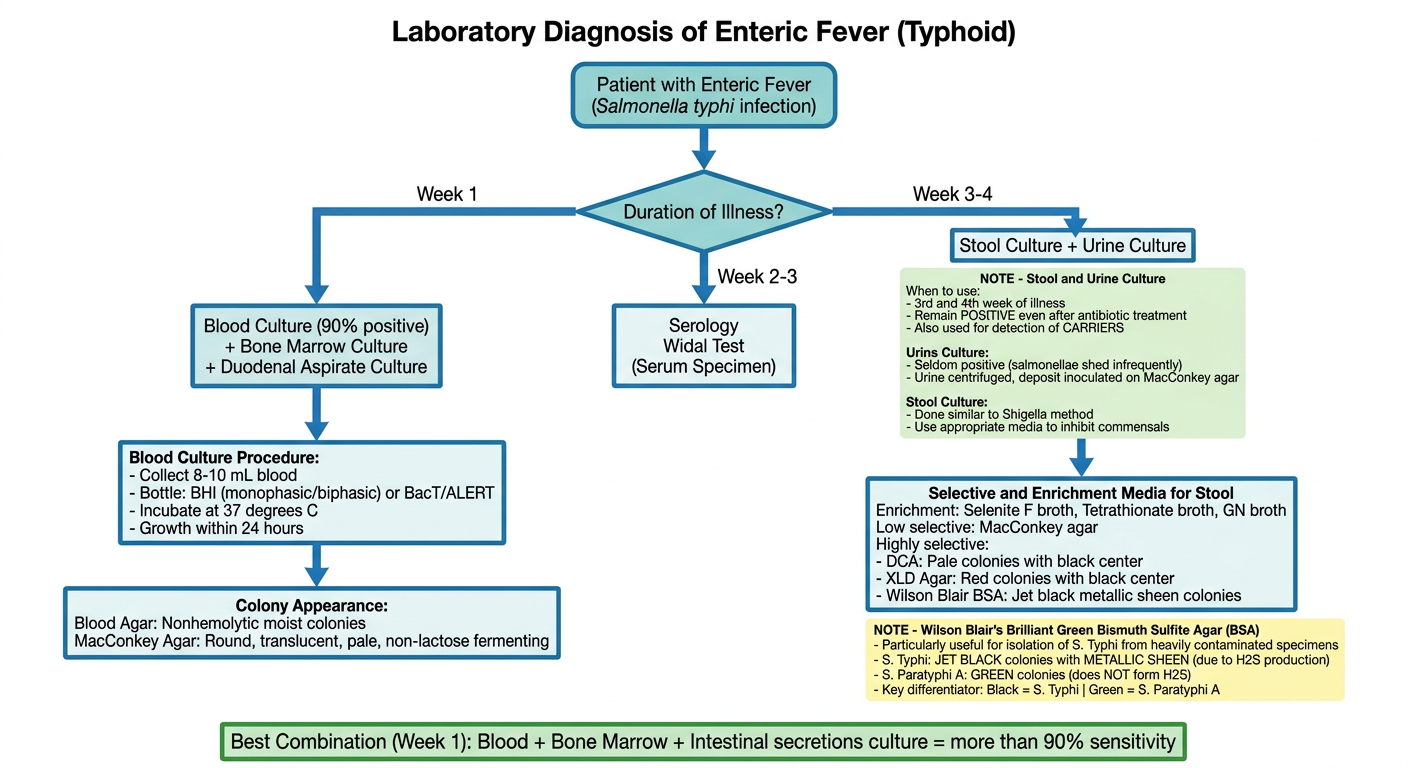
- 3rd and 4th week of illness
- Remain positive even after antibiotic treatment
- Also used for detection of carriers
- Seldom positive - salmonellae are shed in urine infrequently
- Urine is centrifuged and deposit inoculated onto MacConkey agar
- Done similar to the Shigella method
- Appropriate media used to inhibit commensals in the stool
Add note on other specimens that can be collected from give image and generate new image
A detailed, well-spaced, professional medical flowchart titled "Laboratory Diagnosis of Enteric Fever (Typhoid)" on a clean white background. Use blue and teal color scheme for main boxes, yellow for Wilson Blair note, green for stool/urine note, and orange/pink for Other Specimens note. TOP: Rounded blue box "Patient with Enteric Fever (Salmonella typhi infection)" Arrow down to diamond: "Duration of Illness?" THREE BRANCHES: LEFT - "Week 1": Teal box: "Blood Culture (90% positive) + Bone Marrow Culture + Duodenal Aspirate Culture" Arrow down to blue box: "Blood Culture Procedure: - 8-10 mL blood collected - Bottle: BHI (monophasic/biphasic) or BacT/ALERT - Incubate at 37C, growth within 24 hours" Arrow down to blue box: "Colony Appearance: Blood Agar: Nonhemolytic moist colonies MacConkey Agar: Round, translucent, pale, non-lactose fermenting" CENTER - "Week 2-3": Teal box: "Serology Widal Test (Serum Specimen)" RIGHT - "Week 3-4": Teal box: "Stool Culture + Urine Culture" Below the Stool/Urine box, LIGHT GREEN note box with dark green border: "NOTE - Stool and Urine Culture When to use: - 3rd and 4th week of illness - Remain POSITIVE even after antibiotic treatment - Used for detection of CARRIERS Urine: Seldom positive; centrifuge and inoculate deposit on MacConkey agar Stool: Similar to Shigella method; use media to inhibit commensals" Arrow down from stool/urine to blue box: "Selective and Enrichment Media: Enrichment: Selenite F broth, Tetrathionate broth, GN broth Low selective: MacConkey agar Highly selective: DCA: Pale colonies, black center XLD Agar: Red colonies, black center Wilson Blair BSA: Jet black metallic sheen colonies" Below that, YELLOW note box: "NOTE - Wilson Blair's BSA - Best for heavily contaminated specimens - S. Typhi: JET BLACK colonies with METALLIC SHEEN (H2S production) - S. Paratyphi A: GREEN colonies (no H2S) - Black = S. Typhi | Green = S. Paratyphi A" Below that on the LEFT side, LIGHT ORANGE note box with orange border: "NOTE - Other Specimens Bone Marrow Culture: - Used in Week 1 when blood culture is NEGATIVE - 55-90% sensitive - Especially useful when patient is ON ANTIBIOTICS Duodenal Aspirate Culture: - Used in Week 1 if BOTH blood AND bone marrow culture turn negative Other specimens from which Salmonellae can be isolated: - Rose spots - Pus from suppurative lesions - CSF (cerebrospinal fluid) - Sputum - Autopsy specimens: Gallbladder, Liver, Spleen" BOTTOM wide GREEN box: "Best Combination (Week 1): Blood + Bone Marrow + Intestinal Secretions = more than 90% sensitivity" All boxes well-spaced, arrows clearly connecting related boxes, readable font size, no overlapping text.
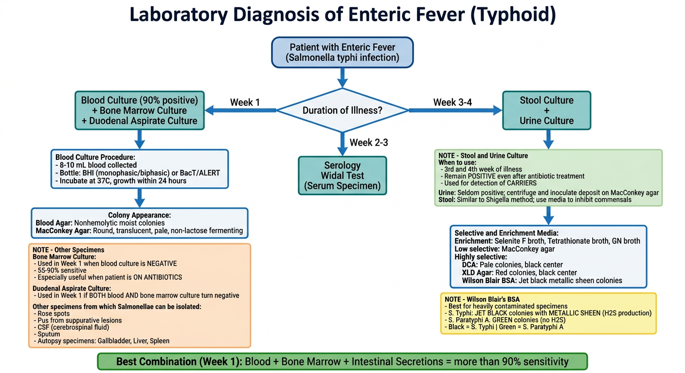
- Used in Week 1 when blood culture is negative
- 55-90% sensitive
- Especially useful when patient is already on antibiotics
- Used in Week 1 only if both blood culture AND bone marrow culture turn negative
- Rose spots
- Pus from suppurative lesions
- CSF (cerebrospinal fluid)
- Sputum
- Autopsy specimens - gallbladder, liver, spleen
- 🟡 Yellow - Wilson Blair's BSA
- 🟢 Green - Stool and Urine Culture
- 🟠 Orange - Other Specimens
- 🟩 Bottom green - Best combination (>90% sensitivity)

Generate a flowchart
A clear, well-spaced professional medical flowchart titled "Identification of Salmonella from Colonies" on a clean white background, blue and teal color scheme. TOP: Rounded blue box: "Salmonella Colonies on Culture Media" Arrow down to: Blue box "Two Methods of Identification" Two branches side by side: LEFT BRANCH - teal box: "Automated Systems" Below it, light blue box: "MALDI-TOF or VITEK" Below that, YELLOW note box: "NOTE - MALDI-TOF Limitation: - Identifies Salmonella up to GENUS level only - Poorly differentiates between serotypes - Reason: Serotypes share same ribosomal proteins" RIGHT BRANCH - teal box: "Conventional Biochemical Tests" Arrow down to: Blue box "Basic Enzyme Tests" Content: "Catalase: POSITIVE Oxidase: NEGATIVE" Arrow down to: Blue box "Other Biochemical Tests" Content: "Indole test: NEGATIVE Citrate test: NEGATIVE Urease test: NEGATIVE" Arrow down to: Blue teal box "TSI - Triple Sugar Iron Test" Content: "Reaction: Alkaline slant / Acid butt" From TSI box, THREE sub-branches: LEFT sub-branch - light blue box: "Gas Production: Most Salmonella: GAS PRESENT Exception: S. Typhi = ANAEROGENIC (no gas)" CENTER sub-branch - light blue box: "H2S Production: Most Salmonella: ABUNDANT H2S" RIGHT sub-branch - RED bordered box: "H2S Exceptions: S. Paratyphi A: H2S NOT produced at all S. Typhi: Only a SPECK of H2S at junction of slant and butt" All three sub-branches merge into bottom box: BOTTOM wide green box: "Serological Confirmation: Slide agglutination with O antisera Then serotype identified by type-specific O antigen" All boxes well-spaced, arrows clearly connecting, readable fonts, no overlapping text, professional clean style.
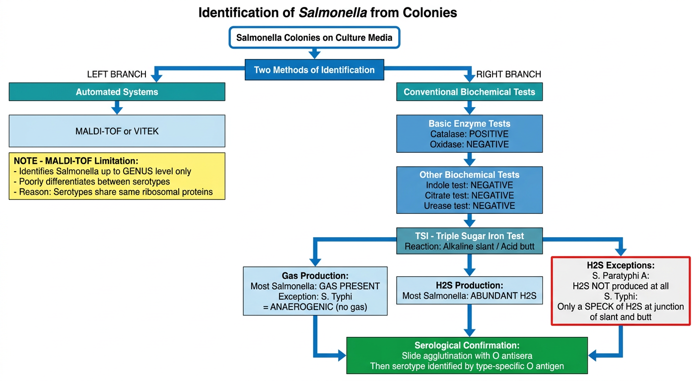
- Automated: MALDI-TOF or VITEK
- Conventional: Biochemical tests
- Catalase: Positive | Oxidase: Negative
- Indole, Citrate, Urease: All Negative
- Alkaline slant / Acid butt
- Gas: Present in most (except S. Typhi which is anaerogenic)
- H2S: Abundant in most, with two key exceptions:
- S. Paratyphi A - no H2S at all
- S. Typhi - only a speck at the junction of slant and butt


Explain widal test according to give images in form of a flowchart or diagram give in form of a image
A comprehensive, detailed, well-spaced professional medical flowchart titled "WIDAL TEST - Serological Diagnosis of Enteric Fever" on a clean white background. Use a clear top-to-bottom flow with color-coded sections. TOP BOX (dark blue rounded): "WIDAL TEST Oldest and most widely used serological test for Enteric Fever Discovered by Fernand Widal, 1896" Arrow down to TEAL box: "PRINCIPLE Agglutination test - H and O antibodies against S. Typhi and S. Paratyphi A and B detected and measured in patient's serum using O and H antigens" Arrow down to BLUE box: "ANTIGENS USED (4 antigens) 1. TO - O antigen of S. Typhi (Blue colored suspension) 2. TH - H antigen of S. Typhi (Pink colored suspension) 3. AH - H antigen of S. Paratyphi A 4. BH - H antigen of S. Paratyphi B NOTE: Paratyphoid O antigens NOT used (cross-react with typhoid O due to shared factor 12)" Arrow down to TEAL box: "PROCEDURE 1. Patient serum serially diluted in normal saline: 1/10 to 1/640 2. Four sets of dilutions made 3. Respective antigen suspensions (TO, TH, AH, BH) added to each set 4. Incubated in water bath at 37 degree C overnight" Arrow down to BLUE box: "RESULTS O agglutination: Compact granular CHALKY CLUMPS (disk-like), clear supernatant H agglutination: Large loose FLUFFY COTTON-WOOLLY clumps, clear supernatant No agglutination: BUTTON formation (antigen deposits), supernatant remains HAZY TITER: Highest dilution at which agglutination occurs = antibody titer" Arrow down splits into two boxes side by side: LEFT LIGHT BLUE box: "SIGNIFICANT TITERS (India) H agglutinin titer: more than 200 O agglutinin titer: more than 100 Low titers = ignored as baseline in endemic areas Cut-off varies by endemicity Fourfold rise in paired sera (1 week apart) = more meaningful than single high titer" RIGHT LIGHT BLUE box: "O vs H ANTIBODY KINETICS O agglutinins: Appear EARLY, disappear EARLY = recent infection H agglutinins: Appear LATE, disappear LATE O antibodies: NONSPECIFIC - raised in S. Typhi, S. Paratyphi A and B H antibodies: SPECIFIC - TH for S. Typhi, AH for S. Paratyphi A, BH for S. Paratyphi B" Below both, two colored note boxes side by side: LEFT RED bordered box: "FALSE POSITIVE causes: - Anamnestic response: Transient rise due to unrelated infections (malaria, dengue) in persons with prior enteric fever - falls after 1 week - Bacterial antigens not free from fimbriae - Inapparent infection - Prior immunization with TAB vaccine" RIGHT ORANGE bordered box: "FALSE NEGATIVE causes: - Early stage: 1st week of illness - Late stage: after 4th week - Carriers - Patients on antibiotics - Prozone phenomenon (antibody excess) - overcome by serial dilution of sera" All boxes well-spaced, no text overlap, clean arrows, readable fonts, professional medical style.
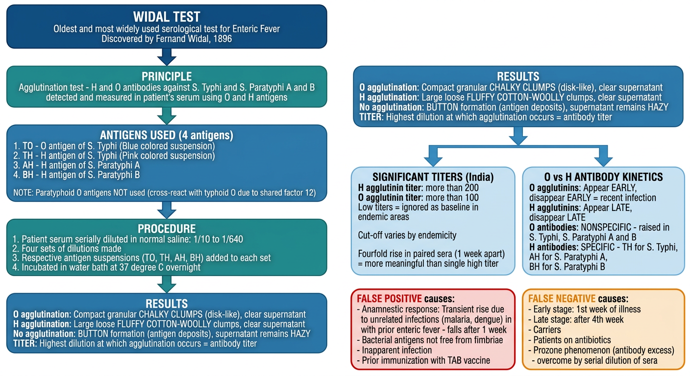
- O agglutination: chalky compact disc-like clumps
- H agglutination: fluffy cotton-woolly clumps
- No agglutination: button formation, hazy supernatant
Make it more systemic
A highly systematic, structured, step-by-step medical flowchart titled "WIDAL TEST" as a vertical sequential flow on a clean white background. Each step is clearly numbered and color-coded. Use a strict top-to-bottom single flow with numbered steps and branching only where needed. HEADER BOX (dark navy blue, wide): "WIDAL TEST Serological Diagnosis of Enteric Fever Oldest and most widely used test | Discovered by Fernand Widal, 1896" STEP 1 BOX (dark teal, left-numbered badge "1"): "PRINCIPLE An agglutination test that detects and measures H and O antibodies against S. Typhi and S. Paratyphi A and B in the patient's serum" STEP 2 BOX (blue, left-numbered badge "2"): "ANTIGENS USED — 4 Antigens TO — O antigen of S. Typhi (Blue suspension) TH — H antigen of S. Typhi (Pink suspension) AH — H antigen of S. Paratyphi A BH — H antigen of S. Paratyphi B IMPORTANT: Paratyphoid O antigens NOT used in the test (They cross-react with typhoid O antigen — share factor 12)" STEP 3 BOX (teal, left-numbered badge "3"): "PROCEDURE Step A: Patient serum serially diluted in normal saline (1/10 to 1/640) Step B: Four separate sets of dilution tubes prepared Step C: TO, TH, AH, BH antigen suspensions added to respective sets Step D: Incubated in water bath at 37°C overnight" STEP 4 BOX (blue, left-numbered badge "4"): "RESULTS — Reading the Tubes O agglutination: Compact granular CHALKY CLUMPS (disk-like pattern) — clear supernatant H agglutination: Large loose FLUFFY COTTON-WOOLLY CLUMPS — clear supernatant No agglutination: BUTTON FORMATION (antigen deposits at bottom) — supernatant is HAZY TITER DEFINITION: Highest dilution at which agglutination still occurs = antibody titer" STEP 5 BOX (indigo, left-numbered badge "5"): "INTERPRETATION Significant titer in India: H agglutinin titer > 200 | O agglutinin titer > 100 Low titers = baseline in endemic areas — IGNORE Fourfold rise in paired sera tested 1 week apart = MORE MEANINGFUL than a single high titer" STEP 6 BOX (slate blue, left-numbered badge "6"): "O vs H ANTIBODY KINETICS O antibodies: Appear EARLY — Disappear EARLY — indicate RECENT infection — NONSPECIFIC (raised in S. Typhi, S. Paratyphi A and B) H antibodies: Appear LATE — Disappear LATE — SPECIFIC: TH = S. Typhi | AH = S. Paratyphi A | BH = S. Paratyphi B" Then two side-by-side boxes at the bottom: LEFT BOX (red background with white text, badge "FP"): "FALSE POSITIVE — Causes 1. Anamnestic response — transient rise in titer from unrelated illness (malaria, dengue) in person with prior enteric fever — titer FALLS after 1 week 2. Antigen suspensions not free from fimbriae 3. Inapparent infection 4. Prior immunization with TAB vaccine" RIGHT BOX (orange background with white text, badge "FN"): "FALSE NEGATIVE — Causes 1. Early stage — 1st week of illness (antibodies not yet formed) 2. Late stage — after 4th week 3. Carriers 4. Patients on antibiotics 5. Prozone phenomenon (antibody excess) — overcome by serial dilution of serum" FOOTER BOX (dark green, wide): "KEY TAKEAWAY Fourfold rise in titer over 1 week in paired sera = confirms true infection Anamnestic response titer = falls after 1 week (NOT a true infection)" Strict vertical numbered flow, clear downward arrows between each step, clean sans-serif fonts, adequate spacing, no overlapping text, professional medical textbook style.
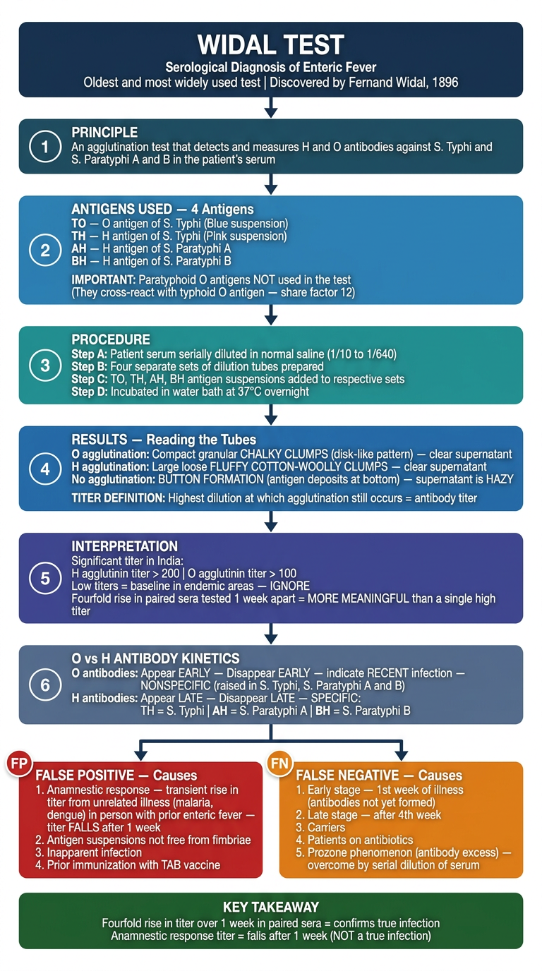
| Step | Section | Key Point |
|---|---|---|
| 1 | Principle | Agglutination test for H and O antibodies |
| 2 | Antigens | 4 antigens (TO, TH, AH, BH) - paratyphoid O excluded |
| 3 | Procedure | Serial dilution 1/10 to 1/640, four sets, water bath 37°C overnight |
| 4 | Results | Chalky clumps (O), fluffy clumps (H), button = no agglutination |
| 5 | Interpretation | H > 200, O > 100 significant in India; fourfold rise > single titer |
| 6 | Kinetics | O = early/nonspecific; H = late/specific per serotype |
| FP | False Positive | Anamnestic response, fimbriae, TAB vaccine, inapparent infection |
| FN | False Negative | Week 1, week 4+, carriers, antibiotics, prozone phenomenon |

Add the information about interpretation of widal test from this table to the above flowchart and make new flocahrt
A highly systematic, structured, step-by-step medical flowchart titled "WIDAL TEST" as a vertical sequential flow on a clean white background. Each step is clearly numbered and color-coded. Strict top-to-bottom single flow with numbered steps. HEADER BOX (dark navy blue, wide, bold large font): "WIDAL TEST Serological Diagnosis of Enteric Fever Oldest and most widely used test | Discovered by Fernand Widal, 1896" STEP 1 BOX (dark teal, left circle badge "1"): "PRINCIPLE An agglutination test detecting H and O antibodies against S. Typhi and S. Paratyphi A and B in patient serum" Arrow down STEP 2 BOX (steel blue, left circle badge "2"): "ANTIGENS USED — 4 Antigens TO — O antigen of S. Typhi (Blue suspension) TH — H antigen of S. Typhi (Pink suspension) AH — H antigen of S. Paratyphi A BH — H antigen of S. Paratyphi B IMPORTANT: Paratyphoid O antigens NOT used — cross-react with typhoid O (shared factor 12)" Arrow down STEP 3 BOX (teal, left circle badge "3"): "PROCEDURE A. Serially dilute patient serum in normal saline: 1/10 to 1/640 B. Prepare four sets of dilution tubes C. Add TO, TH, AH, BH antigen suspensions to respective sets D. Incubate in water bath at 37°C overnight" Arrow down STEP 4 BOX (steel blue, left circle badge "4"): "RESULTS — Reading the Tubes O agglutination: Compact CHALKY GRANULAR CLUMPS (disk-like) — clear supernatant H agglutination: Large loose FLUFFY COTTON-WOOLLY CLUMPS — clear supernatant No agglutination: BUTTON FORMATION at bottom — supernatant remains HAZY TITER: Highest dilution at which agglutination still occurs = antibody titer" Arrow down STEP 5 BOX (indigo blue, left circle badge "5"): "INTERPRETATION — Significant Titers India cutoff: H agglutinin titer > 200 | O agglutinin titer > 100 Low titers = baseline in endemic areas — IGNORE Fourfold rise in paired sera at 1-week interval = MORE MEANINGFUL than single high titer" Arrow down STEP 5B — Wide table-style box (dark maroon header with white text, rows alternating white and light pink): Header row: "TABLE 30.3 — INTERPRETATION OF WIDAL TEST RESULT" Two column headers in maroon: "WIDAL TEST RESULT" | "SUGGESTIVE OF" Row 1: "Rise of TO and TH antibody" | "Enteric fever due to S. Typhi" Row 2: "Rise of TO and AH antibody" | "Enteric fever due to S. Paratyphi A" Row 3: "Rise of TO and BH antibody" | "Enteric fever due to S. Paratyphi B" Row 4: "Rise of only TO antibody" | "Recent infection: S. Typhi or S. Paratyphi A or B" Row 5: "Rise of only TH antibody" | "? Convalescent stage / Anamnestic response" Row 6: "Rise of all TH, AH, BH antibodies" | "Post TAB vaccination" Arrow down STEP 6 BOX (slate blue, left circle badge "6"): "O vs H ANTIBODY KINETICS O antibodies: Appear EARLY — Disappear EARLY — indicate RECENT infection — NONSPECIFIC (S. Typhi + S. Paratyphi A and B) H antibodies: Appear LATE — Disappear LATE — SPECIFIC per serotype: TH = S. Typhi | AH = S. Paratyphi A | BH = S. Paratyphi B" Arrow down to two side-by-side boxes: LEFT BOX (solid red, white text, badge "FP"): "FALSE POSITIVE — Causes 1. Anamnestic response: Transient titer rise from unrelated illness (malaria, dengue) in person with prior enteric fever — FALLS after 1 week 2. Antigen suspensions not free from fimbriae 3. Inapparent infection 4. Prior immunization with TAB vaccine" RIGHT BOX (solid orange, white text, badge "FN"): "FALSE NEGATIVE — Causes 1. Early stage — 1st week (antibodies not yet formed) 2. Late stage — after 4th week 3. Carriers 4. Patients on antibiotics 5. Prozone phenomenon (antibody excess) — overcome by serial dilution" Arrow down from both to: FOOTER BOX (dark green, wide, bold): "KEY TAKEAWAY Fourfold rise in titer over 1 week (paired sera) = TRUE INFECTION Anamnestic titer = FALLS after 1 week (NOT true infection)" Strict vertical numbered flow with clear downward arrows between all steps, clean modern sans-serif fonts, adequate spacing between each section, no overlapping text, professional medical textbook diagram style.
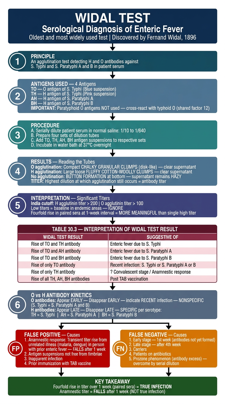
| Widal Test Result | Suggestive of |
|---|---|
| Rise of TO + TH | Enteric fever - S. Typhi |
| Rise of TO + AH | Enteric fever - S. Paratyphi A |
| Rise of TO + BH | Enteric fever - S. Paratyphi B |
| Rise of only TO | Recent infection - S. Typhi or S. Paratyphi A or B |
| Rise of only TH | ? Convalescent stage / Anamnestic response |
| Rise of all TH, AH, BH | Post TAB vaccination |

Explain in form of flowchart give in form of inage
A clean, systematic, well-spaced medical flowchart titled "VACCINES FOR TYPHOID FEVER" on a white background, professional medical style, color-coded sections. HEADER BOX (dark navy blue, wide, large bold font): "VACCINES FOR TYPHOID FEVER Immunization provides SHORT-TERM protection" Arrow down to: TEAL BOX titled "INDICATIONS FOR VACCINATION": "Who should be vaccinated? - Travelers going to endemic areas - People attending melas and yatras - Household contacts - People at increased risk (school children) - People living in endemic areas (optional)" Arrow down to diamond decision box: "Types of Vaccines Available" Two branches going down LEFT and RIGHT: LEFT BRANCH — BLUE header box: "VACCINE 1 Vi Antigen Capsular Polysaccharide (Vi-CPS) Vaccine" Below it, light blue details box: "Composition: Purified Vi capsular polysaccharide antigen derived from S. Typhi strain Ty2 Route: Single dose IM or Subcutaneous containing 25 microgram of Vi antigen Protection: 2 years Booster: Every 2 years Age restriction: Given ONLY after 2 years of age Reason: Capsular antigen is T-independent antigen = poorly immunogenic in children less than 2 years" Below that, a highlighted ORANGE sub-box: "Vi-rEPA VARIANT: Vi antigen conjugated with recombinant Pseudomonas aeruginosa Exotoxin A Conjugation INCREASES immunogenicity Therefore can be given to children LESS THAN 2 years" RIGHT BRANCH — GREEN header box: "VACCINE 2 Typhoral — Oral Live Attenuated S. Typhi Ty21a Vaccine" Below it, light green details box: "Type: Stable live attenuated mutant of S. Typhi strain Ty21a Key feature: LACKS the enzyme UDP-galactose-4-epimerase (Gal E mutant)" Both branches converge to bottom COMPARISON TABLE box (white background, dark borders): Header row (maroon): "QUICK COMPARISON" Row 1 headers: "Feature | Vi-CPS | Typhoral (Ty21a)" Row 2: "Type | Polysaccharide (inactivated) | Live attenuated oral" Row 3: "Route | IM / Subcutaneous | Oral" Row 4: "Age | Above 2 years | —" Row 5: "Protection | 2 years | Short term" Row 6: "Booster | Every 2 years | —" Row 7: "Special | Vi-rEPA variant for under 2 years | Gal E mutant" FOOTER BOX (dark green): "NOTE: Vi-CPS is poor immunogen in children under 2 years Solution: Vi-rEPA conjugate vaccine improves immunogenicity Typhoral lacks UDP-galactose-4-epimerase (Gal E mutant)" Clean arrows, numbered steps where appropriate, adequate spacing, no overlapping text, professional medical diagram.
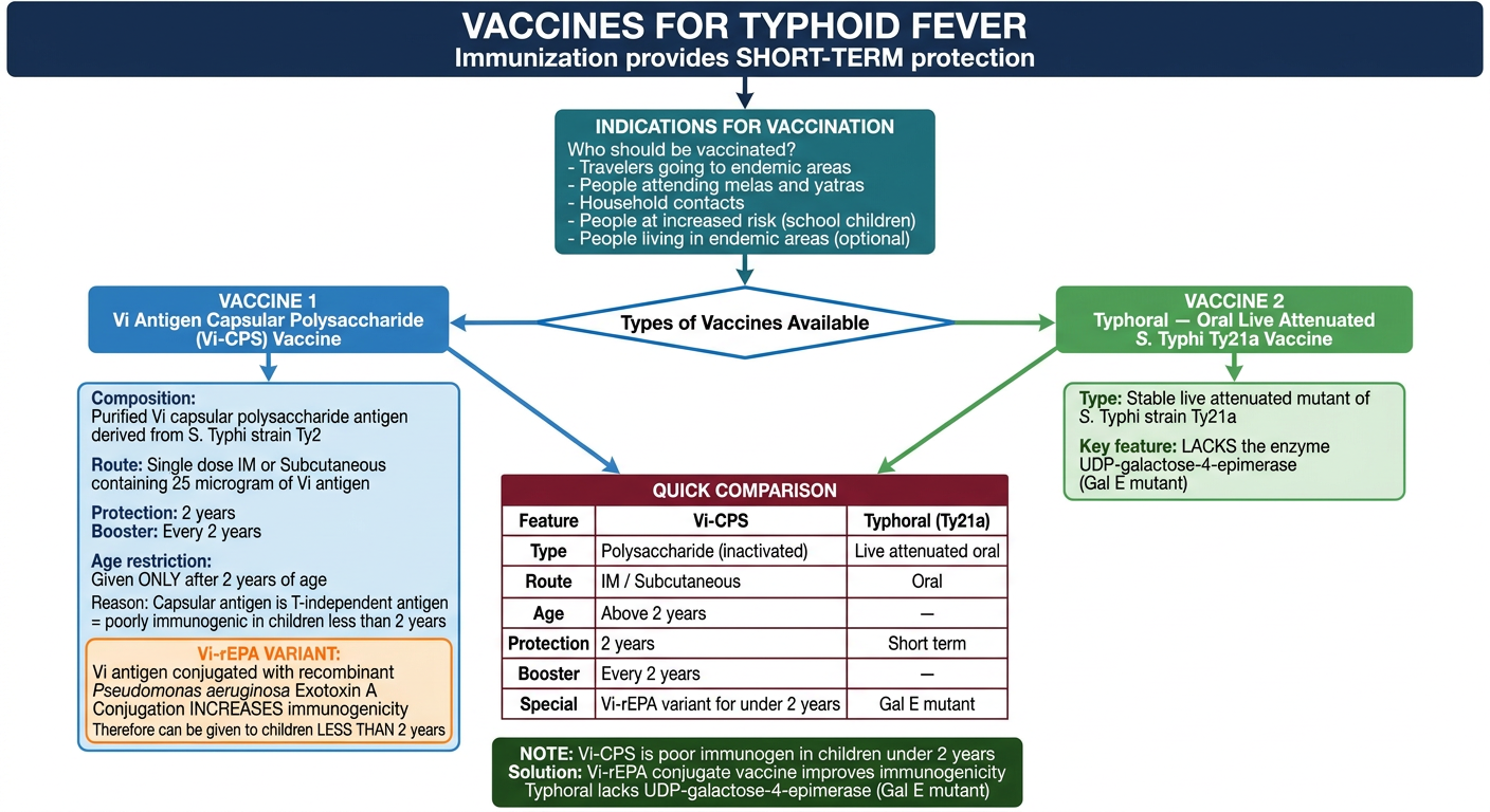
- Purified Vi polysaccharide from S. Typhi strain Ty2
- Single dose 25 mcg IM/Subcutaneous
- Protection for 2 years, booster every 2 years
- Only for above 2 years (T-independent antigen = poor immunogenicity in under 2s)
- Vi-rEPA variant: conjugated with Pseudomonas aeruginosa Exotoxin A - overcomes age restriction, works in children under 2
- Live attenuated oral vaccine
- Stable mutant of S. Typhi strain Ty21a
- Key mechanism: lacks UDP-galactose-4-epimerase (Gal E mutant)

Also add this information in the flowchart and make it more presentable
A highly presentable, clean, professional medical flowchart titled "VACCINES FOR TYPHOID FEVER" on a white background. Systematic top-to-bottom layout with color-coded vaccine cards. Well-spaced, no overlapping text. HEADER (dark navy, wide, large font): "VACCINES FOR TYPHOID FEVER Immunization provides SHORT-TERM protection" Arrow down to TEAL box: "INDICATIONS FOR VACCINATION - Travelers going to endemic areas - People attending melas and yatras - Household contacts - People at increased risk (school children) - People living in endemic areas (optional)" Arrow down to label: "THREE TYPES OF VACCINES" THREE EQUAL COLUMNS side by side, each with a colored header and white body: COLUMN 1 — BLUE header: "VACCINE 1 Vi-CPS Vaccine (Vi Antigen Capsular Polysaccharide)" Body: "Composition: Purified Vi capsular polysaccharide from S. Typhi strain Ty2 Route: Single dose IM or Subcutaneous Dose: 25 microgram Vi antigen Protection: 2 years Booster: Every 2 years Age: ONLY above 2 years Reason: T-independent antigen — poorly immunogenic in children under 2 years Vi-rEPA VARIANT: Vi antigen + recombinant Pseudomonas aeruginosa Exotoxin A Conjugation increases immunogenicity Can be given to children LESS THAN 2 years" COLUMN 2 — GREEN header: "VACCINE 2 Typhoral (Ty21a) Oral Live Attenuated Vaccine" Body: "Type: Stable live attenuated mutant of S. Typhi strain Ty21a Key feature: LACKS UDP-galactose-4-epimerase (Gal E mutant) Mechanism: On ingestion, multiplies stimulating immune response then SELF-DESTRUCTS after 4-5 cell divisions (due to lack of Gal E enzyme) — cannot induce pathogenesis Age: Only after 6 years Form: Lyophilized enteric coated capsules Dosing: Oral, before food, on alternate days — Day 1, 3, 5, 7 No antibiotics during this period Revaccination: Every 5 years (full 4-dose series) Protective immunity: Starts 7 days after last dose Duration: Lasts 4 years" COLUMN 3 — RED header: "VACCINE 3 Parenteral TAB Vaccine (Historical — No longer used)" Body: "Type: Heat-killed whole cell S. Typhi / S. Paratyphi A and B vaccine Status: NO LONGER IN USE Reason: Significant side effects" All three columns same height, clear borders. Below all three columns, a COMPARISON TABLE box (white with dark borders, maroon header row): "COMPARISON OF TYPHOID VACCINES" Headers: Feature | Vi-CPS | Typhoral Ty21a | TAB Vaccine Row 1: Type | Polysaccharide | Live attenuated oral | Killed whole cell Row 2: Route | IM / SC | Oral | Parenteral Row 3: Dose | Single | Days 1,3,5,7 | — Row 4: Age | Above 2 years | Above 6 years | — Row 5: Protection | 2 years | 4 years | — Row 6: Booster | Every 2 years | Every 5 years | — Row 7: Status | In use | In use | DISCONTINUED Row 8: Special note | Vi-rEPA for under 2 yrs | Gal E mutant, no antibiotics | Side effects FOOTER BOX (dark green): "KEY NOTES Vi-CPS is T-independent antigen — poor immunogen in under 2 years — Vi-rEPA conjugate solves this Typhoral self-destructs after 4-5 divisions — stimulates immunity without causing disease TAB vaccine discontinued due to significant side effects" Clean layout, adequate spacing, professional color scheme, readable font, no overlapping text.
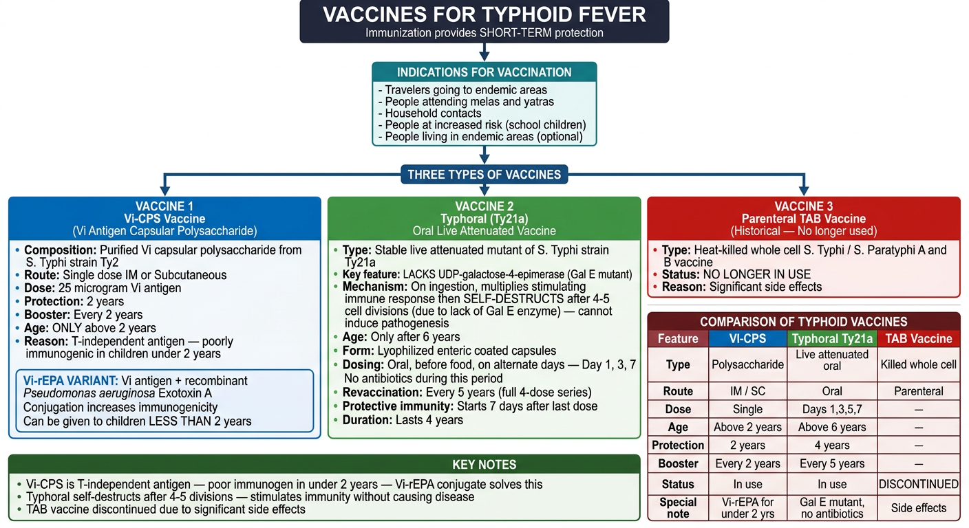
- Mechanism of self-destruction - multiplies 4-5 divisions then dies due to lack of Gal E enzyme - stimulates immunity without causing disease
- Age: Only after 6 years
- Form: Lyophilized enteric coated capsules
- Dosing schedule: Days 1, 3, 5, 7 (alternate days, before food, no antibiotics during this period)
- Revaccination: Every 5 years (full 4-dose series)
- Protective immunity starts 7 days after last dose, lasts 4 years
- Heat-killed whole cell S. Typhi / S. Paratyphi A and B
- No longer in use due to significant side effects