
Discuss bthe causes of hematuria
hematuria causes diagram urology kidney bladder
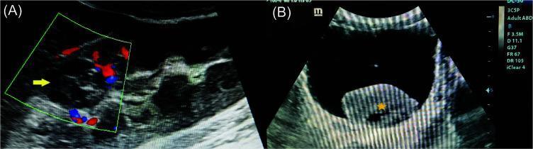
This dual-panel diagnostic image presents ultrasonographic findings of the urinary system in a case of renal oncocytoma. Image (A) is a Color Doppler ultrasonography of the right kidney showing a well-circumscribed, heteroechoic, cortical-based lesion (yellow arrow) in the upper pole. The Doppler overlay reveals no significant internal vascularity within the mass, a feature useful in differentiating renal lesions. Image (B) is a high-resolution transverse ultrasonography of the urinary bladder. It demonstrates a large, well-defined, heteroechoic mass (yellow star) settled in the dependent portion of the bladder lumen, characteristic of a giant retained blood clot secondary to gross hematuria. The clot shows mixed echogenicity with internal hypoechoic and hyperechoic regions, distinct from the anechoic urine and the uniform echogenicity of the bladder wall. These images illustrate the primary renal tumor and its downstream clinical complication of bladder clot retention, relevant for radiology and urology training.
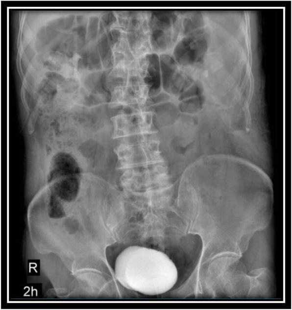
This diagnostic image is an anterior-posterior (AP) abdominal radiograph depicting an Intravenous Pyelogram (IVP) taken 2 hours after the administration of intravenous iodine contrast. The image captures the lower abdomen and pelvic region, focusing on the urinary system. Significant radiopaque contrast medium is concentrated within the urinary bladder, appearing as a dense, ovoid, well-defined structure in the midline pelvis. Faint traces of contrast are visible in the collecting system of the left kidney, indicating renal excretion. Skeletal landmarks are clearly visualized, including the lumbar vertebrae, sacroiliac joints, iliac crests, and pubic symphysis. Intestinal gas patterns are superimposed over the abdominal cavity. This modality is used in urology to evaluate the structural integrity and functional drainage of the kidneys, ureters, and bladder, particularly in cases of hematuria or suspected trauma. The 2-hour delay allows for the assessment of the bladder filling phase and ensures no significant obstruction or extravasation is present.
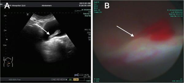
This composite image illustrates diagnostic findings in a patient with gross hematuria, categorized under Urology and Clinical Imaging. Image A is a transverse pelvic ultrasonography showing the urinary bladder. An arrow indicates a long, strip-like, hypoechoic mass measuring approximately 4.2 x 1.2 cm. The mass is seen floating within the bladder lumen, with its pedicle attached to the bladder base. This visual appearance is characteristic of an organized blood clot or a pedunculated intravesical lesion. Image B is a cystoscopic photograph providing an internal view of the bladder wall. The arrow points to a specific site at the right bladder base demonstrating active, pulsatile hemorrhage with fresh red blood. Together, these images demonstrate the correlation between non-invasive ultrasound findings (identifying the intravesical mass/clot) and invasive endoscopic confirmation (identifying the vascular source of bleeding). These materials are suitable for medical education regarding the diagnostic workup of hematuria and the identification of bladder pathology via ultrasound and cystoscopy.
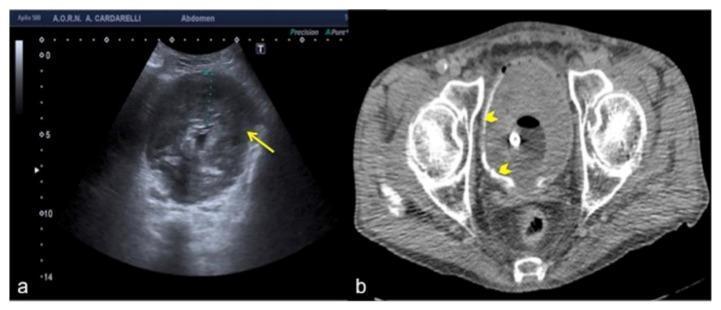
This dual-modality clinical imaging comparison presents a transverse ultrasound (US) scan and an axial Computed Tomography (CT) scan of the urinary bladder in a 78-year-old male presenting with hematuria. Image (a) is a transverse ultrasound showing a markedly distended bladder with diffuse, irregular wall thickening (indicated by a yellow arrow). The bladder lumen is filled with heterogeneous, low-to-medium level echoes, which can mimic a pseudo-mass or urothelial malignancy. Image (b) is a non-contrast axial CT scan of the same patient, revealing that the bladder lumen is entirely occupied by a large, hyperdense, homogenous mass (indicated by yellow arrowheads), identified as a massive intravesical blood clot. The CT scan clarifies the ultrasound findings by distinguishing the intraluminal clot from the true bladder wall. This comparison highlights a diagnostic pitfall where extensive blood clots can simulate bladder wall pathology or solid tumors on ultrasound. These images are clinically significant for emergency radiology and urology, illustrating the importance of cross-sectional imaging correlation in the workup of gross hematuria.
Causes of Hematuria
- Gross (macroscopic) hematuria: visibly red or brown urine, sometimes with clots - always pathologic
- Microscopic (non-visible) hematuria: detected only on urinalysis
Pseudohematuria (False-Positive Dipstick)
| Category | Examples |
|---|---|
| Medications | Rifampin, nitrofurantoin, phenazopyridine, doxorubicin, phenytoin |
| Foods/dyes | Beets, blackberries, food coloring |
| Metabolites | Bilirubin, porphyrins, myoglobin (rhabdomyolysis), hemoglobin (hemolysis), melanin |
True Causes of Hematuria
I. RENAL CAUSES
A. Glomerular Causes
- IgA Nephropathy (Berger's disease) - the most common glomerular cause of asymptomatic hematuria worldwide; often presents with episodic gross hematuria following upper respiratory infections
- Thin Basement Membrane Nephropathy (TBMN) - benign familial hematuria; a common cause of isolated microscopic hematuria
- Alport Syndrome - X-linked hereditary nephritis with sensorineural deafness and ocular abnormalities
- Postinfectious (Poststreptococcal) Glomerulonephritis - follows streptococcal throat or skin infection; low complement, cola-colored urine
- Systemic Lupus Erythematosus (SLE) - lupus nephritis; associated with ANA, low complement
- Goodpasture Syndrome - anti-GBM antibodies; pulmonary-renal syndrome
- Henoch-Schönlein Purpura (HSP) / IgA Vasculitis - small vessel vasculitis with palpable purpura, arthritis, abdominal pain
- Hemolytic Uremic Syndrome (HUS) - microangiopathic hemolytic anemia + thrombocytopenia + renal failure
- Other vasculitides - ANCA-associated (granulomatosis with polyangiitis, microscopic polyangiitis)
B. Tubulointerstitial Causes
- Nephrolithiasis (Kidney stones) - one of the most common causes of both gross and microscopic hematuria; associated with colicky flank pain
- Pyelonephritis - upper UTI; fever, costovertebral angle tenderness, pyuria
- Acute Interstitial Nephritis (AIN) - often drug-induced (NSAIDs, antibiotics); triad of fever, rash, eosinophilia (not always present)
- Papillary Necrosis - seen in sickle cell disease, diabetes, analgesic nephropathy, obstructive uropathy
- Acute Tubular Necrosis (ATN)
- Polycystic Kidney Disease (PKD) - autosomal dominant or recessive; bilateral enlarged cystic kidneys
- Medullary Sponge Kidney - ectatic collecting ducts; associated with hypercalciuria and recurrent stones
C. Renal Vascular Causes
- Renal Artery Embolism or Thrombosis - acute flank pain + hematuria; elevated LDH
- Renal Vein Thrombosis - associated with nephrotic syndrome
- Arteriovenous Malformation (AVM) or Fistula - may be congenital or post-traumatic/post-biopsy
- Nutcracker Syndrome - compression of the left renal vein between the aorta and superior mesenteric artery; left flank pain and hematuria, more common in thin young women
- Malignant Hypertension - severe hypertension causing renal arteriolar fibrinoid necrosis
D. Renal Metabolic Causes
- Hypercalciuria - a common cause of hematuria, particularly in children; often idiopathic
- Hyperuricosuria - uric acid crystal deposition
E. Renal Neoplasms
- Renal Cell Carcinoma (RCC) - classic triad: hematuria, flank pain, palpable mass (now rarely all three together)
- Transitional Cell Carcinoma (TCC) of the Renal Pelvis/Ureter - painless hematuria; associated with smoking and analgesic abuse
II. EXTRARENAL (UROLOGICAL) CAUSES
A. Ureteral Causes
- Ureteral Calculi - stone obstruction causing colicky pain radiating to the groin + hematuria
- Ureteral TCC / Benign Polyp
- Ureteral Stricture
B. Bladder Causes
- Bladder Cancer (TCC / Squamous Cell Carcinoma) - most common cause of gross hematuria in patients >50 years; painless gross hematuria is the hallmark. Risk factors: smoking, industrial chemical/aniline dye exposure, alkylating chemotherapy (cyclophosphamide), schistosomiasis (for SCC) - Campbell Walsh Wein Urology
- Urinary Tract Infection / Cystitis - the most frequent cause of hematuria overall; associated with dysuria, frequency, urgency
- Hemorrhagic Cystitis - caused by cyclophosphamide, ifosfamide, radiation
- Noninfectious Cystitis - interstitial cystitis, radiation cystitis
- Bladder Stones - urinary stasis, foreign body
- Bladder Trauma / Foreign Body
- Schistosomiasis (S. haematobium) - endemic cause of bladder SCC in Africa/Middle East
C. Prostatic Causes
- Benign Prostatic Hyperplasia (BPH) - a common cause of hematuria in older men
- Prostate Cancer - usually late-stage when causing hematuria
- Prostatitis - acute bacterial prostatitis; perineal pain, fever, hematuria
D. Urethral Causes
- Urethritis - gonococcal, chlamydial (particularly in HIV patients)
- Urethral Stricture
- Meatal Stenosis / Posterior Urethral Valves (in children)
- Urethral Caruncle (benign urethral lesion in postmenopausal women)
III. SYSTEMIC / OTHER CAUSES
- Coagulopathy / Anticoagulation - warfarin, heparin, DOACs; hematuria from anticoagulation should still prompt evaluation for underlying urological lesion
- Thrombocytopenia - ITP, TTP, bone marrow failure
- Sickle Cell Disease / Trait - papillary necrosis, medullary carcinoma
- Trauma - blunt or penetrating abdominal/renal trauma; also recent urologic instrumentation
- Exercise-Induced Hematuria - strenuous exercise (marathon runners); resolves within 72 hours of stopping
- Endometriosis - cyclical hematuria in women
- HIV - reported incidence of hematuria 18-50% in HIV-infected patients; causes include UTI, chlamydial/gonococcal urethritis - Tintinalli's Emergency Medicine
- Pregnancy - associated with UTI, nephrolithiasis, or preeclampsia
Localizing the Source: Key Clinical Clues
| Feature | Interpretation |
|---|---|
| Initial-stream hematuria | Prostatic or urethral source |
| Terminal hematuria | Bladder neck / trigone |
| Total (throughout stream) hematuria | Upper urinary tract (kidney, ureter) |
| Wormlike (vermiform) clots | Upper tract origin |
| Cuboid/amorphous clots | Bladder origin |
| Clots present (any shape) | Never glomerular; always lower/upper tract |
| Dysmorphic RBCs + RBC casts | Glomerular origin |
| Proteinuria + hematuria | Glomerular disease |
| Painless gross hematuria | Urological malignancy until proven otherwise |
| Colicky flank pain + hematuria | Nephrolithiasis |
A Practical Anatomical Summary
Kidney  →  Glomerular (IgA, PSGN, SLE, Alport)
           Tubulointerstitial (stones, pyelonephritis, AIN, PKD)
           Vascular (RVT, AVM, nutcracker, RCC)

Ureter  →  Calculi, TCC, stricture

Bladder →  Cancer (TCC), cystitis, BPH-related, hemorrhagic cystitis

Prostate → BPH, prostate cancer, prostatitis

Urethra →  Urethritis, stricture, trauma