
Teach me Neuroanatomy / Brain Anatomy of its parts and relations
Reading File
Now let me search the medical library for comprehensive neuroanatomy content across multiple topics simultaneously.
Finding Sources
Finding Sources
Excellent! Great sources found. Let me now search for specific brain anatomy sections across these top books simultaneously.
Finding Sources
Finding Sources
Finding Sources
Let me search the TOC of the neuroanatomy book and the neuroscience book to find the best sections.
Finding Sources
Finding Sources
Let me read key content from the Neuroanatomy Through Clinical Cases and the Neuroscience Exploring the Brain textbooks - reading the core brain anatomy blocks.
Reading File
Reading File
Let me read deeper into the clinical neuroanatomy content and also fetch anatomical brain images from the Color Atlas.
Reading File
Finding Sources
Excellent content found! Let me now read more from the neuroanatomy book covering deeper structures (brainstem, cerebellum, diencephalon, limbic system) and fetch the brain lobe diagram image.
Reading File
Loading Image
Excellent! Great lobe diagram obtained. Now let me read the deeper structures content (brainstem, thalamus, cerebellum, basal ganglia, limbic system) from block1 and also fetch more key images.
Reading File
Loading Image
I now have very rich content. Let me fetch a few more critical brain anatomy images - thalamus diagram and the brainstem/cerebellum diagram - then compile the comprehensive response.
Loading Image
Loading Image
I now have comprehensive content from the textbook plus several excellent diagrams. Let me read a bit more content about brainstem, cerebellum, and limbic system from the textbook.
Reading File
Loading Image
I have enough rich content to now write a comprehensive neuroanatomy lesson. Let me compile everything into a thorough, well-organized response with the fetched images.
Neuroanatomy: The Brain - Parts, Structure, and Relations
Based on Neuroanatomy through Clinical Cases, 3rd Ed. | Neuroscience: Exploring the Brain, 5th Ed. | Adams and Victor's Principles of Neurology, 12th Ed.
Overview: Divisions of the Nervous System
Before the brain itself, understand the big picture. The human nervous system is divided into:
| Division | Components |
|---|---|
| Central Nervous System (CNS) | Brain + Spinal cord |
| Peripheral Nervous System (PNS) | Cranial nerves, spinal nerves, ganglia |
| Autonomic Nervous System (ANS) | Sympathetic, parasympathetic, enteric divisions |
The brain itself is organized into three major embryological divisions that persist in the adult:
| Embryo | Adult Structure |
|---|---|
| Prosencephalon (forebrain) | Cerebral hemispheres + Diencephalon (thalamus/hypothalamus) |
| Mesencephalon (midbrain) | Midbrain (part of brainstem) |
| Rhombencephalon (hindbrain) | Pons + Medulla + Cerebellum |
1. The Cerebral Hemispheres (Telencephalon)
The cerebral cortex is not a smooth sheet - it has folds called gyri (ridges) and sulci (grooves). This folding massively increases surface area. Two hemispheres are separated by the interhemispheric (longitudinal) fissure and connected by the corpus callosum, a C-shaped band of white matter that links homologous and heterologous regions across both sides.
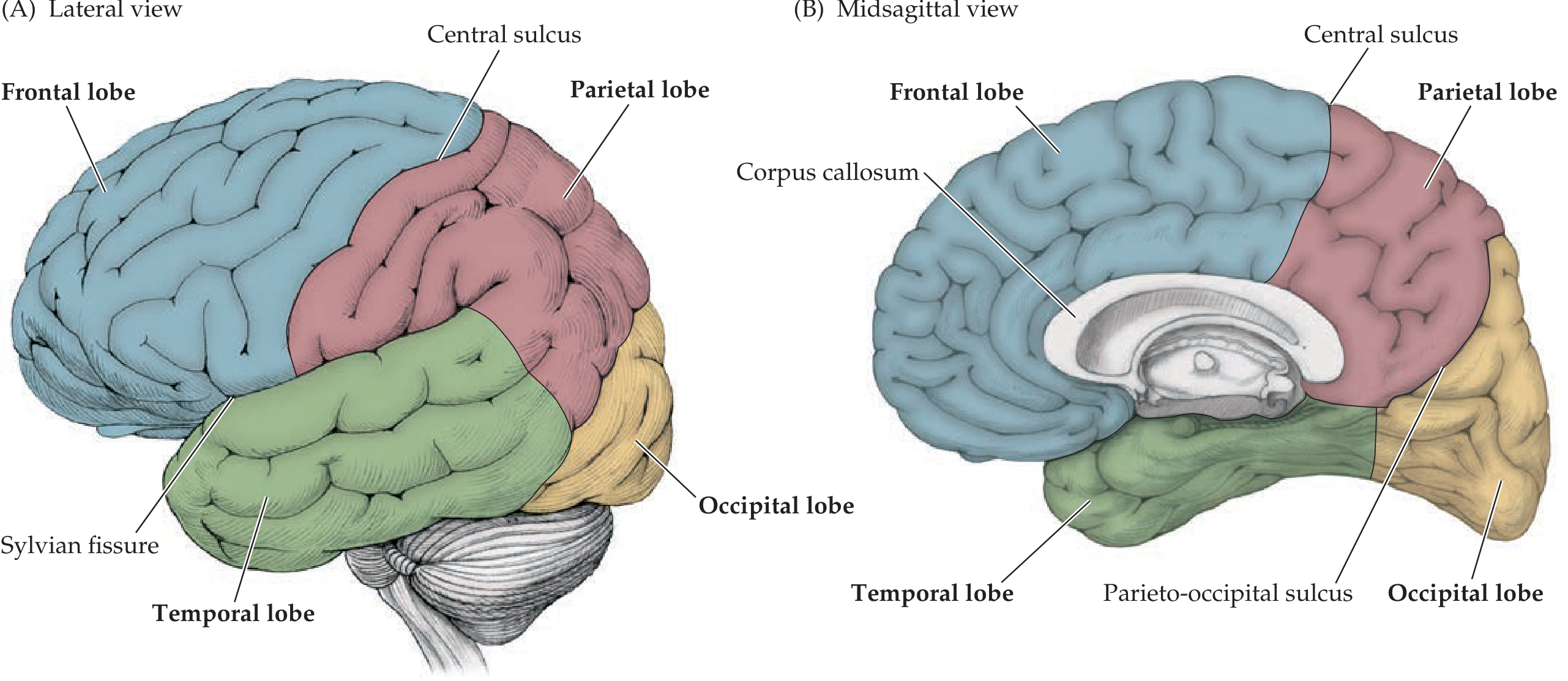
The Four Major Lobes
Frontal Lobe (blue in diagram)
- Location: Anterior brain, from the frontal pole back to the central sulcus of Rolando. Separated from the temporal lobe inferiorly by the Sylvian (lateral) fissure
- Key gyri: Precentral gyrus (primary motor cortex, M1), superior/middle/inferior frontal gyri
- Functions:
- Primary motor cortex (precentral gyrus) - voluntary movement, contralateral body representation
- Premotor cortex - motor planning
- Prefrontal cortex - executive function, planning, personality, working memory, social behavior
- Broca's area (inferior frontal gyrus, dominant hemisphere, usually left) - expressive (motor) speech
- Frontal eye fields - voluntary eye movements
Parietal Lobe (pink/red in diagram)
- Location: Bounded anteriorly by the central sulcus, antero-inferiorly by the Sylvian fissure. No sharp line from occipital lobe laterally; the parieto-occipital sulcus defines the boundary on the medial surface
- Key gyri: Postcentral gyrus, superior/inferior parietal lobules, supramarginal gyrus, angular gyrus
- Functions:
- Primary somatosensory cortex (postcentral gyrus) - processes touch, proprioception, pain, temperature from contralateral body
- Association cortex - spatial awareness, visuospatial processing
- Dominant hemisphere: language comprehension (angular/supramarginal gyri), reading/writing
- Non-dominant (right): left-sided neglect, dressing apraxia, constructional tasks
Temporal Lobe (green in diagram)
- Location: Below the Sylvian fissure, wraps around the lateral and inferior brain
- Key gyri: Superior, middle, inferior temporal gyri; parahippocampal gyrus; fusiform gyrus; transverse gyri of Heschl (primary auditory cortex, buried in the Sylvian fissure)
- Functions:
- Primary auditory cortex (Heschl's gyri)
- Wernicke's area (posterior superior temporal gyrus, dominant hemisphere) - receptive language comprehension
- Memory - medial temporal lobe: hippocampus + entorhinal cortex = declarative (episodic/semantic) memory
- Face recognition (fusiform face area, fusiform gyrus)
- Olfaction (uncus, parahippocampal gyrus = primary olfactory cortex)
Occipital Lobe (yellow in diagram)
- Location: Posterior pole of the brain; parieto-occipital sulcus separates it from parietal lobe on medial surface
- Key structure: Calcarine sulcus on medial surface - primary visual cortex (V1/striate cortex) lies along its banks
- Functions:
- V1 (primary visual cortex) - processes raw visual input from contralateral visual field
- V2-V5 - higher visual processing (motion, color, depth, object recognition)
- Ventral stream ("what pathway"): V1 → temporal lobe = object/face recognition
- Dorsal stream ("where/how pathway"): V1 → parietal lobe = spatial location, eye-hand coordination
The Insula (Hidden Lobe)
Buried within the Sylvian fissure, covered by frontal (frontal operculum) and parietal cortex (parietal operculum). Functions include: visceral sensation, taste, autonomic processing, empathy, body awareness, and pain integration.
2. White Matter and Deep Connections
The cerebral white matter carries three types of fibers:
| Fiber Type | Function | Examples |
|---|---|---|
| Commissural | Connect the two hemispheres | Corpus callosum, anterior commissure |
| Association | Connect different cortical areas in the same hemisphere | Arcuate fasciculus (language), uncinate fasciculus, cingulum |
| Projection | Connect cortex to subcortical structures | Corticospinal, corticobulbar tracts (via internal capsule) |
The internal capsule is a critical V-shaped band of projection fibers (posterior limb carries motor and sensory tracts; anterior limb carries frontal-thalamic fibers). Strokes here cause contralateral hemiplegia.
3. Diencephalon
Located deep in the cerebral white matter just above the brainstem, behind the basal ganglia.
Thalamus - "The Gateway to Consciousness"
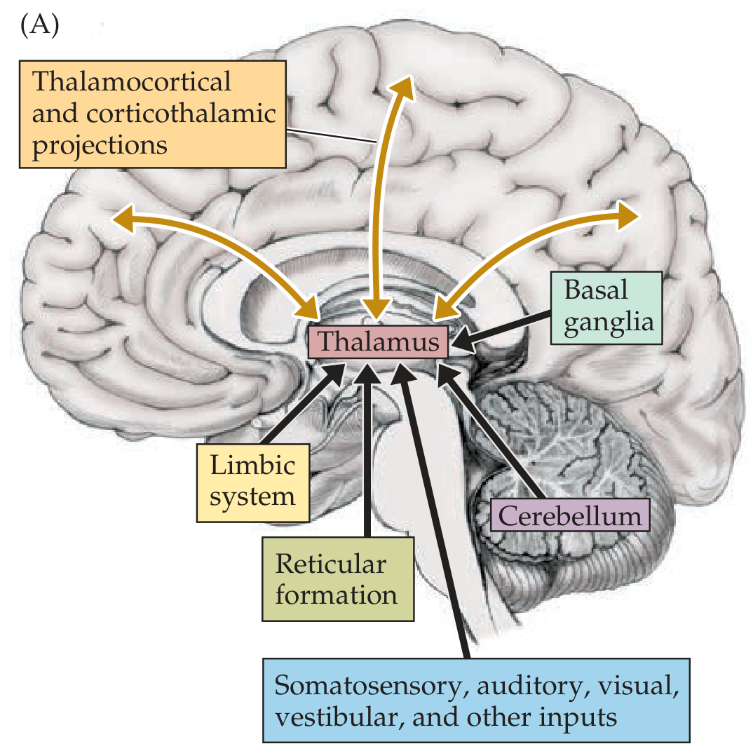
The thalamus is the brain's central relay station. Nearly ALL pathways projecting to the cerebral cortex synapse here first (olfaction is the major exception). It consists of multiple named nuclei:
| Thalamic Nucleus | Relay For |
|---|---|
| VPL (ventral posterolateral) | Somatosensory from body (spinothalamic + dorsal columns) |
| VPM (ventral posteromedial) | Somatosensory from face (trigeminal) + taste |
| LGN (lateral geniculate nucleus) | Vision (from optic tract → visual cortex) |
| MGN (medial geniculate nucleus) | Hearing (from inferior colliculus → auditory cortex) |
| VL/VA (ventral lateral/anterior) | Motor signals from cerebellum and basal ganglia → motor cortex |
| MD (mediodorsal) | Limbic/prefrontal connections |
| Pulvinar | Higher visual/association areas |
| Intralaminar nuclei | Arousal, consciousness (reticular formation input) |
Key anatomical relations: thalami sit as a paired egg-shaped structure, forming an inverted V in horizontal sections, flanking the third ventricle.
Hypothalamus
Sits below the thalamus, forming the floor and walls of the third ventricle. Small but controls enormous functions:
- Autonomic regulation - master controller of sympathetic and parasympathetic tone
- Endocrine - controls the pituitary gland via portal vessels (anterior pituitary) and direct axons (posterior pituitary releases ADH/oxytocin)
- Temperature regulation (anterior hypothalamus = heat loss; posterior = heat conservation)
- Hunger/satiety (lateral = feeding center; ventromedial = satiety center)
- Circadian rhythms - suprachiasmatic nucleus (SCN)
- Thirst, water balance (osmoregulation)
- Sleep-wake cycle (lateral hypothalamus, orexin/hypocretin neurons)
- Emotional behavior via connections to limbic system
4. Basal Ganglia
A group of deep gray matter nuclei embedded in the white matter of the cerebral hemispheres and upper brainstem. Key components:
Basal Ganglia = Caudate + Putamen + Globus Pallidus + Subthalamic nucleus + Substantia nigra
Striatum = Caudate + Putamen (phylogenetically newer)
Lenticular nucleus = Putamen + Globus Pallidus
Relations: Caudate nucleus wraps around the thalamus in a C-shape. Putamen and globus pallidus lie lateral to the internal capsule. The subthalamic nucleus (STN) sits at the diencephalon-midbrain junction. Substantia nigra is in the midbrain.
Function - Motor control circuit:
- Cortex sends excitatory input → striatum
- Direct pathway (Go): Striatum → inhibits GPi/SNr → releases thalamus → activates cortex (facilitates movement)
- Indirect pathway (No-Go): Striatum → GPe → STN → GPi → inhibits thalamus (suppresses unwanted movements)
- Dopamine from substantia nigra (nigrostriatal pathway) facilitates direct and inhibits indirect pathway
Clinical relevance:
- Parkinson's disease = loss of dopaminergic neurons in substantia nigra → bradykinesia, rigidity, tremor
- Huntington's disease = loss of striatal neurons → chorea, psychiatric symptoms
- Basal ganglia also involved in habit learning, reward circuits (via nucleus accumbens)
5. Brainstem
The brainstem connects the cerebral hemispheres to the spinal cord and is divided into three parts (from top to bottom):
Midbrain (Mesencephalon)
- Dorsal surface (tectum): Superior colliculi (visual reflexes - pupillary light reflex, eye movements) + Inferior colliculi (auditory relay)
- Ventral surface (tegmentum): Reticular formation, red nucleus, substantia nigra, cranial nerve nuclei
- Cranial nerves: CN III (oculomotor) - from interpeduncular fossa; CN IV (trochlear) - only CN exiting dorsally
- Cerebral peduncles carry corticospinal and corticobulbar tracts descending to spinal cord
Pons
- Contains: Large bulge (basilar part) with corticospinal tracts, pontine nuclei (relay to cerebellum via middle cerebellar peduncle), tegmentum with reticular formation
- Cranial nerves: CN V (trigeminal - largest CN), CN VI (abducens), CN VII (facial), CN VIII (vestibulocochlear)
- Key structures: Locus coeruleus (norepinephrine), pontine respiratory group, medial longitudinal fasciculus (MLF - coordinates eye movements)
Medulla Oblongata
- Location: Most inferior brainstem, merges with spinal cord at foramen magnum
- Ventral surface: Pyramids (corticospinal tracts) - pyramidal decussation occurs here (fibers cross to contralateral side, explaining why left brain controls right body)
- Olivary complex (inferior olive) - relay for cerebellar learning
- Cranial nerves: CN IX (glossopharyngeal), CN X (vagus), CN XI (accessory), CN XII (hypoglossal)
- Vital centers: Respiratory rhythm (pre-Botzinger complex), cardiovascular center, vomiting center
Clinical pearl: Brainstem lesions produce "crossed" deficits - ipsilateral cranial nerve signs + contralateral hemiplegia/sensory loss. This is because cranial nerve nuclei are at the level of the lesion (ipsilateral) but motor/sensory tracts are already crossed.
6. Cerebellum - "The Little Brain"
Located in the posterior fossa, posterior to the brainstem and below the tentorium cerebelli (the dural fold separating it from the occipital lobes).
Anatomy
- Two hemispheres + central vermis
- Surface has narrow folds called folia (not gyri)
- Fissures divide it into lobes: anterior, posterior, and flocculonodular lobes
- Three layers of cortex: molecular, Purkinje cell, granule cell layers - Purkinje cells are the sole output neurons of the cerebellar cortex (inhibitory, GABA)
- Deep cerebellar nuclei (from lateral to medial): Dentate, Emboliform, Globose, Fastigial (mnemonic: Don't Eat Greasy Food)
Three Functional Divisions and Their Connections
| Division | Anatomy | Input | Function |
|---|---|---|---|
| Vestibulocerebellum | Flocculonodular lobe | Vestibular nuclei | Balance, eye movements |
| Spinocerebellum | Vermis + intermediate zone | Spinal cord (proprioception) | Coordination of limb/axial movement |
| Cerebrocerebellum | Lateral hemispheres | Cerebral cortex (via pons) | Motor planning, timing, cognitive functions |
Connections (Peduncles)
- Superior cerebellar peduncle (SCP): Main OUTPUT - dentate nucleus → (crosses in midbrain) → VL thalamus → motor cortex
- Middle cerebellar peduncle (MCP): INPUT from contralateral pontine nuclei (corticopontocerebellar)
- Inferior cerebellar peduncle (ICP): INPUT from spinal cord (spinocerebellar), vestibular nuclei, inferior olive
The cerebellar circuit corrects movement in real time. Crucially, cerebellar lesions produce ipsilateral deficits (the outputs cross twice - once in the SCP, once via cortex → spinal cord), unlike cerebral lesions which produce contralateral deficits.
Signs of cerebellar dysfunction: Ataxia (gait/limb), dysmetria (past-pointing), intention tremor, dysdiadochokinesia (impaired rapid alternating movements), dysarthria (scanning/slurred speech), nystagmus.
7. Limbic System
The limbic system is a ring of cortex and subcortical structures on the medial aspect of the hemispheres, sometimes called the "emotional brain." Key components:
- Hippocampus (within medial temporal lobe) - declarative memory formation; H.M.'s case (bilateral hippocampectomy) showed inability to form new long-term memories (anterograde amnesia)
- Amygdala (anterior temporal lobe, almond-shaped) - fear conditioning, emotional memory, threat detection, social behavior
- Cingulate gyrus (above corpus callosum) - attention, pain processing, emotion regulation; anterior cingulate = motivation/conflict monitoring
- Entorhinal cortex - interface between hippocampus and neocortex; gateway for memory consolidation
- Parahippocampal gyrus - spatial context, scene recognition
- Fornix - major white matter bundle from hippocampus to mammillary bodies/septal nuclei
- Mammillary bodies - part of Papez circuit; destroyed in Wernicke-Korsakoff syndrome (thiamine deficiency)
- Septal nuclei / nucleus accumbens - reward, motivation (dopamine target)
Papez Circuit (memory/emotion circuit):
Hippocampus → Fornix → Mammillary bodies → Anterior thalamus → Cingulate gyrus → Entorhinal cortex → Hippocampus
8. Ventricular System and CSF
The brain floats in cerebrospinal fluid (CSF), produced by the choroid plexus inside the ventricles.
LATERAL VENTRICLES (2, one in each hemisphere)
↓ via Foramina of Monro
THIRD VENTRICLE (diencephalon)
↓ via Cerebral Aqueduct of Sylvius
FOURTH VENTRICLE (pons/medulla)
↓ via Foramina of Magendie (median) + Luschka (lateral)
SUBARACHNOID SPACE
↓ absorbed at Arachnoid granulations → dural venous sinuses
Clinical relevance: Obstruction at any point causes hydrocephalus (pressure buildup). The cerebral aqueduct is the narrowest point and most commonly blocked.
9. Blood Supply to the Brain
The brain receives blood from two systems that communicate via the Circle of Willis:
| Artery | Territories |
|---|---|
| ACA (anterior cerebral artery) | Medial frontal + parietal lobe - leg area of motor/sensory cortex |
| MCA (middle cerebral artery) | Lateral frontal, parietal, temporal - arm/face motor-sensory, language |
| PCA (posterior cerebral artery) | Occipital lobe, medial temporal (hippocampus), thalamus |
| Basilar artery | Brainstem, cerebellum (via AICA, SCA, PICA), thalamus |
| PICA (posterior inferior cerebellar) | Lateral medulla + inferior cerebellum - Wallenberg syndrome if occluded |
10. Summary: Key Functional-Anatomical Correlations
| Function | Primary Brain Region |
|---|---|
| Voluntary movement | Primary motor cortex (precentral gyrus) |
| Sensation | Primary somatosensory cortex (postcentral gyrus) |
| Vision | Occipital lobe (calcarine cortex) |
| Hearing | Superior temporal gyrus (Heschl's gyri) |
| Language - expression | Broca's area (left inferior frontal gyrus) |
| Language - comprehension | Wernicke's area (left posterior superior temporal) |
| Memory formation | Hippocampus + medial temporal lobe |
| Emotion/Fear | Amygdala |
| Movement coordination | Cerebellum |
| Movement initiation/suppression | Basal ganglia |
| Sensory relay | Thalamus |
| Autonomic/endocrine control | Hypothalamus |
| Arousal/consciousness | Reticular activating system (brainstem) + thalamus |
Quick Visual Summary of Major Connections
Sources
- Neuroanatomy through Clinical Cases, 3rd Ed. (Blumenfeld) - the gold-standard clinical neuroanatomy text
- Neuroscience: Exploring the Brain, 5th Ed. (Bear, Connors, Paradiso)
- Adams and Victor's Principles of Neurology, 12th Ed.
- Localization in Clinical Neurology, 8th Ed. (Brazis et al.)
Want me to go deeper on any specific part? For example:
- Cranial nerves (all 12, origins, functions, clinical tests)
- Cortical maps (Brodmann areas, homunculus)
- Specific pathways (visual, auditory, motor, sensory tracts in detail)
- Clinical syndromes (stroke syndromes, herniation syndromes, raised ICP)
- Ventricular system and CSF in depth
- Limbic system and memory circuits in detail
For neet PG and with Diagram and picture easy to remember
Finding Sources
Finding Sources
Loading Image
Finding Sources
Loading Image
Loading Image
Finding Sources
Loading Image
Loading Image
Finding Sources
Here is your NEET PG Neuroanatomy Master Guide - high-yield, image-rich, mnemonic-packed. Every section is tagged with what NEET PG loves to ask.
🧠 NEET PG Neuroanatomy - Brain Anatomy Complete Guide
Source: Neuroanatomy through Clinical Cases 3rd Ed. | Neuroscience: Exploring the Brain 5th Ed.
PART 1 - THE 4 LOBES OF THE CEREBRAL CORTEX
Quick Reference - Boundaries
| Lobe | Anterior Boundary | Posterior Boundary | Inferior Boundary |
|---|---|---|---|
| Frontal | Frontal pole | Central sulcus (Rolando) | Sylvian fissure |
| Parietal | Central sulcus | Parieto-occipital sulcus (medial) | Sylvian fissure |
| Temporal | Sylvian fissure (above) | No sharp line | - |
| Occipital | Parieto-occipital sulcus | Occipital pole | - |
NEET TRICK: Central sulcus of Rolando = moRotor in front, sensoRy behind. Sylvian fissure = separates frontal/parietal ABOVE from temporal BELOW.
PART 2 - PRIMARY CORTICAL AREAS (HIGH YIELD!)
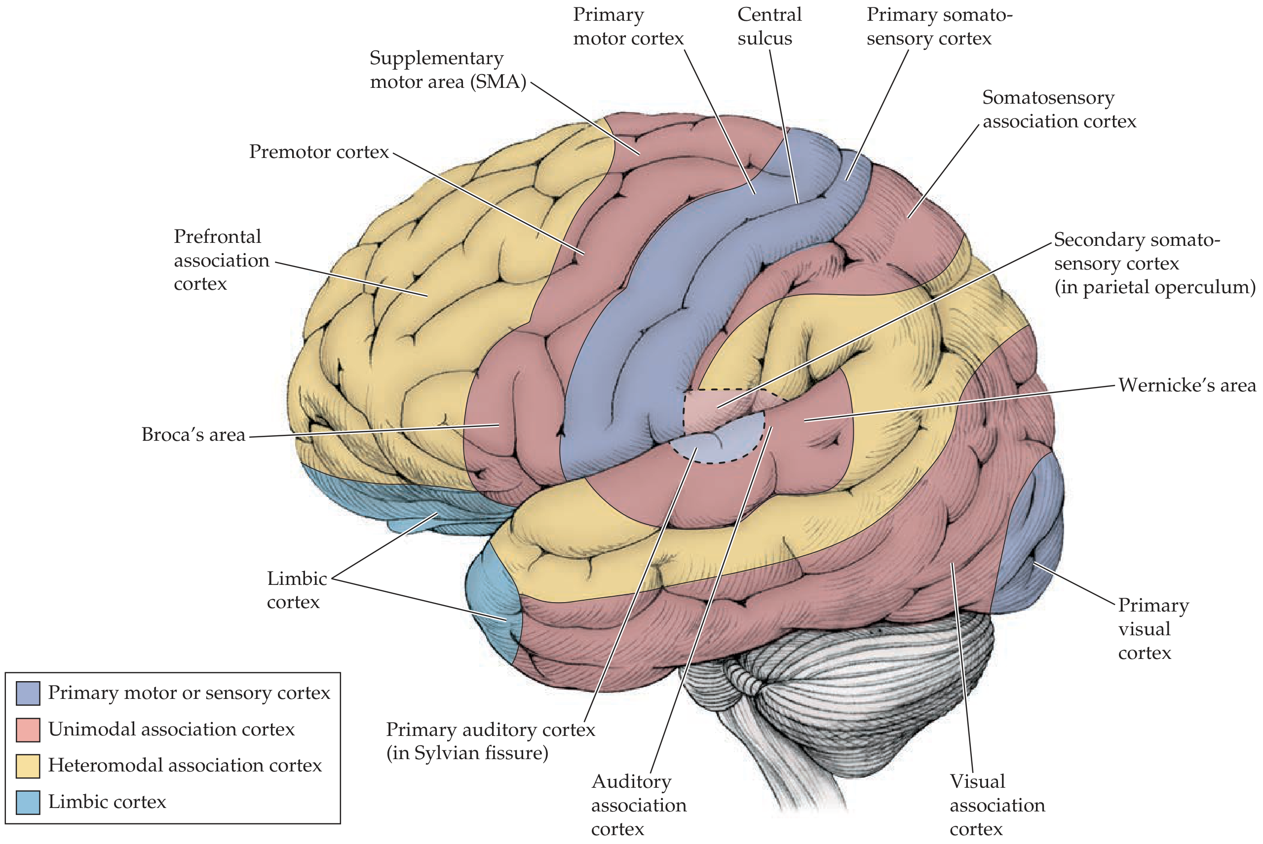
The 6 Primary Cortical Areas - NEET Favorites
| Cortical Area | Gyrus/Location | Brodmann Area | Function |
|---|---|---|---|
| Primary Motor | Precentral gyrus | BA 4 | Voluntary movement (contralateral) |
| Primary Somatosensory | Postcentral gyrus | BA 1, 2, 3 | Touch/pain/proprioception (contralateral) |
| Primary Visual | Banks of calcarine fissure | BA 17 | Vision (contralateral visual field) |
| Primary Auditory | Heschl's transverse gyri (in Sylvian fissure) | BA 41, 42 | Hearing (bilateral, contralateral stronger) |
| Primary Olfactory | Uncus + parahippocampal gyrus | BA 28 | Smell (IPSILATERAL - only one!) |
| Primary Gustatory | Parietal operculum / insula | BA 43 | Taste |
MNEMONIC for Brodmann Areas:
- 4 = Motor (4 limbs to move)
- 1,2,3 = Sensory (SSS)
- 17 = Visual (1 + 7 = 17, 2 eyes)
- 41/42 = Auditory (2 ears = 4+1, 4+2)
- 44/45 = Broca's (B = 4+4=8... remember "44 is Broca's door")
- 22 = Wernicke's (WW = double meaning)
PART 3 - LANGUAGE AREAS (REPEATED NEET TOPIC)
Broca's Area vs Wernicke's Area
| Feature | Broca's Area | Wernicke's Area |
|---|---|---|
| Location | Inferior frontal gyrus (pars triangularis + opercularis) | Posterior superior temporal gyrus |
| Brodmann | BA 44 & 45 | BA 22 |
| Hemisphere | Dominant (Left in 95% right-handers) | Dominant (Left) |
| Function | Motor/expressive speech production | Receptive/sensory language comprehension |
| Lesion → | Broca's aphasia = Non-fluent, effortful speech; comprehension preserved; patient AWARE of deficit | Wernicke's aphasia = Fluent but nonsensical ("word salad"); poor comprehension; patient UNAWARE |
| Associated | Apraxia of speech | Paraphasias (word substitutions) |
MNEMONIC: "Broca BROKe his speech" (can't speak fluently). "Wernicke doesn't WORK in understanding."
Connection between them: Arcuate fasciculus (superior longitudinal fasciculus) connects Broca's to Wernicke's. Lesion → Conduction aphasia (can't repeat, but fluent speech + good comprehension).
PART 4 - MOTOR HOMUNCULUS & SENSORY HOMUNCULUS
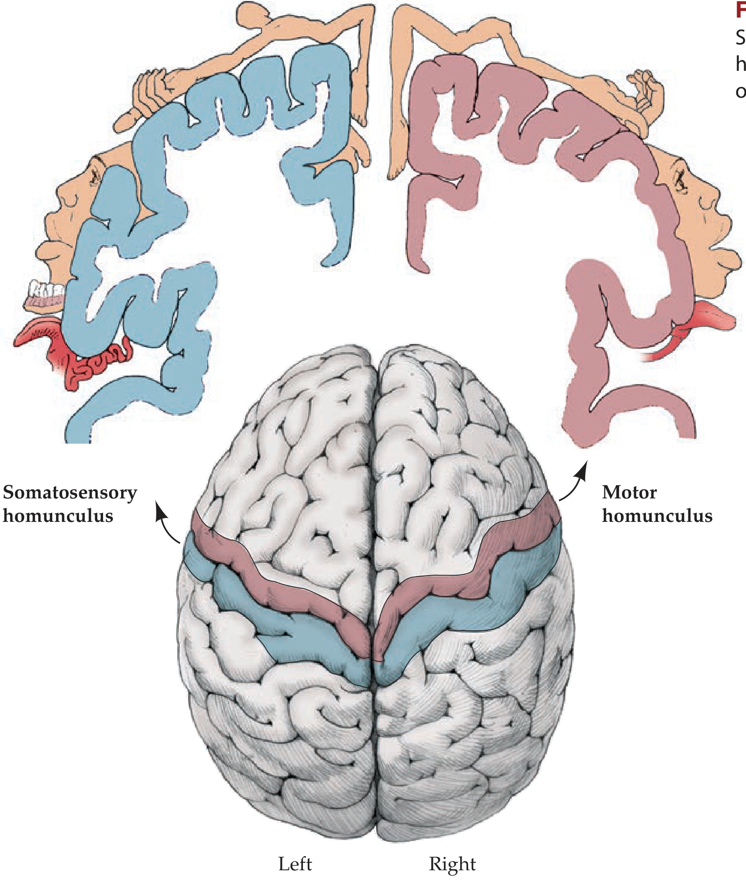
Key Points for NEET PG
- Body is mapped upside down - leg at top (medial surface), face at bottom (lateral surface)
- Lips, tongue, hands have largest representation (most cortical area per body part)
- Leg area is supplied by ACA - that's why ACA stroke = leg weakness
- Face + Arm area is supplied by MCA - MCA stroke = face + arm weakness
- Motor cortex is anterior to central sulcus; Sensory cortex is posterior
NEET TRICK: ACA = Anterior = Leg (leg moves forward when walking). MCA = Middle = Arms + Face.
PART 5 - KEY BRODMANN AREAS (High Yield Table)
| Brodmann Area | Name | Location | Clinical Lesion |
|---|---|---|---|
| 4 | Primary motor cortex | Precentral gyrus | Contralateral UMN weakness |
| 6 | Premotor + SMA | Anterior to precentral | Apraxia, loss of complex movements |
| 8 | Frontal eye field | Middle frontal gyrus | Eyes deviate TOWARD lesion (ipsilateral) |
| 1,2,3 | Primary somatosensory | Postcentral gyrus | Contralateral sensory loss |
| 17 | Primary visual (striate) | Calcarine cortex | Contralateral homonymous hemianopia |
| 18, 19 | Visual association | Occipital lobe | Visual agnosia |
| 41, 42 | Primary auditory | Heschl's gyri | Mild hearing loss (bilateral input) |
| 22 | Wernicke's area | Posterior sup temporal | Receptive aphasia |
| 44, 45 | Broca's area | Inf frontal gyrus | Expressive aphasia |
| 39 | Angular gyrus | Inf parietal lobule | Gerstmann syndrome (dominant) |
| 40 | Supramarginal gyrus | Inf parietal lobule | Ideomotor apraxia (dominant) |
Gerstmann Syndrome - NEET FAVOURITE
Lesion of Angular gyrus (BA 39), dominant (left) parietal lobe. Features = 4 Fingers:
MNEMONIC: "A Girl Writes A Calculation"
- Agraphia (can't write)
- Ger (finger agnosia - can't name fingers)
- Word... no - Acalculia (can't calculate)
- L/R disorientation (can't tell left from right)
PART 6 - THALAMUS (Gateway to Cortex)
Thalamic Nuclei - NEET PG High Yield
| Nucleus | Abbreviation | Relay For | Lesion |
|---|---|---|---|
| Ventral posterolateral | VPL | Somatosensory from BODY (spinothalamic + dorsal column) | Contralateral hemisensory loss |
| Ventral posteromedial | VPM | Somatosensory from FACE (trigeminal) + TASTE | Contralateral face sensory loss |
| Lateral geniculate nucleus | LGN | VISION (optic tract → visual cortex) | Contralateral hemianopia |
| Medial geniculate nucleus | MGN | HEARING (inferior colliculus → auditory cortex) | Hearing changes |
| Ventral anterior/lateral | VA/VL | Motor signals from cerebellum + basal ganglia → motor cortex | Movement disorders |
| Anterior nucleus | AN | Limbic/Papez circuit (memory) | Memory deficits |
| Mediodorsal | MD | Prefrontal + limbic | Cognitive/personality changes |
| Pulvinar | - | Higher visual/parietal association | - |
| Intralaminar (CM, PF) | - | Arousal, consciousness (reticular formation) | Decreased consciousness |
MNEMONIC for lateral nuclei: "Very Pretty Moles Love Visiting Anthill Pubs" VPL - VPM - LGN - MGN - VL/VA - AN - Pulvinar
PART 7 - BASAL GANGLIA CIRCUIT
Components
STRIATUM = Caudate + Putamen
LENTIFORM/LENTICULAR = Putamen + Globus Pallidus (GP)
CORPUS STRIATUM = Striatum + GP
BASAL GANGLIA = Striatum + GP + Subthalamic nucleus (STN) + Substantia nigra (SN)
The Motor Circuit - DIRECT vs INDIRECT Pathway
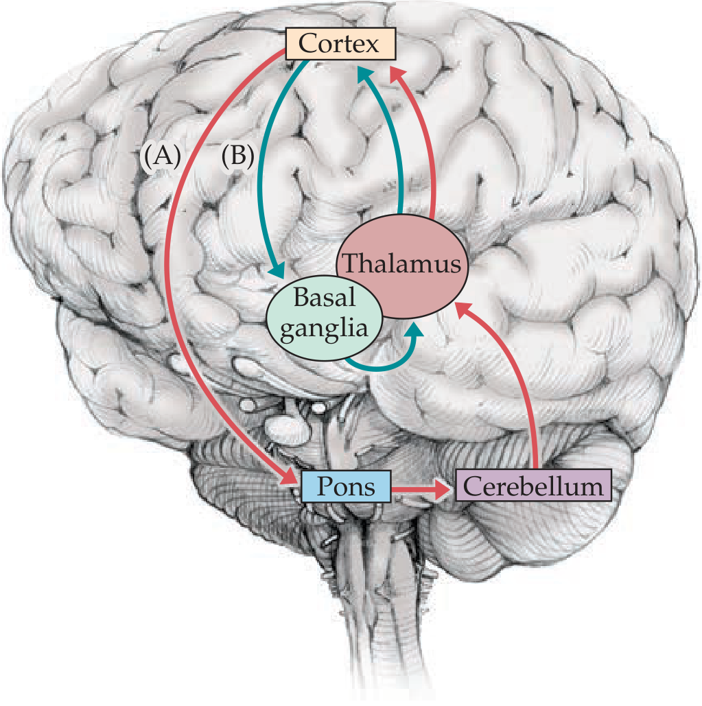
DIRECT PATHWAY (GO - facilitates movement):
Cortex → Striatum → GPi/SNr (inhibited) → Thalamus RELEASED → Cortex ✓
Dopamine (D1 receptor) FACILITATES direct pathway
INDIRECT PATHWAY (STOP - suppresses movement):
Cortex → Striatum → GPe (inhibited) → STN released → GPi/SNr activated → Thalamus INHIBITED → Cortex ✗
Dopamine (D2 receptor) INHIBITS indirect pathway
Diseases of Basal Ganglia
| Disease | Pathology | Movement Type | Features |
|---|---|---|---|
| Parkinson's | Loss of dopamine neurons in Substantia nigra pars compacta | Hypokinetic | TRAP: Tremor (resting, pill-rolling), Rigidity (cogwheel), Akinesia/bradykinesia, Postural instability |
| Huntington's | Loss of GABA neurons in striatum (esp. caudate) | Hyperkinetic | Chorea, dementia, psychiatric; CAG repeat (Chr 4); caudate atrophy on CT ("box-car ventricles") |
| Hemiballismus | Lesion of subthalamic nucleus (STN) | Violent flinging (proximal) | Contralateral side |
| Wilson's disease | Copper accumulation; basal ganglia + liver | Mixed | Kayser-Fleischer rings; low ceruloplasmin |
MNEMONIC: "TRAP Parkinson's": T=Tremor (resting), R=Rigidity, A=Akinesia, P=Postural instability
PART 8 - CEREBELLUM
Functional Zones - NEET Favourite
| Zone | Anatomy | Input | Function | Lesion |
|---|---|---|---|---|
| Vestibulocerebellum | Flocculonodular lobe | Vestibular nuclei | Balance, eye movements | Truncal ataxia, nystagmus |
| Spinocerebellum | Vermis + intermediate | Spinal cord (proprioception) | Gait + limb coordination | Gait ataxia (vermis lesion) |
| Cerebrocerebellum | Lateral hemispheres | Cerebral cortex via pons | Motor planning, timing | Ipsilateral limb ataxia, dysmetria |
MNEMONIC for Cerebellar Peduncles: "SIC" = Superior (output to thalamus), Inferior (vestibular input), Middle (largest - pontine input)
Deep Cerebellar Nuclei (Lateral → Medial)
"Don't Eat Greasy Food" = Dentate → Emboliform → Globose → Fastigial
- Dentate = largest; receives from cerebrocerebellum; projects via SCP to VL thalamus → motor cortex
- Fastigial = receives from vestibulocerebellum; projects to vestibular nuclei
Cerebellar Signs - "DANISH"
D - Dysdiadochokinesia (can't do rapid alternating movements) A - Ataxia (gait, truncal) N - Nystagmus (horizontal, fast phase toward lesion) I - Intention tremor (tremor WORSE on approaching target) S - Scanning (staccato) dysarthria H - Hypotonia
KEY NEET POINT: Cerebellar lesions = IPSILATERAL signs (output crosses twice and cancels out). Opposite of cerebral lesions!
PART 9 - BRAINSTEM OVERVIEW
Level-by-Level Summary
MIDBRAIN
├── CN III (Oculomotor) - exits interpeduncular fossa
├── CN IV (Trochlear) - ONLY CN exiting DORSALLY
├── Superior colliculi - visual reflexes, pupillary light reflex
├── Inferior colliculi - auditory relay
├── Substantia nigra (dopamine)
└── Red nucleus (motor relay)
PONS
├── CN V (Trigeminal) - largest CN, motor + sensory
├── CN VI (Abducens) - exits at pontomedullary junction
├── CN VII (Facial)
├── CN VIII (Vestibulocochlear)
├── Locus coeruleus (norepinephrine)
└── PPRF (paramedian pontine reticular formation) - horizontal gaze
MEDULLA
├── CN IX (Glossopharyngeal)
├── CN X (Vagus) - cardiorespiratory
├── CN XI (Accessory)
├── CN XII (Hypoglossal)
├── Pyramidal DECUSSATION (corticospinal fibers cross)
├── Nucleus gracilis + cuneatus (dorsal column relay)
└── Nucleus tractus solitarius (NTS) - taste + visceral sensory
Crossed Syndromes - NEET GOLD
Brainstem rule = CROSSED findings:
- IPSILATERAL cranial nerve signs (nucleus is at the level of lesion)
- CONTRALATERAL hemiplegia + sensory loss (long tracts already crossed or yet to cross)
Wallenberg Syndrome (Lateral Medullary Syndrome) - PICA occlusion:
| Finding | Side | Why |
|---|---|---|
| Pain/temp loss face | Ipsilateral | Trigeminal spinal nucleus |
| Pain/temp loss body | Contralateral | Spinothalamic tract |
| Horner's syndrome | Ipsilateral | Descending sympathetic fibers |
| Dysphagia, dysarthria | Ipsilateral | CN IX, X nuclei |
| Ataxia | Ipsilateral | Inferior cerebellar peduncle |
| NO hemiplegia | - | Pyramids are ANTERIOR (spared) |
PART 10 - LIMBIC SYSTEM
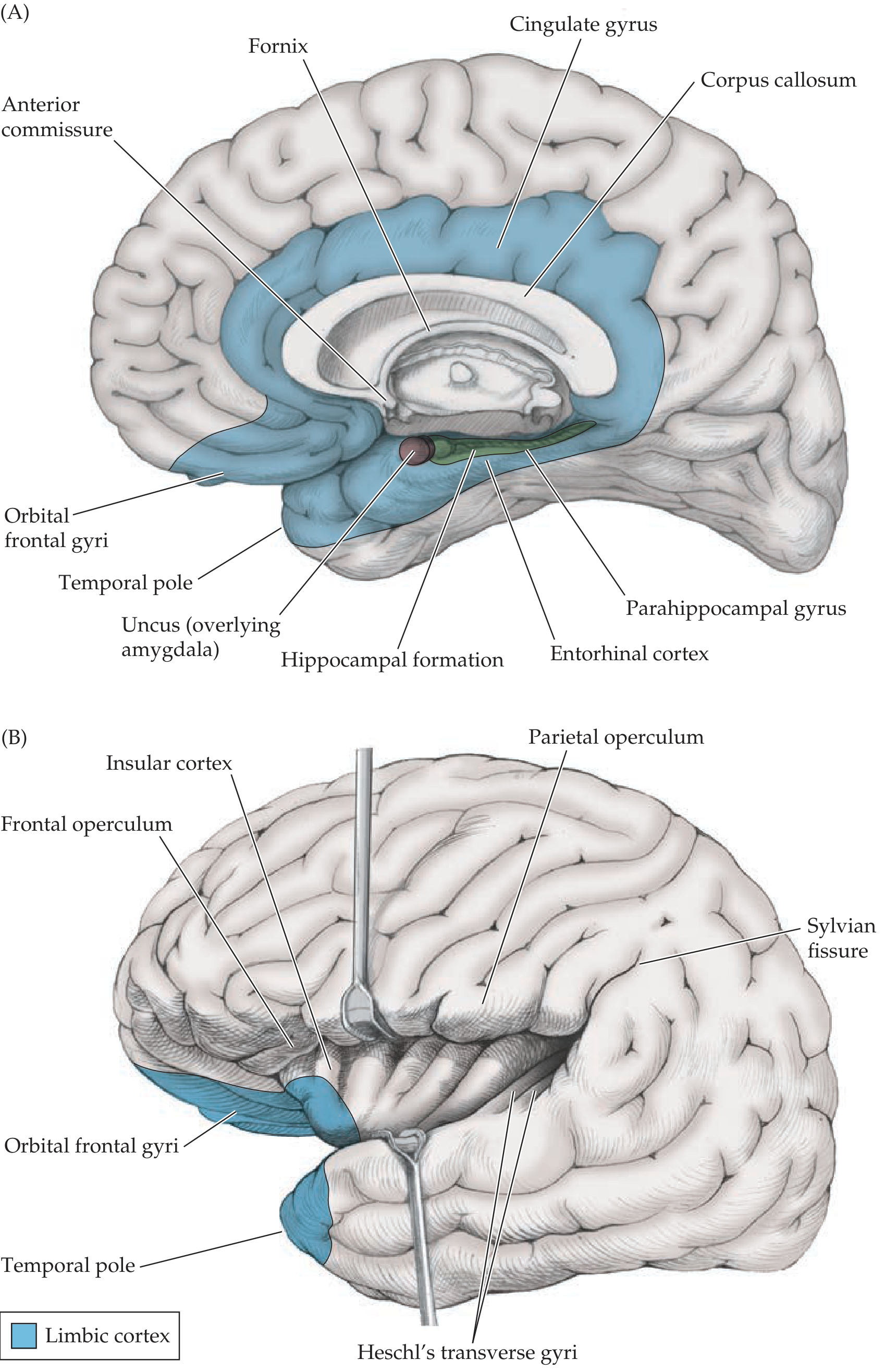
Papez Circuit (Memory Circuit) - NEET Favourite
Mnemonic: "Happy Fornication Makes Anterior Cortical Excitement"Hippocampus → Fornix → Mammillary bodies → Anterior thalamus → Cingulate gyrus → Entorhinal cortex → Hippocampus
Lesions in Papez circuit:
- Hippocampus damage → Anterograde amnesia (can't form NEW memories; old memories preserved)
- Mammillary bodies damage → Wernicke-Korsakoff syndrome (thiamine B1 deficiency in alcoholics) → Confabulation + anterograde amnesia
- Amygdala damage → Kluver-Bucy syndrome = hyperorality, hypersexuality, visual agnosia, placidity (no fear)
Limbic Structures Quick Summary
| Structure | Location | Function | Lesion Syndrome |
|---|---|---|---|
| Hippocampus | Medial temporal lobe | New declarative memory formation | Anterograde amnesia |
| Amygdala | Anterior temporal lobe (under uncus) | Fear, emotional memory, threat | Kluver-Bucy syndrome |
| Cingulate gyrus | Above corpus callosum | Attention, pain, emotion | Akinetic mutism (anterior) |
| Mammillary bodies | Hypothalamus | Papez circuit relay | Wernicke-Korsakoff |
| Fornix | White matter arch | Hippocampus → mammillary bodies | Memory loss if sectioned |
PART 11 - SENSORY PATHWAYS (High Yield!)
Two Main Pathways - NEET Must Know
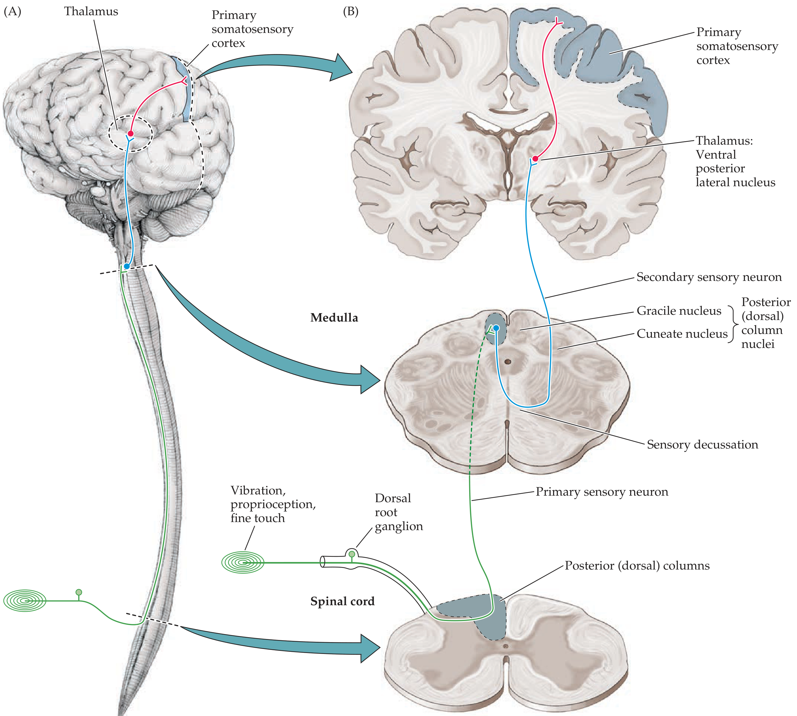
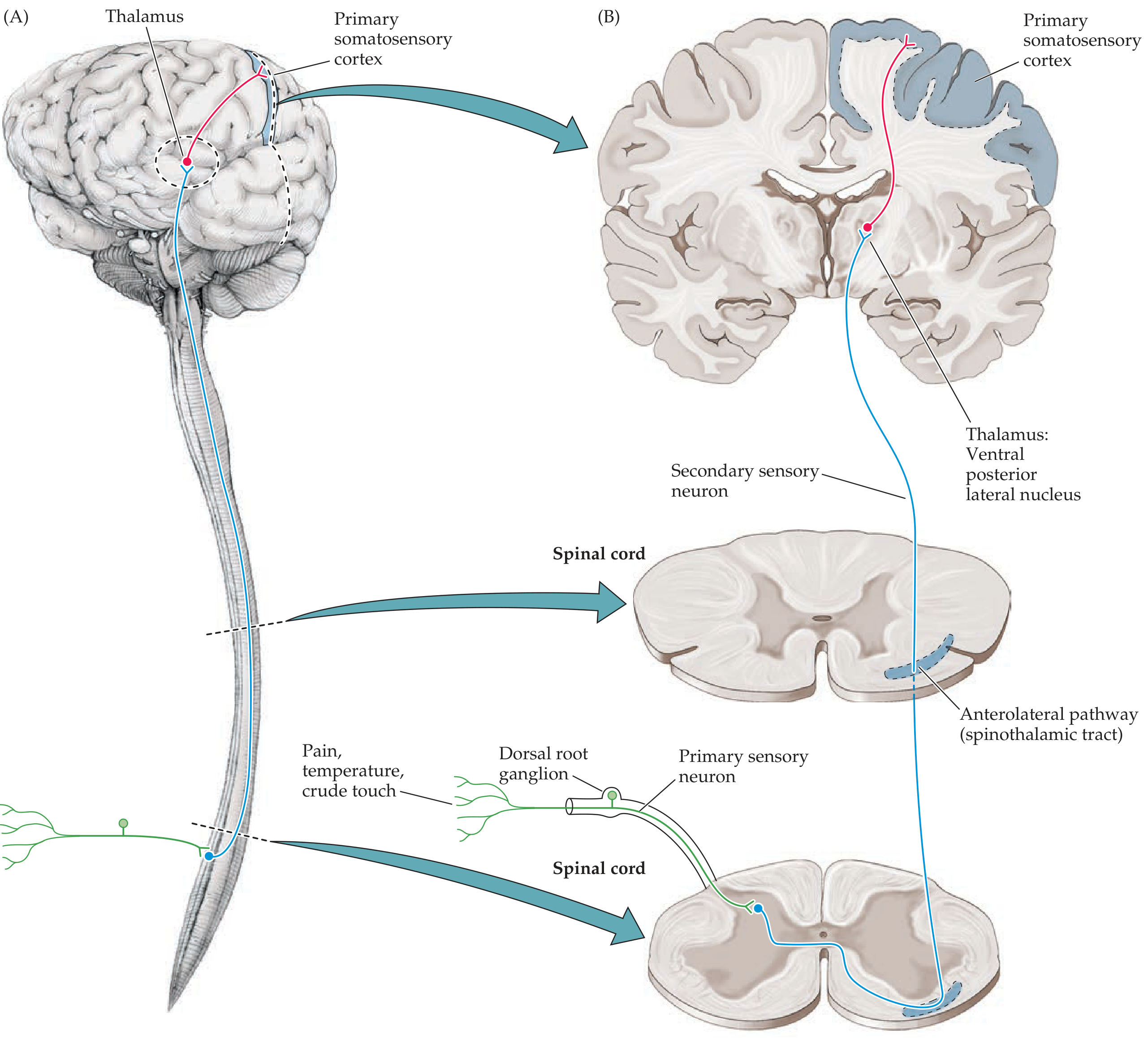
| Feature | Dorsal Column-Medial Lemniscal | Spinothalamic (Anterolateral) |
|---|---|---|
| Sensation | Vibration, proprioception, fine/discriminative touch, 2-point discrimination | Pain, temperature, crude touch |
| 1st neuron | DRG → enters spinal cord → ascends IPSILATERAL in posterior column | DRG → enters spinal cord → synapses in dorsal horn IMMEDIATELY |
| Where does it cross? | MEDULLA (at gracile/cuneate nuclei) | SPINAL CORD (within 1-2 segments, via anterior white commissure) |
| 2nd neuron | Gracile nucleus (legs) or Cuneate nucleus (arms) → decussates → medial lemniscus | Crosses → ascends anterolateral column |
| Thalamus relay | VPL nucleus | VPL nucleus |
| 3rd neuron | → Primary somatosensory cortex (postcentral gyrus) | → Primary somatosensory cortex |
NEET TRICK - Brown-Sequard Syndrome (hemisection of spinal cord):
- IPSILATERAL: loss of vibration + proprioception (dorsal column hasn't crossed yet)
- CONTRALATERAL: loss of pain + temperature (spinothalamic already crossed)
- IPSILATERAL: UMN signs (corticospinal tract)
PART 12 - VENTRICULAR SYSTEM
LATERAL VENTRICLES (C-shaped, in each hemisphere)
Frontal horn → Body → Occipital horn → Temporal horn (contains choroid plexus)
↓ Foramen of Monro (interventricular foramen)
THIRD VENTRICLE (between thalami)
↓ Cerebral Aqueduct of Sylvius (NARROWEST - most common site of obstruction)
FOURTH VENTRICLE (between pons/medulla and cerebellum)
↓ Foramen of Magendie (medial, 1) + Foramina of Luschka (lateral, 2)
SUBARACHNOID SPACE
↓ Absorbed at Arachnoid granulations → Superior sagittal sinus
MNEMONIC for openings of 4th ventricle: "One Magician, Two Luckys" = 1 Magendie (medial) + 2 Luschka (lateral)
PART 13 - BLOOD SUPPLY (Stroke Localization)
| Artery | Territory | Classic Stroke Syndrome |
|---|---|---|
| ACA | Medial frontal + parietal (LEG area) | Contralateral LEG weakness > arm; abulia, incontinence |
| MCA | Lateral frontal, parietal, temporal (ARM + FACE + language) | Contralateral face + arm weakness > leg; aphasia (dominant); neglect (non-dominant) |
| PCA | Occipital lobe + medial temporal + thalamus | Contralateral hemianopia (macular sparing); alexia without agraphia |
| PICA | Lateral medulla + inferior cerebellum | Wallenberg syndrome (see above) |
| Basilar | Entire brainstem | Locked-in syndrome (quadriplegia, preserved consciousness and vertical gaze only) |
| Lenticulostriate (from MCA) | Basal ganglia + internal capsule | Pure motor stroke (posterior limb of internal capsule) |
MASTER REVISION TABLE - Lesion → Deficit
| Lesion Site | Classic NEET Finding |
|---|---|
| Precentral gyrus (BA 4) | Contralateral UMN weakness |
| Postcentral gyrus (BA 1,2,3) | Contralateral hemisensory loss |
| Broca's area (BA 44/45) | Non-fluent expressive aphasia |
| Wernicke's area (BA 22) | Fluent receptive aphasia ("word salad") |
| Angular gyrus (BA 39) | Gerstmann syndrome |
| Frontal eye fields (BA 8) | Eyes deviate TOWARD lesion |
| Calcarine cortex (BA 17) | Contralateral homonymous hemianopia |
| Hippocampus | Anterograde amnesia |
| Amygdala | Kluver-Bucy syndrome |
| Mammillary bodies | Wernicke-Korsakoff |
| Subthalamic nucleus | Contralateral hemiballismus |
| Substantia nigra | Parkinson's disease |
| Caudate nucleus | Huntington's disease |
| Cerebellar hemisphere | Ipsilateral limb ataxia |
| Cerebellar vermis | Truncal/gait ataxia |
| Lateral medulla (PICA) | Wallenberg syndrome |
| Posterior limb internal capsule | Pure motor hemiplegia |
| Cerebral aqueduct (obstruction) | Obstructive hydrocephalus |
TOP 10 NEET PG MNEMONICS SUMMARY
| # | Mnemonic | What it covers |
|---|---|---|
| 1 | TRAP | Parkinson's features (Tremor, Rigidity, Akinesia, Postural instability) |
| 2 | DANISH | Cerebellar signs (Dysdiadochokinesia, Ataxia, Nystagmus, Intention tremor, Scanning speech, Hypotonia) |
| 3 | Don't Eat Greasy Food | Deep cerebellar nuclei (Dentate, Emboliform, Globose, Fastigial) |
| 4 | "Broca BROKe his speech" | Broca's = expressive aphasia |
| 5 | Happy Fornication Makes Anterior Cortical Excitement | Papez circuit |
| 6 | "ACA = Anterior = Leg" | ACA stroke causes leg weakness |
| 7 | "One Magician, Two Luckys" | 4th ventricle openings (1 Magendie, 2 Luschka) |
| 8 | BA 4 = Motor; 1,2,3 = Sensory; 17 = Vision; 44/45 = Broca's; 22 = Wernicke's | Top Brodmann areas |
| 9 | Wallenberg = No Hemiplegia | Lateral medullary lesion spares pyramids |
| 10 | Brown-Sequard: same side vibration, opposite side pain | Spinal cord hemisection |
Sources: Neuroanatomy through Clinical Cases 3rd Ed. (Blumenfeld) | Neuroscience: Exploring the Brain 5th Ed. | Adams & Victor's Principles of Neurology 12th Ed.
Want me to go deeper on any one topic? Popular NEET PG areas I can expand:
- All 12 Cranial Nerves with exit foramina, nuclei, and clinical palsies
- Visual pathway in detail (field defects at each level - huge NEET topic)
- Complete stroke syndromes with MCQs
- Ventricular system + CSF (hydrocephalus types)
- UMN vs LMN signs table
For each part give me images showing all details given in that parts also can give multiple images for a part also to include all info of that part.
Finding Sources
Finding Sources
Finding Sources
Finding Sources
I now have all the image URLs. Let me fetch all of them in parallel - getting every image for every part simultaneously.
Loading Image
Loading Image
Loading Image
Loading Image
Loading Image
Loading Image
Loading Image
Loading Image
Finding Sources
Loading Image
Loading Image
Here is the complete image-rich NEET PG Neuroanatomy guide, every part with all its diagrams:
🧠 NEET PG Neuroanatomy - Full Image Guide
Source: Neuroanatomy through Clinical Cases, 3rd Ed. (Blumenfeld)
🔷 FOUNDATION: CNS vs PNS Overview
Image 1 - The whole nervous system in the body:
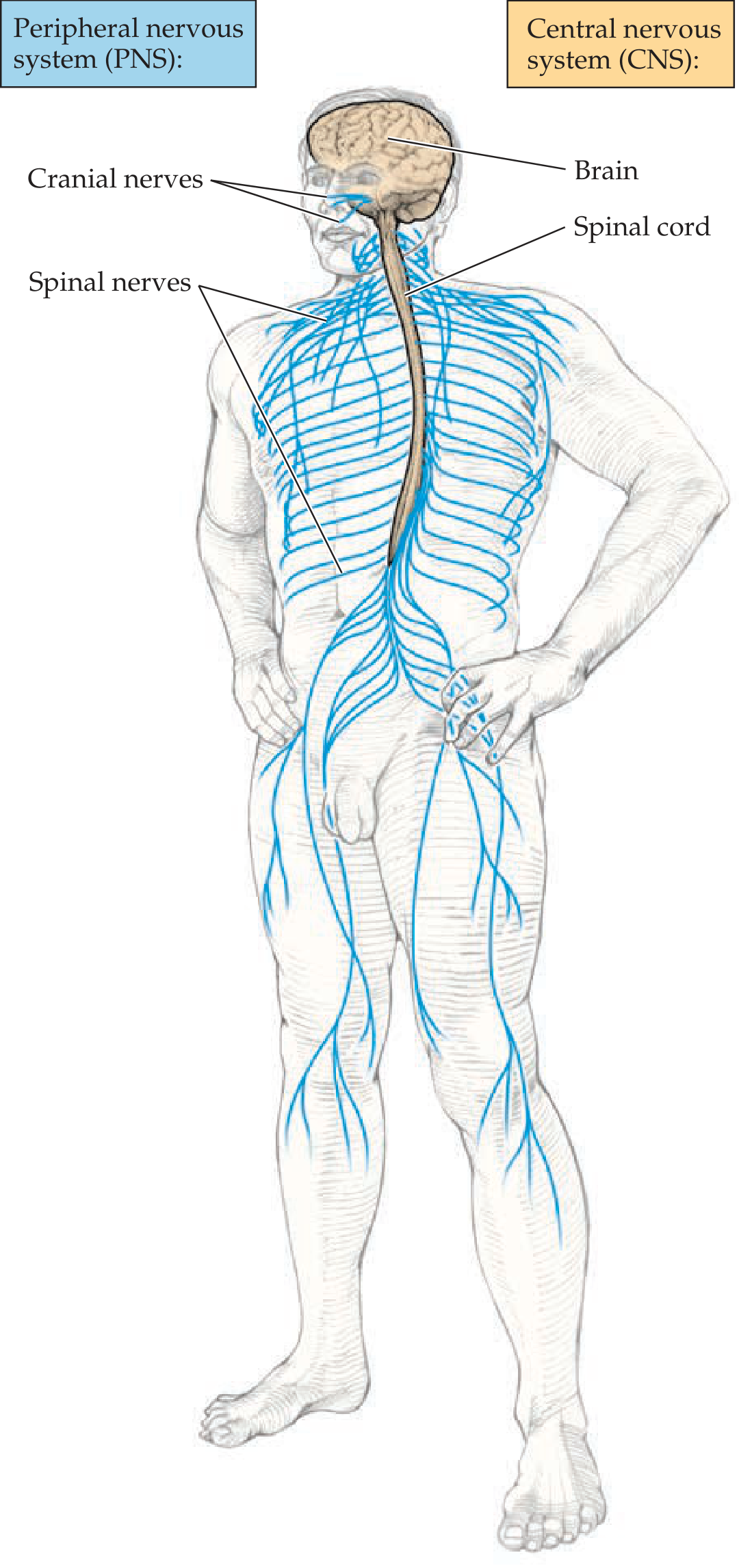
Image 2 - Embryological development → adult brain divisions (HIGH YIELD):
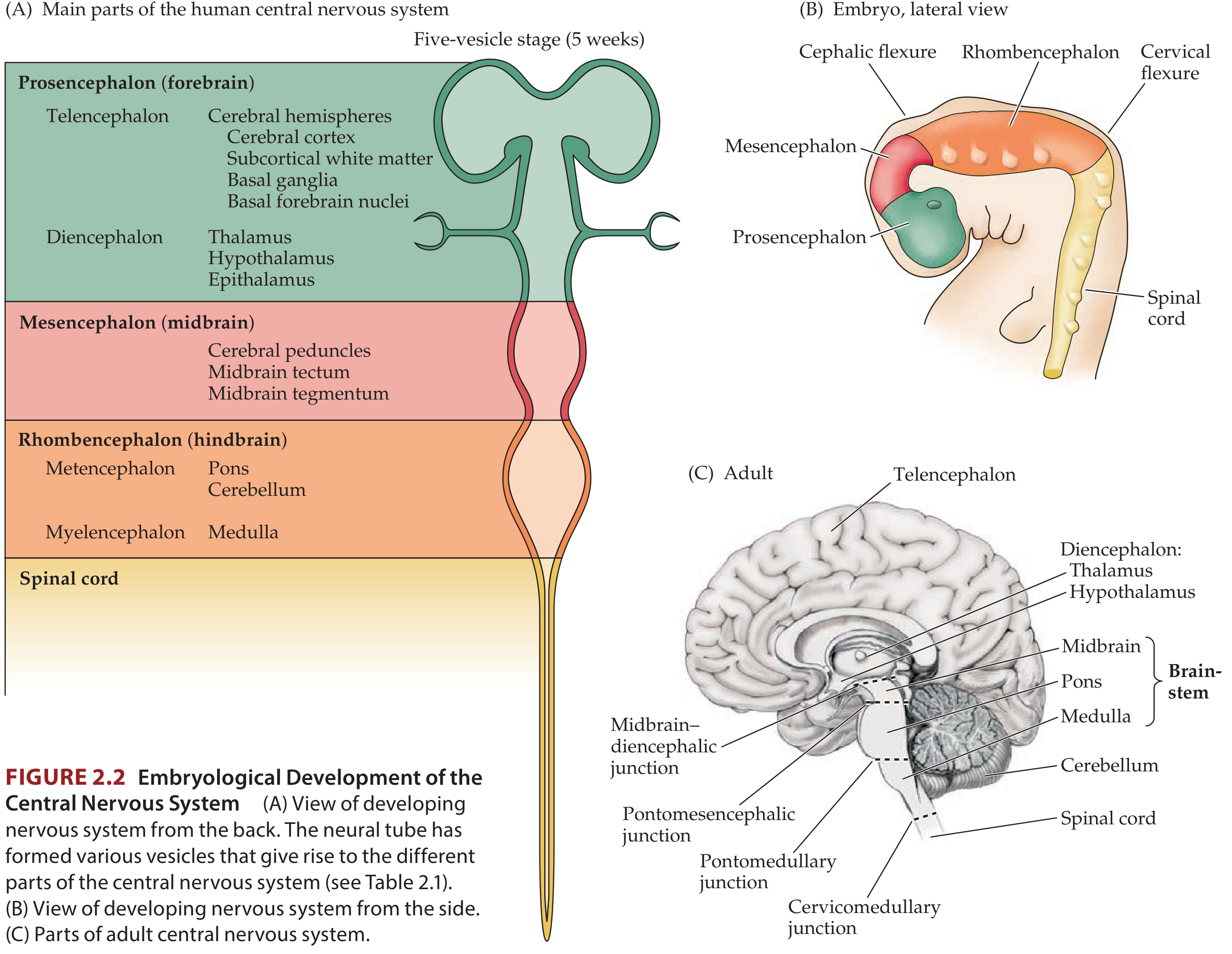
NEET TRICK: Embryo → Adult
- Prosencephalon → Forebrain (cerebrum + thalamus/hypothalamus)
- Mesencephalon → Midbrain
- Rhombencephalon → Hindbrain (pons + cerebellum + medulla)
🔷 PART 1 - THE 4 LOBES + MAJOR SULCI & GYRI
Image 1 - Color-coded lobes (lateral + midsagittal):
Image 2 - Fully labeled lateral + medial views (ALL gyri and sulci named):
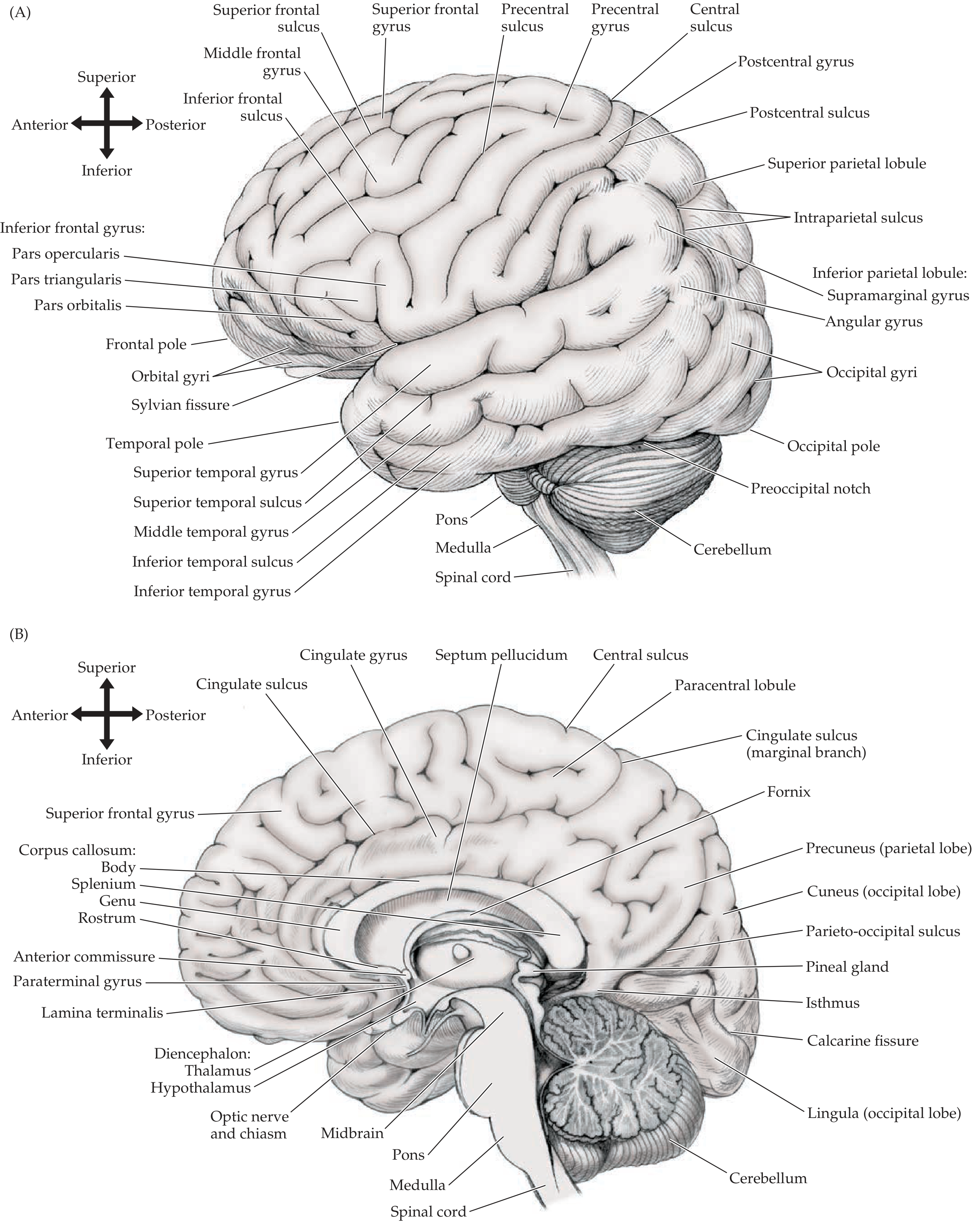
Image 3 - Inferior view + superior view (C and D):
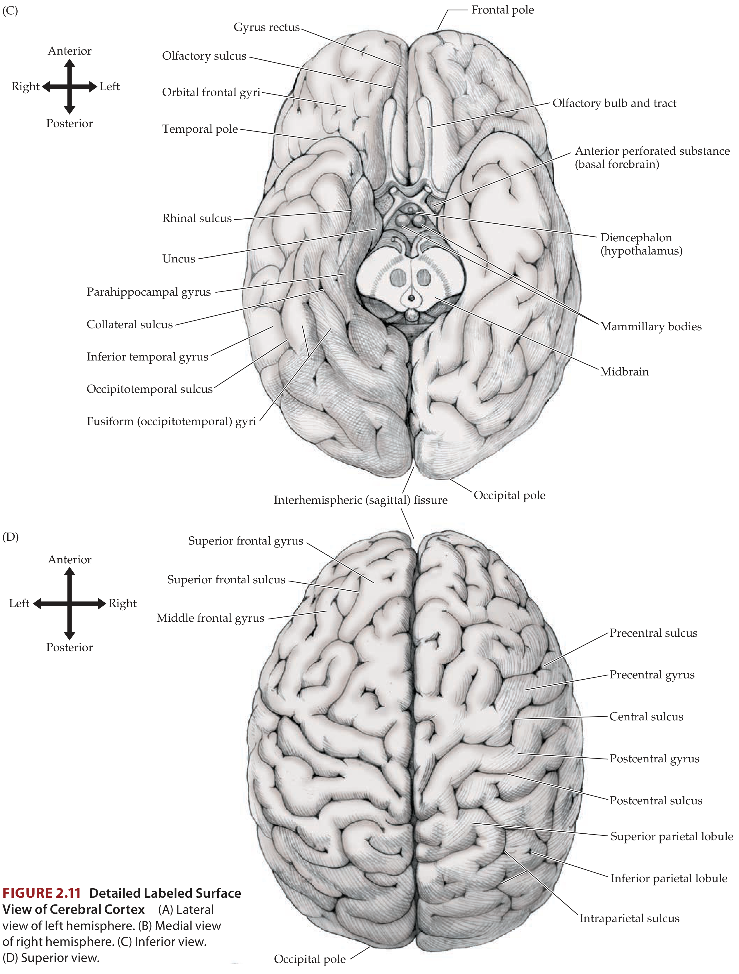
Key NEET Points from these images:
- Pars triangularis + pars opercularis of inferior frontal gyrus = Broca's area (BA 44/45)
- Supramarginal gyrus (BA 40) + Angular gyrus (BA 39) = inferior parietal lobule
- Uncus on inferior surface overlies the amygdala - uncal herniation compresses CN III
- Calcarine fissure on medial occipital = primary visual cortex (BA 17)
🔷 PART 2 - PRIMARY CORTICAL AREAS + FUNCTIONAL MAP
Image 1 - Primary cortical areas labeled on lateral brain:
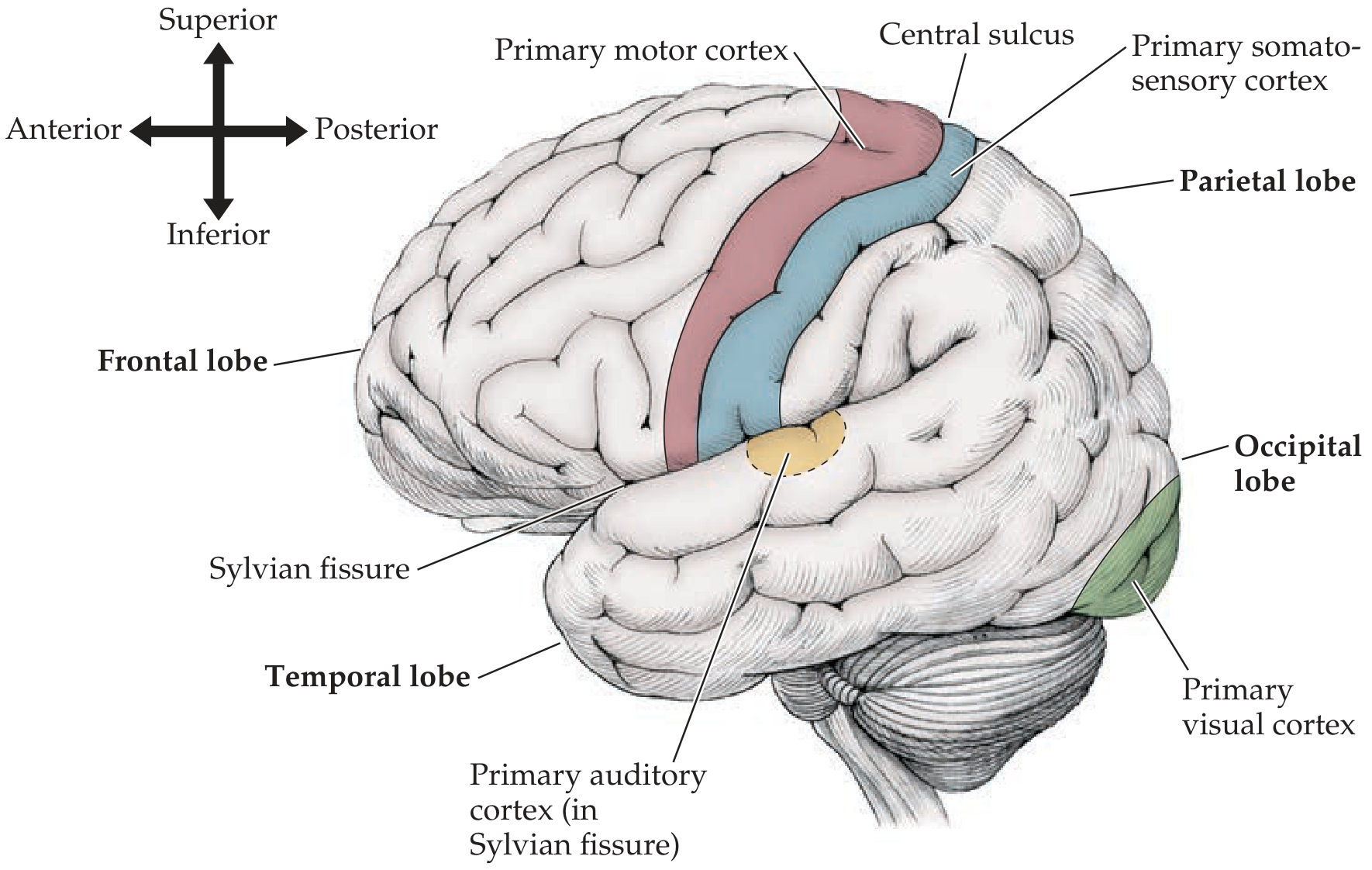
Image 2 - Complete functional cortical map with ALL areas named:
Image 3 - Brodmann areas map (original Brodmann illustration + cortical layers of BA 4, 46, 17):
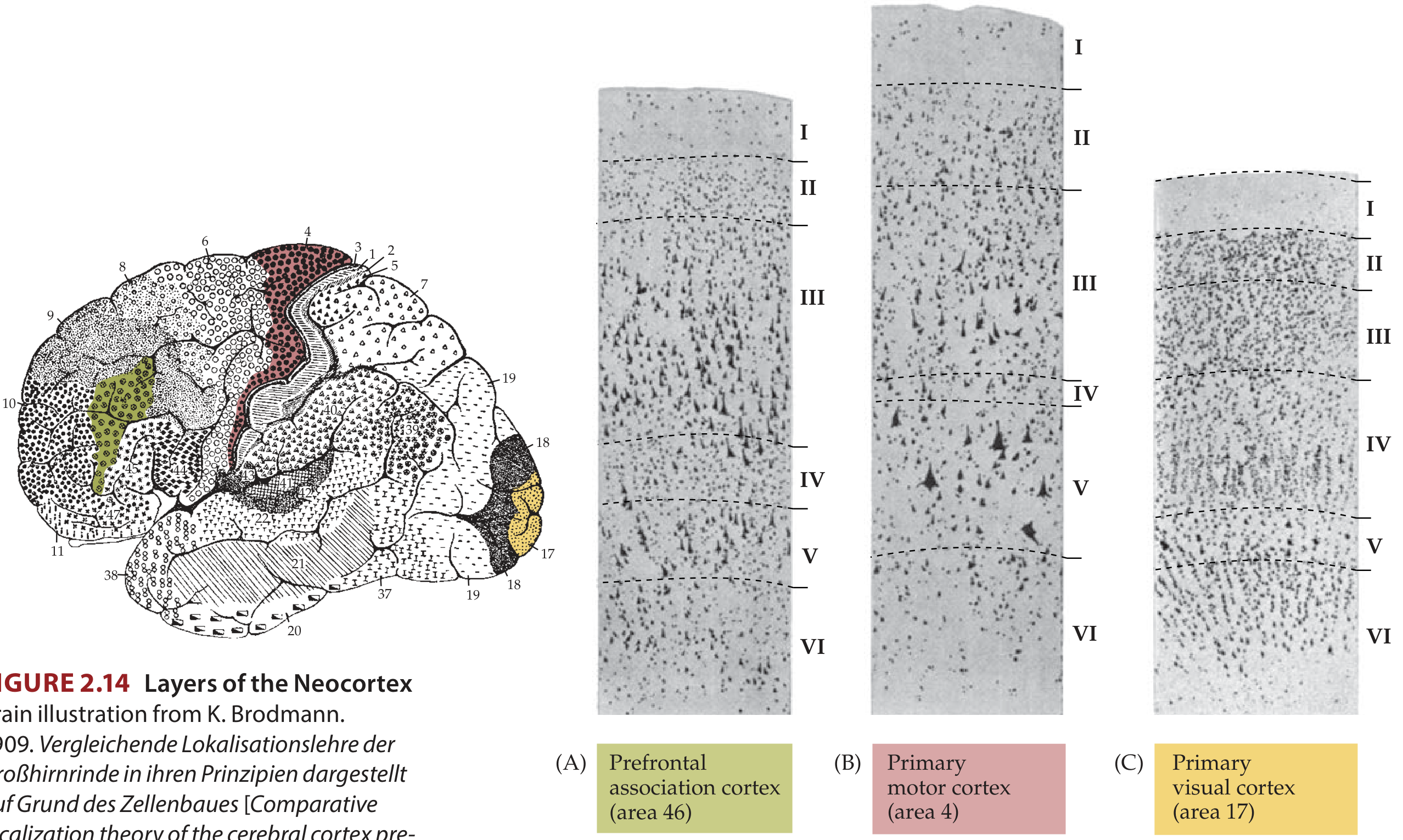
NEET KEY: Motor cortex (BA 4) has huge Betz cells in layer V (sends output to spinal cord). Visual cortex (BA 17) has thickest layer IV (line of Gennari - visible to naked eye, hence "striate cortex").
🔷 PART 3 & 4 - HOMUNCULUS (Motor + Sensory Body Maps)
Image - Motor homunculus (right, pink) + Somatosensory homunculus (left, blue) on brain:
NEET High Yield from this image:
- Leg area = MEDIAL surface → supplied by ACA → ACA stroke = leg weakness
- Face + arm = LATERAL surface → supplied by MCA → MCA stroke = face + arm weakness
- Large lips/tongue = important for speech and swallowing (massive sensory feedback needed)
- Sensory (postcentral) is BEHIND central sulcus; Motor (precentral) is IN FRONT
🔷 PART 5 - THALAMUS (Relay Nuclei)
Image 1 - Thalamus as central relay hub (connections diagram):
Image 2 - Thalamic nuclear groups (3D colored dissection view):
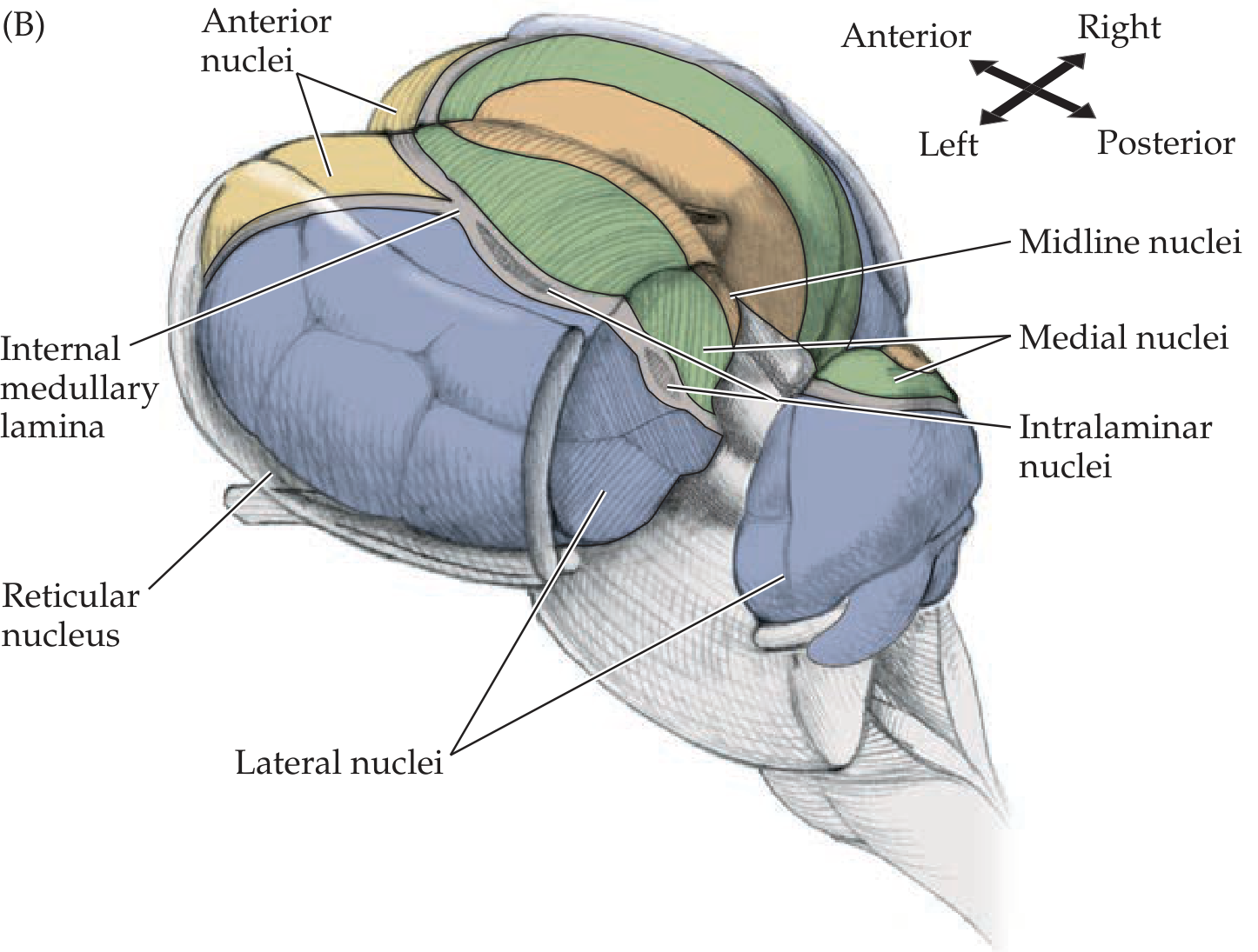
Image 3 - Spinothalamic pathway (pain/temp) showing thalamus VPL relay:
Image 4 - Dorsal column pathway (vibration/proprioception) showing thalamus VPL relay:
🔷 PART 6 - BASAL GANGLIA + CEREBELLAR MOTOR CIRCUITS
Image - Motor feedback loops (basal ganglia + cerebellar circuits together):
NEET Key Points from this image:
- Both cerebellum AND basal ganglia feedback through the thalamus to reach motor cortex
- Neither directly synapses on lower motor neurons
- Cerebellar output: Dentate nucleus → SCP → VL thalamus → Motor cortex
- Basal ganglia output: GPi/SNr → VA/VL thalamus → Motor cortex
- Lesion in basal ganglia = movement disorders (Parkinson's, Huntington's, hemiballismus)
- Lesion in cerebellum = ataxia/coordination disorders (ipsilateral signs)
🔷 PART 7 - BRAINSTEM (All 3 levels + Cranial Nerves)
Image 1 - Ventral (anterior) view of brainstem with ALL cranial nerves labeled:
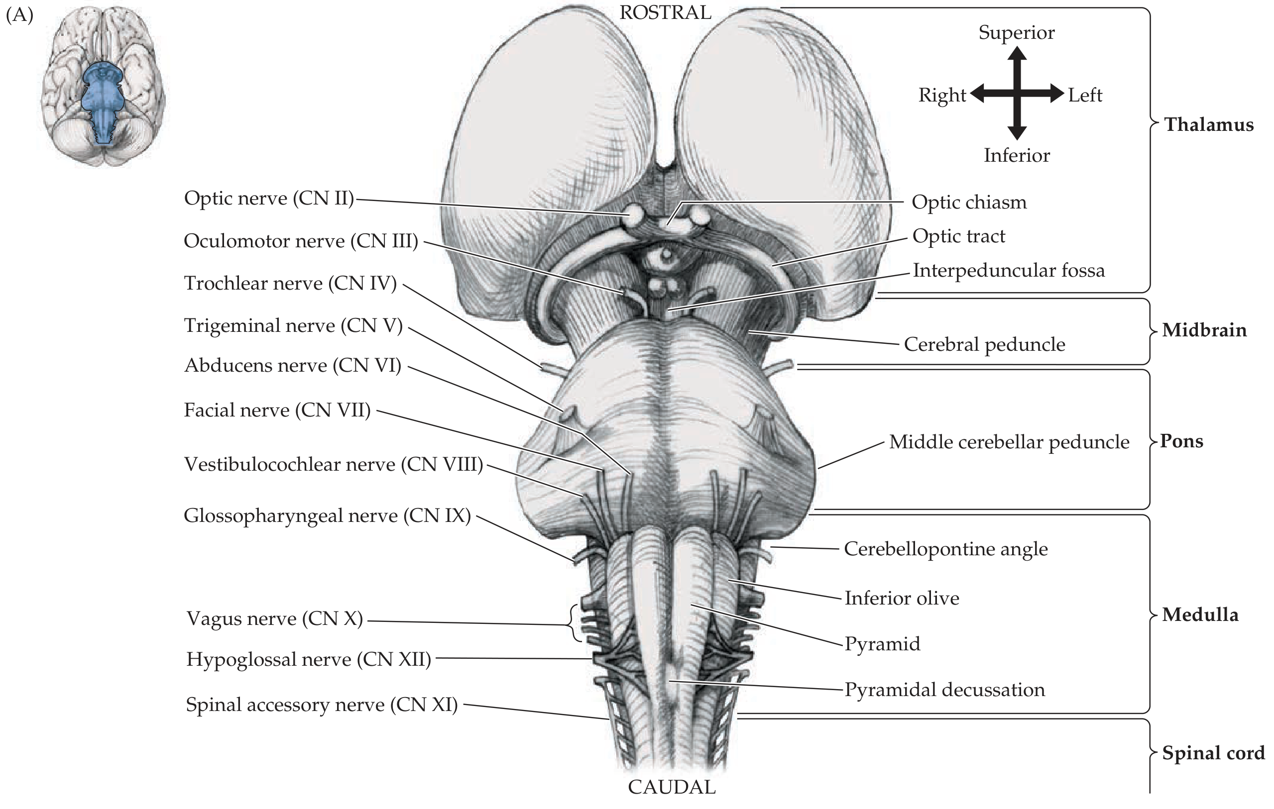
Image 2 - Dorsal view + lateral view showing CN IV (dorsal exit), colliculi, cerebellar peduncles, and dorsal column nuclei:
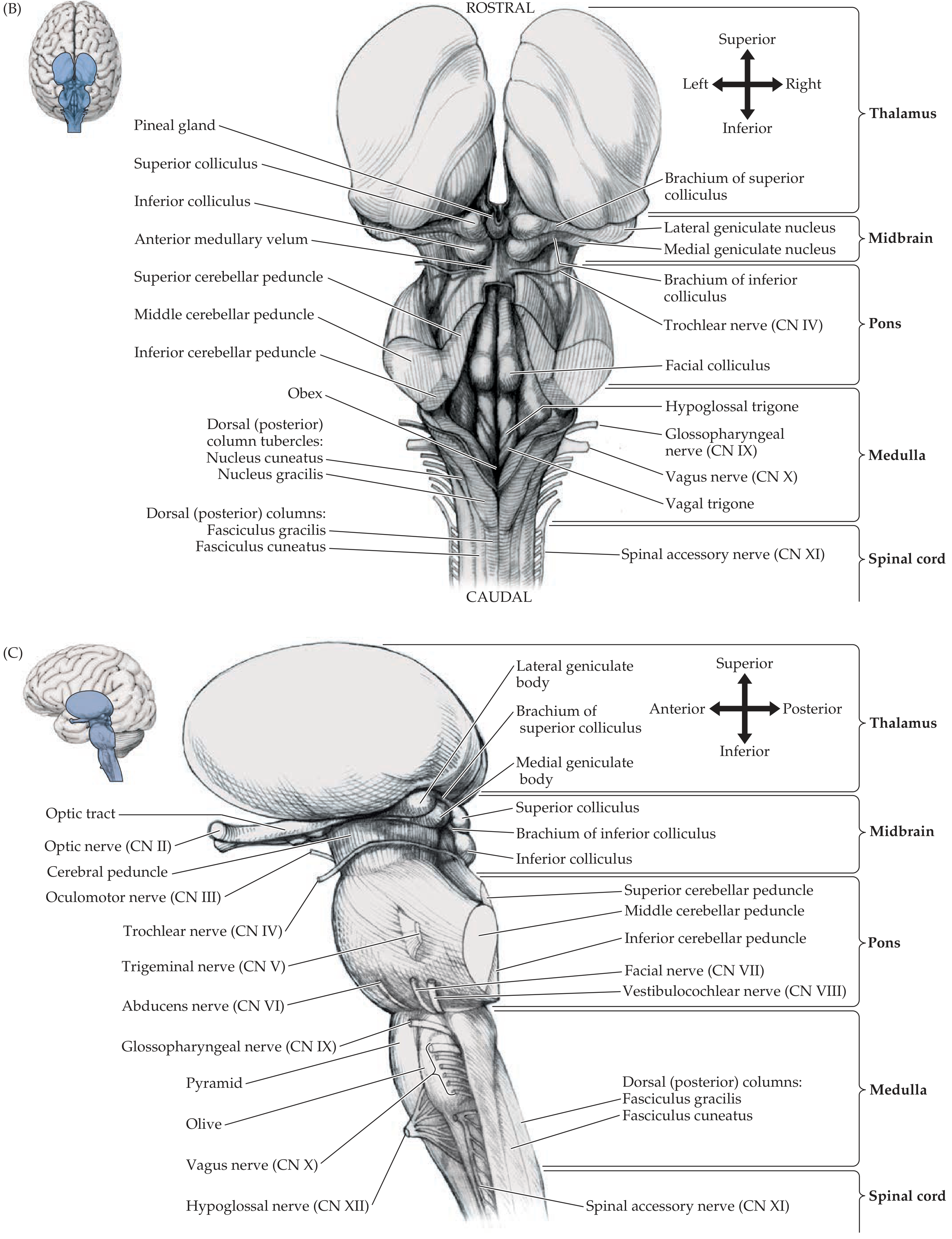
NEET Keys from brainstem images:
- CN IV (trochlear) = only CN exiting dorsally + only CN that decussates before exiting
- CN V (trigeminal) = largest CN, exits lateral pons
- CN VI (abducens) exits at the pontomedullary junction (vulnerable to raised ICP - false localizing sign)
- Pyramidal decussation in medulla = where corticospinal fibers cross (left brain controls right body)
- Inferior olive = prominent bulge on medulla, relay for cerebellar learning
- Cerebellopontine angle = site of acoustic neuromas (CN VIII schwannoma)
🔷 PART 8 - LIMBIC SYSTEM (Memory + Emotion Circuit)
Image 1 - Medial view (Papez circuit structures) + Lateral view (insula + Heschl's gyri):
NEET Keys from limbic image:
- Hippocampus = in medial temporal lobe; damage → anterograde amnesia (can't form new memories)
- Amygdala = under the uncus on inferior surface; damage → Kluver-Bucy syndrome
- Fornix connects hippocampus to mammillary bodies (Papez circuit white matter tract)
- Cingulate gyrus = emotion/attention; anterior cingulate lesion → akinetic mutism
- Entorhinal cortex (parahippocampal gyrus, BA 28) = primary olfactory cortex; gateway to hippocampus
- Insula = hidden in Sylvian fissure; visceral sensation, taste, empathy, pain
- Heschl's gyri = primary auditory cortex (BA 41/42) hidden inside Sylvian fissure
🔷 PART 9 - SENSORY PATHWAYS
Image 1 - Dorsal column-medial lemniscal pathway (vibration/proprioception/fine touch):
Image 2 - Spinothalamic pathway (pain/temperature/crude touch) for comparison:
Image 3 - Stretch reflex circuit (UMN vs LMN concept):
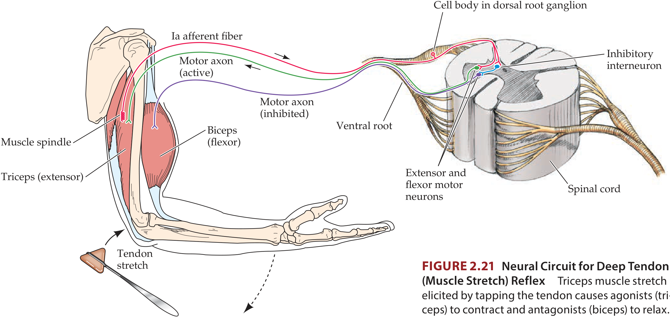
Summary comparison table with images in mind:
| Feature | Dorsal Column | Spinothalamic |
|---|---|---|
| Sensation | Vibration, proprioception, fine touch | Pain, temperature, crude touch |
| Crosses at | Medulla (image 1) | Spinal cord (image 2) |
| Spinal location | Posterior column | Anterolateral column |
| 1st synapse | Medulla (gracile/cuneate) | Dorsal horn (ipsilateral) |
| Thalamic nucleus | VPL | VPL |
🔷 PART 10 - BRAINSTEM (VENTRAL + DORSAL) WITH ALL CNs AT A GLANCE
The two brainstem images together cover everything:
Ventral view - use to remember: CN exits + levels + pyramidal decussation + inferior olive
Dorsal view - use to remember: CN IV (only dorsal exit), colliculi (superior = vision, inferior = hearing), cerebellar peduncles (SCP/MCP/ICP), dorsal column nuclei in medulla
MNEMONIC for brainstem CN levels:
- Midbrain: CN III, IV ("3 and 4 in the midbrain floor")
- Pons: CN V, VI, VII, VIII ("5 to 8, pontine great")
- Medulla: CN IX, X, XI, XII ("9 to 12 in the medulla's time")
🔷 QUICK RECALL - All Images at a Glance
| Part | Image shows |
|---|---|
| CNS overview | Whole body nervous system + embryological brain divisions |
| Lobes | Color-coded 4 lobes (lateral + medial) + full gyri/sulci labels (4 views) |
| Cortical areas | Primary areas on brain + complete functional map with Broca's/Wernicke's |
| Brodmann areas | Original map + microscopic layers comparing BA 4 (motor) vs 17 (visual) vs 46 (prefrontal) |
| Homunculus | Motor + somatosensory homunculi showing body map upside-down |
| Thalamus | Relay hub connections + 3D nuclear groups + sensory pathways through VPL |
| Basal ganglia | Motor feedback loops with cerebellar circuit side by side |
| Brainstem | Ventral (all CNs) + dorsal (CN IV, colliculi, cerebellar peduncles) + lateral views |
| Limbic system | Medial view (Papez circuit) + lateral view (insula + Heschl's gyri) |
| Sensory pathways | Dorsal column + spinothalamic full pathway diagrams + stretch reflex arc |
All images from: Neuroanatomy through Clinical Cases, 3rd Ed. (Blumenfeld) - the top neuroanatomy textbook for clinical medicine and NEET PG.
Want me to now add images for cranial nerves detail, cerebellum sections, ventricular system, or visual pathway field defects - each of which is a heavy NEET PG topic?
This is a shared conversation. Sign in to Orris to start your own chat.