
focus only on the ascending and descending tracts
python3 << 'EOF' import pdfplumber pdf_path = "/tmp/workspace/attachments/7da64047-e557-4984-a739-c78934c3a6f8/19-20. Ascending & Descending Tracts-2.pdf" with pdfplumber.open(pdf_path) as pdf: print(f"Total pages: {len(pdf.pages)}") for i, page in enumerate(pdf.pages): text = page.extract_text() print(f"\n{'='*60}") print(f"PAGE {i+1}") print('='*60) if text: print(text) else: print("[No text extracted]") EOF
python3 << 'EOF' from pypdf import PdfReader, PdfWriter import os pdf_path = "/tmp/workspace/attachments/7da64047-e557-4984-a739-c78934c3a6f8/19-20. Ascending & Descending Tracts-2.pdf" out_dir = "/tmp/workspace/tracts_pages" os.makedirs(out_dir, exist_ok=True) reader = PdfReader(pdf_path) writer_list = [] for i, page in enumerate(reader.pages): w = PdfWriter() w.add_page(page) out_path = f"{out_dir}/page_{i+1}.pdf" with open(out_path, "wb") as f: w.write(f) print("Done splitting. Files created:") for f in sorted(os.listdir(out_dir)): print(f) EOF
python3 << 'EOF' # Try to convert PDF pages to PNG using pypdf's page rendering or PIL # Check what image libraries are available import subprocess result = subprocess.run(['python3', '-c', 'import pdf2image; print("pdf2image available")'], capture_output=True, text=True) print(result.stdout, result.stderr) result2 = subprocess.run(['python3', '-c', 'from pypdf import PdfReader; r=PdfReader("/tmp/workspace/tracts_pages/page_1.pdf"); p=r.pages[0]; print(dir(p))'], capture_output=True, text=True) print(result2.stdout[:500]) # Try pymupdf (fitz) result3 = subprocess.run(['python3', '-c', 'import fitz; print("fitz available")'], capture_output=True, text=True) print(result3.stdout, result3.stderr) EOF
python3 << 'EOF' from pdf2image import convert_from_path import os pdf_path = "/tmp/workspace/attachments/7da64047-e557-4984-a739-c78934c3a6f8/19-20. Ascending & Descending Tracts-2.pdf" out_dir = "/tmp/workspace/tracts_images" os.makedirs(out_dir, exist_ok=True) images = convert_from_path(pdf_path, dpi=150) print(f"Converted {len(images)} pages") for i, img in enumerate(images): path = f"{out_dir}/page_{i+1:02d}.jpg" img.save(path, "JPEG", quality=85) print(f"Saved {path}") EOF
python3 << 'EOF' from PIL import Image import os img_dir = "/tmp/workspace/tracts_images" out_dir = "/tmp/workspace/tracts_composites" os.makedirs(out_dir, exist_ok=True) # Load all pages pages = [] for i in range(1, 21): path = f"{img_dir}/page_{i:02d}.jpg" img = Image.open(path) pages.append(img) print(f"Loaded {len(pages)} pages") print(f"Page 1 size: {pages[0].size}") # Create composites of 4 pages each (2x2 grid), 5 composites total def make_composite(imgs, outpath, cols=2): # Resize all to same width target_w = 800 resized = [] for img in imgs: ratio = target_w / img.width new_h = int(img.height * ratio) resized.append(img.resize((target_w, new_h), Image.LANCZOS)) rows = (len(resized) + cols - 1) // cols # Pad to fill grid while len(resized) < rows * cols: resized.append(Image.new('RGB', (target_w, resized[0].height), (255, 255, 255))) row_imgs = [] for r in range(rows): row_parts = resized[r*cols:(r+1)*cols] combined = Image.new('RGB', (target_w * cols, row_parts[0].height)) for c, part in enumerate(row_parts): combined.paste(part, (c * target_w, 0)) row_imgs.append(combined) total_h = sum(img.height for img in row_imgs) final = Image.new('RGB', (target_w * cols, total_h)) y = 0 for row_img in row_imgs: final.paste(row_img, (0, y)) y += row_img.height final.save(outpath, "JPEG", quality=85) print(f"Saved {outpath} ({final.size})") # 5 composites of 4 pages each for i in range(5): batch = pages[i*4:(i+1)*4] make_composite(batch, f"{out_dir}/composite_{i+1:02d}.jpg") print("All composites created") EOF
ascending descending spinal cord tracts diagram
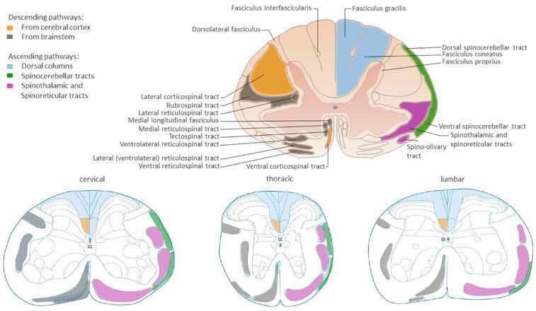
Anatomical diagrams illustrating the organization of major white matter tracts in the human spinal cord across cervical, thoracic, and lumbar cross-sections. A primary large-scale diagram provides detailed labeling of pathways, while three comparative smaller diagrams show regional variations. Descending (motor) pathways are color-coded in orange (corticospinal tracts from the cerebral cortex) and grey/black (brainstem-derived tracts including rubrospinal, reticulospinal, and tectospinal tracts). Ascending (sensory) pathways are highlighted in blue for dorsal columns (fasciculus gracilis and cuneatus), green for dorsal and ventral spinocerebellar tracts, and pink for the spinothalamic and spinoreticular tracts. The diagrams illustrate key neuroanatomical landmarks such as the central canal (CC), dorsal horns, and ventral horns. Educational focus is placed on the somatotopic arrangement and the relative change in white matter volume versus grey matter across different spinal levels, highlighting the clinical relevance for localized spinal cord injury and neurological deficit mapping.
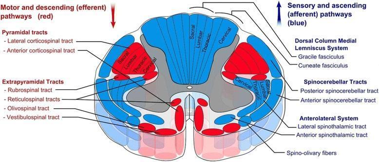
This medical anatomical diagram illustrates a transverse section of the human spinal cord, specifically detailing the organization of white matter tracts. The visual uses a color-coded schema to differentiate between motor and sensory pathways: descending (efferent) motor tracts are highlighted in red, while ascending (afferent) sensory tracts are shown in blue. Key anatomical structures identified include the Pyramidal tracts (lateral and anterior corticospinal tracts), Extrapyramidal tracts (rubrospinal, reticulospinal, olivospinal, and vestibulospinal), the Dorsal Column-Medial Lemniscus system (gracile and cuneate fasciculi), Spinocerebellar tracts, and the Anterolateral system (lateral and anterior spinothalamic tracts). A significant educational feature of the diagram is the depiction of somatotopic organization, showing the concentric arrangement of sacral, lumbar, thoracic, and cervical fibers within the lateral corticospinal and dorsal column tracts. The image provides a comprehensive overview of spinal cord neuroanatomy, useful for understanding clinical localization of spinal cord lesions and the functional topography of the central nervous system.
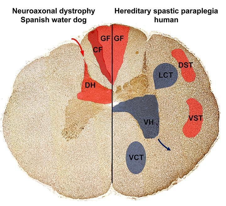
This Comparison Diagram presents a cross-sectional illustration of the spinal cord, contrasting the localization of axonal spheroids and neuronal loss in Neuroaxonal Dystrophy (NAD) and Hereditary Spastic Paraplegia (HSP). The diagram is split into two halves: the left represents a Spanish Water Dog with NAD, and the right represents human HSP. Ascending sensory pathways are highlighted in red, including the Gracile Fasciculus (GF), Cuneate Fasciculus (CF), and Dorsal Horn (DH) on the NAD side, and GF, Dorsal Spinocerebellar Tract (DST), and Ventral Spinocerebellar Tract (VST) on the HSP side. Descending motor pathways, affected only in the HSP model, are highlighted in blue, depicting the Lateral Corticospinal Tract (LCT), Ventral Corticospinal Tract (VCT), and Ventral Horn (VH). The visual demonstrates that in NAD, pathology is restricted to sensory, ascending tracts in the dorsal funiculus and dorsal horn. In contrast, human HSP shows involvement of both sensory pathways and descending motor pathways (pyramidal tracts and ventral horns). This diagram illustrates the pathophysiology of upper motor neuron and sensory system degeneration relevant to neurology and neurohistology.
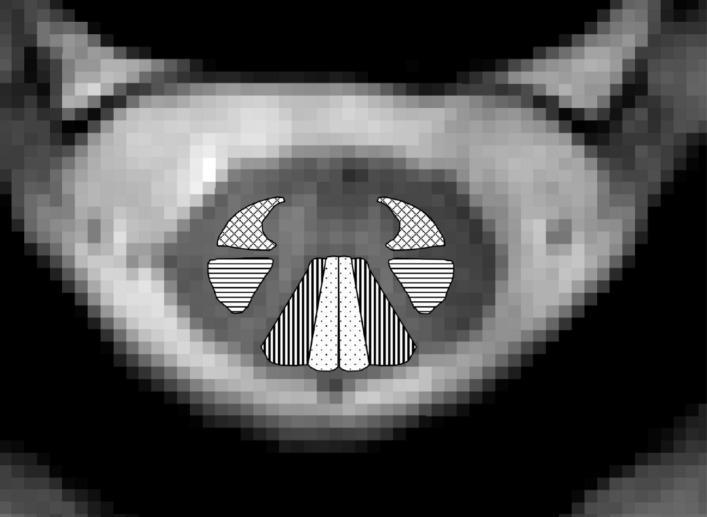
This diagnostic image is an axial cross-sectional Magnetic Resonance (MR) image of the human spinal cord at the C2/C3 cervical level. The image serves as an anatomical diagram by overlaying specific patterns on the white matter to identify major ascending and descending tracts. Centrally located within the spinal cord's white matter, four bilateral regions are highlighted: 1) The lateral corticospinal tracts are indicated by horizontal lines, showing their posterior-lateral position. 2) The cuneate fasciculi are marked with vertical lines in the dorsal column. 3) The gracile fasciculi are represented by dotted patterns, located medially in the dorsal column adjacent to the posterior median sulcus. 4) The combined spinothalamic and spinoreticular tracts are shown with crosshatched patterns in the anterolateral region. This visual aid is designed for advanced medical education to illustrate neuroanatomical localization and the functional organization of spinal cord pathways, providing clinical relevance for assessing regional white matter integrity in spinal cord pathologies or injuries.
Ascending & Descending Tracts of the Spinal Cord
Note: Your uploaded PDF is an image-based (scanned) file, so direct text extraction was not possible. The notes below are drawn from authoritative textbook sources (Neuroanatomy through Clinical Cases 3e, Localization in Clinical Neurology 8e, and Neuroscience: Exploring the Brain 5e) and cover the standard content of a lecture on this topic.
Overview: The Three Most Important Long Tracts
| Pathway | Type | Function |
|---|---|---|
| Lateral corticospinal tract | Descending (motor) | Voluntary movement of extremities |
| Posterior (dorsal) columns | Ascending (sensory) | Vibration, proprioception, fine touch |
| Anterolateral pathways (spinothalamic) | Ascending (sensory) | Pain, temperature, crude touch |
ASCENDING TRACTS (Sensory)
1. Posterior Column - Medial Lemniscal Pathway
- 1st-order neuron: Primary sensory neuron enters via dorsal root → travels ipsilaterally up the posterior (dorsal) columns all the way to the dorsal column nuclei in the medulla (nucleus gracilis for lower body; nucleus cuneatus for upper body/arms)
- 2nd-order neuron: Decussates in the medulla → ascends as the medial lemniscus → synapses in the ventral posterior lateral (VPL) nucleus of the thalamus
- 3rd-order neuron: Projects via thalamic somatosensory radiations → primary somatosensory cortex (postcentral gyrus, Brodmann areas 3, 1, 2)
2. Anterolateral Pathways (Spinothalamic Tract)
| Tract | Termination | Function |
|---|---|---|
| Spinothalamic tract | VPL of thalamus → somatosensory cortex | Discriminative pain/temperature (location, intensity) |
| Spinoreticular tract | Medullary-pontine reticular formation → intralaminar thalamus (centromedian nucleus) | Emotional/arousal aspects of pain (phylogenetically older) |
| Spinomesencephalic tract | Periaqueductal gray (PAG) | Modulation of pain |
- 1st-order neuron: Small-diameter and unmyelinated axons enter via dorsal root entry zone → immediately synapse in dorsal horn (lamina I - marginal zone; lamina V) - some collaterals ascend/descend a few segments in Lissauer's tract first
- 2nd-order neuron: Axons cross in the anterior (ventral) commissure of the spinal cord (takes 2-3 spinal segments to reach opposite side) → ascend in the anterolateral white matter (contralateral)
- 3rd-order neuron: VPL thalamus → primary somatosensory cortex
3. Spinocerebellar Tracts
| Tract | Origin | Crossing | Location in cord |
|---|---|---|---|
| Dorsal (posterior) spinocerebellar | Clarke's nucleus (T1-L2) | Uncrossed (ipsilateral) | Lateral funiculus, posterior part |
| Ventral (anterior) spinocerebellar | Lumbar spinal cord | Crosses twice (net ipsilateral) | Lateral funiculus, anterior part |
| Cuneocerebellar | Lateral cuneate nucleus (medulla) | Uncrossed | - |
DESCENDING TRACTS (Motor)
LATERAL PATHWAYS (Fine voluntary movement)
1. Lateral Corticospinal Tract (Most Clinically Important)
-
50% from primary motor cortex (Brodmann area 4, precentral gyrus, layer 5 pyramidal neurons, including giant Betz cells)
- Remainder from premotor/supplementary motor cortex (area 6) and parietal lobe (areas 3, 1, 2, 5, 7)
- Axons enter the corona radiata (cerebral white matter)
- Descend through posterior limb of the internal capsule
- Pass through the cerebral peduncles (ventral midbrain), then ventral pons
- Converge in the ventral medulla to form the pyramids (hence "pyramidal tract")
- At the caudal medulla: ~85-90% of fibers cross at the pyramidal decussation → descend in the lateral funiculus of the contralateral spinal cord = lateral corticospinal tract
- Remaining ~10-15% do NOT cross → form the ventral (anterior) corticospinal tract (crosses later in the ventral white commissure at cervical/upper thoracic levels)
- Synapse on lower motor neurons (LMNs) in the anterior horn (laminae IV-VII and IX)
2. Rubrospinal Tract
- Origin: Red nucleus in the midbrain tegmentum (receives input from cortex areas 4, 6, 3, 1, 2 via corticorubrospinal pathway)
- Crosses: In the midbrain (decussates immediately)
- Course: Descends in the lateral funiculus of the spinal cord
- Function: Voluntary limb movement (important in animals; less so in humans, where corticospinal takes over)
VENTROMEDIAL (MEDIAL) PATHWAYS (Posture, balance, head/neck)
3. Vestibulospinal Tracts
| Component | Origin | Course | Function |
|---|---|---|---|
| Lateral vestibulospinal | Lateral vestibular nucleus (Deiters') | Ipsilateral, full cord length; anterior lateral funiculus | Facilitates extensor/antigravity muscles - maintains upright posture |
| Medial vestibulospinal | Medial vestibular nucleus | Bilateral; cervical and upper thoracic only; ventral funiculus | Controls neck/back muscles, head stabilization |
- Localization in Clinical Neurology 8e; Neuroscience: Exploring the Brain 5e
4. Reticulospinal Tracts
| Tract | Origin | Crossing | Spinal Location | Effect |
|---|---|---|---|---|
| Pontine (medial) reticulospinal | Pontine reticular formation | Ipsilateral (uncrossed) | Ventral funiculus | Facilitates extensor (antigravity) stretch reflexes - aids standing posture |
| Medullary (lateral) reticulospinal | Medullary reticular formation | Ipsilateral, descends in ventrolateral funiculus | Ventrolateral funiculus | Inhibits antigravity muscles (liberates them from reflex control) |
5. Tectospinal Tract
- Origin: Superior colliculus of the midbrain (receives input from retina, visual, auditory, somatosensory, and motor cortex)
- Crosses: Immediately after leaving the colliculus (decussates in midbrain)
- Course: Descends close to the midline; limited to cervical cord only
- Function: Orienting head and eyes toward new stimuli; controls neck, upper trunk, and shoulder muscles
Summary Diagram
Decussation Points - Quick Reference
| Tract | Where it Crosses |
|---|---|
| Lateral corticospinal | Pyramidal decussation (caudal medulla) |
| Anterior corticospinal | Ventral white commissure (each spinal segment) |
| Posterior columns | Internal arcuate fibers (medulla) |
| Spinothalamic | Anterior commissure (spinal cord, 2-3 segments above entry) |
| Rubrospinal | Midbrain tegmentum (ventral tegmental decussation) |
| Tectospinal | Dorsal tegmental decussation (midbrain) |
| Vestibulospinal (lateral) | Uncrossed (ipsilateral) |
| Reticulospinal (pontine) | Predominantly uncrossed |
UMN vs LMN Signs
| Feature | UMN Lesion | LMN Lesion |
|---|---|---|
| Tone | Increased (spasticity) | Decreased (flaccidity) |
| Reflexes | Hyperreflexia | Hyporeflexia / areflexia |
| Babinski sign | Present (extensor plantar) | Absent |
| Atrophy | Mild (disuse) | Severe (denervation) |
| Fasciculations | Absent | Present |
| Weakness | Present (contralateral to lesion above decussation) | Present (ipsilateral, segmental) |
Key Clinical Correlations
- Brown-Sequard syndrome (hemisection of cord): Ipsilateral UMN signs + ipsilateral loss of proprioception/vibration (posterior columns) + contralateral loss of pain/temperature (spinothalamic) - all below the level
- Anterior cord syndrome: Loss of motor function + pain/temperature bilaterally (corticospinal + spinothalamic affected), but proprioception/vibration preserved (posterior columns spared)
- Syringomyelia: Bilateral loss of pain/temperature at the level of the syrinx (crossing spinothalamic fibers in anterior commissure destroyed first), "cape" distribution
- Tabes dorsalis / B12 deficiency: Posterior column degeneration - loss of vibration, proprioception, sensory ataxia