
Pneumothorax Davidson
I'll look up Pneumothorax from Davidson's textbook right away.
Searching sources...
Reading File
Finding Sources
Finding Sources
Finding Sources
Finding Sources
Davidson's Principles and Practice of Medicine is not in this library. The closest authoritative respiratory/internal medicine sources available are Harrison's Principles of Internal Medicine 22E and Murray & Nadel's Textbook of Respiratory Medicine. Let me pull the pneumothorax sections from both, plus the emergency medicine source.
Finding Sources
Reading File
Reading File
Reading File
Reading File
Finding Sources
Reading File
Reading File
Reading File
Reading File
I now have sufficient material from Murray & Nadel's and Harrison's. Let me compile the comprehensive Davidson-style answer (Davidson's itself isn't in the library, but the content from these authoritative respiratory medicine texts covers the same clinical content that Davidson's covers on pneumothorax).
Pneumothorax
Note: Davidson's Principles and Practice of Medicine is not in this textbook library. The following is a comprehensive clinical overview drawn from Murray & Nadel's Textbook of Respiratory Medicine and Harrison's Principles of Internal Medicine 22E (2025) - both cover the same content as Davidson's on this topic.
Definition
Pneumothorax is the presence of gas (air) in the pleural space. It ranges in severity from asymptomatic to life-threatening tension pneumothorax.
Classification
1. Spontaneous Pneumothorax
Primary Spontaneous Pneumothorax (PSP)
- Occurs without underlying lung disease or antecedent trauma
- Typical patient: tall, thin young male, often a smoker
- Due to rupture of apical pleural blebs - small cystic spaces (outpouchings of visceral pleura, <1 cm) that lie within or immediately under the visceral pleura
- Blebs vs. Bullae: A bleb (<1 cm) is an outpouching between the lamina elastica interna and externa; a bulla (>1 cm) is an airspace demarcated by a thin wall (Reid classified bullae into 3 types)
- Microscopic abnormalities include respiratory bronchiolitis and pleural fibroelastosis
- Smoking is a major risk factor (airway inflammation weakens the visceral pleura)
- ~50% of patients will have a recurrence after the first episode
Secondary Spontaneous Pneumothorax (SSP)
- Occurs in the context of underlying lung disease
- More dangerous because of reduced pulmonary reserve
- Causes (Murray & Nadel's):
| Frequency | Causes |
|---|---|
| Common | COPD, pulmonary tuberculosis |
| Uncommon | Cystic fibrosis, lung cancer, Pneumocystis jirovecii infection |
| Rare | IPF, sarcoidosis, Langerhans cell histiocytosis, LAM, rheumatoid arthritis, scleroderma, ankylosing spondylitis, Marfan syndrome, Birt-Hogg-Dube syndrome, Ehlers-Danlos syndrome, catamenial pneumothorax |
- Catamenial pneumothorax: Recurrent pneumothorax in women of childbearing age - consider thoracic endometriosis
- LAM: Spontaneous pneumothorax in younger women (30-40 years) may be the first manifestation
2. Traumatic Pneumothorax
- Results from penetrating or nonpenetrating chest injuries
- Present in 40% of blunt and 20% of penetrating thoracic trauma cases
- Iatrogenic causes: transthoracic needle aspiration, thoracentesis, central venous catheter insertion (most common iatrogenic causes)
- Open pneumothorax ("sucking chest wound"): chest wall defect allows air entry through the wound; if wound approaches the size of the trachea, ventilation is severely compromised
- Management: cover with a one-way flutter (Heimlich) valve or occlusive dressing with one side open before operative repair
3. Tension Pneumothorax
- A medical emergency
- One-way valve mechanism: air enters but cannot escape - progressive intrapleural pressure rise
- Compresses the right ventricle, kinks the SVC/IVC, reduces venous return and cardiac output
- Causes obstructive shock: hypotension + hypoxia
- Hypoxemia is nearly universal (92%) in ventilated patients; present in only 50% breathing unassisted
- Treatment must not wait for radiologic confirmation - immediate thoracic decompression
Clinical Features
- Symptoms: Sudden onset ipsilateral pleuritic chest pain and breathlessness; may be minimal in small PSP
- Signs:
- Reduced chest expansion on affected side
- Hyperresonant percussion note
- Diminished/absent breath sounds
- Tracheal deviation away (in tension - a late sign)
- In tension: hypotension, tachycardia, raised JVP, cyanosis
Diagnosis
Chest X-Ray
- Gold standard for diagnosis
- Visible lung edge with absent lung markings peripherally
- BTS size classification: small (<2 cm rim of air at the hilum), large (≥2 cm)
- Note: Three different guidelines classify size differently with agreement in only 47% of cases
CT Chest
- More accurate for size estimation
- Identifies underlying lung disease, blebs, bullae
- Useful before surgical intervention
Ultrasound (POCUS)
- Pleural sliding (sea-shore sign on M-mode): when present, effectively excludes pneumothorax at that location
- Absence of sliding is not specific (also seen in apnea, pleural adhesions)
- "Lung point" (transition from sliding to no sliding at the edge of the pneumothorax): highly specific for pneumothorax
- B-lines: if present, excludes pneumothorax at that site
- "Bar code" / "stratosphere" sign (M-mode): linear pattern throughout depth - seen when sliding is absent
- More sensitive than CXR for occult pneumothorax, particularly in trauma
Management
Primary Spontaneous Pneumothorax
| Situation | Management |
|---|---|
| Asymptomatic / minimally symptomatic, small | Conservative: observation +/- supplemental O₂ (accelerates reabsorption ~4x); outpatient option for low-risk patients |
| Symptomatic or large | Needle aspiration (first-line) or tube drainage |
| Failure to expand / recurrence | Thoracoscopy with stapling of blebs + pleurodesis |
- Supplemental O₂ accelerates resolution of pneumothorax
- Needle aspiration vs. chest tube: Multiple RCTs show comparable initial success rates (aspiration 48-80%; chest tube 64-100%)
- Ambulatory management: One-way (Heimlich/flutter) valve attached to chest drain; effective in 85.8% - reduces hospital stay vs. standard chest drain (median 8 days hospitalized vs. outpatient)
Secondary Spontaneous Pneumothorax
- Nearly all patients require tube drainage
- Many will need thoracoscopy/thoracotomy with stapling + surgical pleurodesis
- If not a good operative candidate: chemical pleurodesis
Tension Pneumothorax
- Immediate decompression: needle thoracostomy or tube thoracostomy
- ATLS recommendations: insert needle at 4th-5th intercostal space, midaxillary line (higher success rate than traditional 2nd ICS midclavicular line due to thinner chest wall, avoids internal mammary/subclavian artery injury)
- A 5-cm angiocatheter at the anterior axillary line 4th-5th ICS has significantly lower failure rate (13% vs. 38% at midclavicular 2nd ICS)
- Follow immediately with chest tube
Traumatic Pneumothorax
- Usually treated with tube drainage unless very small
- Hemopneumothorax: one tube superiorly (air) + one inferiorly (blood)
- Iatrogenic: often managed with supplemental O₂ or aspiration; tube drainage if these fail
Prevention of Recurrence / Pleurodesis
| Method | Recurrence Rate |
|---|---|
| Drainage alone | High (~30-50%) |
| Thoracoscopic talc poudrage | ~5-10% |
| VATS + bulectomy | ~5% |
| Surgical pleurodesis (thoracoscopy/thoracotomy) | Nearly 100% effective |
- Talc poudrage: 2-4 g sufficient; no long-term sequelae (lung function near-normal 22-35 years later); relative contraindication if future lung transplant is anticipated (but not absolute)
- VATS: Important disadvantage - long-term postoperative pain (>30% patients, lasting 3-18 months)
- Mechanical pleurodesis: abrasion or partial parietal pleurectomy
- Bulectomy alone has unacceptably high recurrence (27.5% at 10 years) - must add pleurodesis
Complications
- Re-expansion pulmonary edema (RPE): Unilateral pulmonary edema after rapid lung reinflation; incidence ~1-3% symptomatic; treat with supplemental O₂ and diuretics
- Haemopneumothorax: Blood + air in pleural space (from torn adhesions or vascularized bullae rupture)
- Persistent air leak (bronchopleural fistula)
- Tension pneumothorax (if untreated)
Special Situations
- Air travel: Pneumothorax may expand at altitude (reduced cabin pressure) - patients should not fly until fully resolved
- Diving: Absolute contraindication until definitive treatment (surgical pleurodesis) given risk of rapid expansion at depth
Sources: Murray & Nadel's Textbook of Respiratory Medicine (Chapter 110 - Pneumothorax); Harrison's Principles of Internal Medicine 22E (2025, p. 2313-2314)
With figures explain like teacher
Let me pull all the figures from the textbook chapters on pneumothorax simultaneously.
Reading File
Reading File
Good - I can see image URLs in the text. Let me extract all the image URLs and fetch the most relevant ones in parallel.
Searching Content
"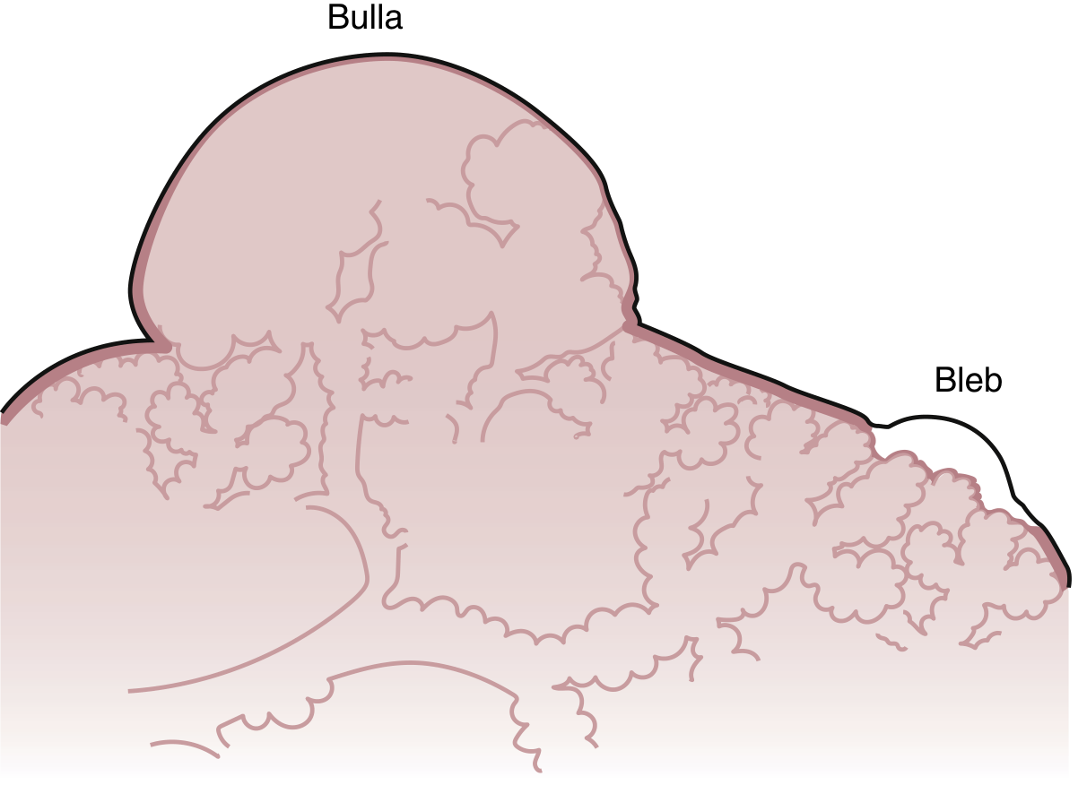
Figure 110.1 - Anatomy of blebs and bullae (Murray & Nadel's, Ch. 110)
Now here is how to tell them apart - a question that comes up in exams:
| Feature | Bleb | Bulla |
|---|---|---|
| Size | < 1 cm | > 1 cm |
| Location | Within/under visceral pleura (between lamina elastica interna and externa) | Sharply demarcated airspace, thin wall ≤1 mm |
| Contents | No lung parenchyma | Minimal/no parenchyma (Reid Type III has most) |
| Analogy | A blister under the skin of the lung | A bubble sitting on the surface of the lung |
And here is what a real bleb looks like through a thoracoscope:
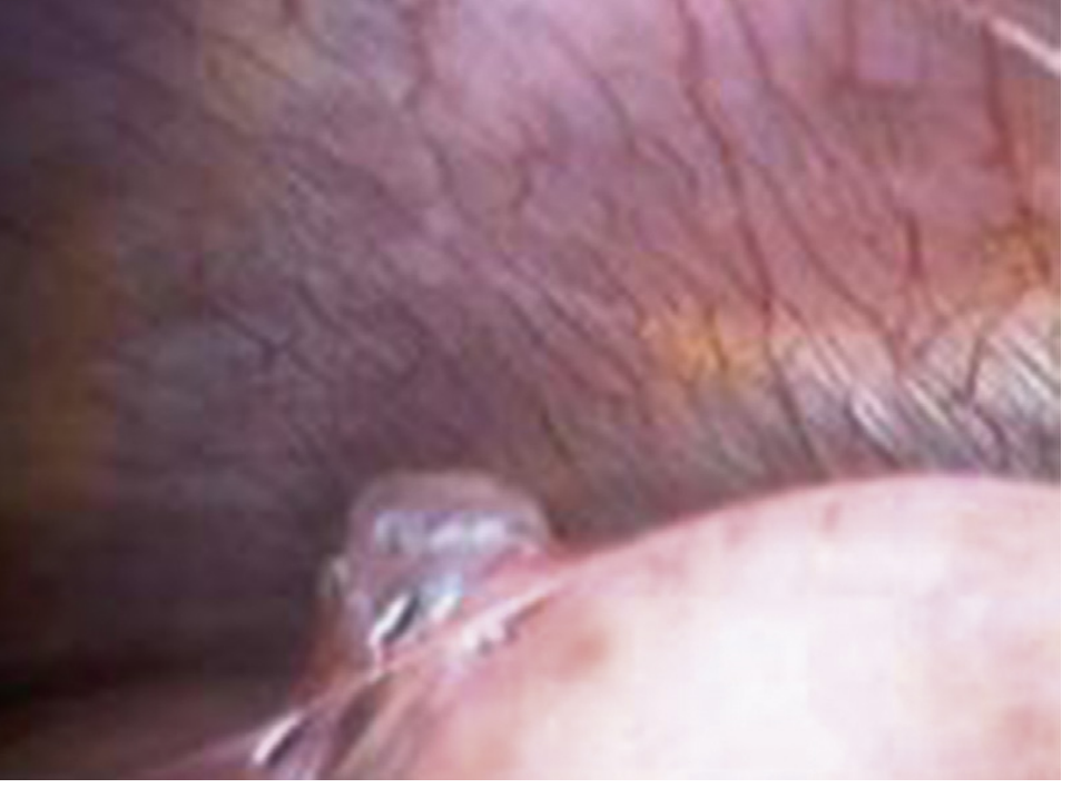
Figure 110.2 - Thoracoscopic view: small, whitish bleb sitting on the visceral pleural surface (Murray & Nadel's, Ch. 110)
Why Do Blebs Form?
- Smoking causes small airways inflammation → weakens the visceral pleura or dilates distal airways → increased airway pressure
- Matrix metalloproteinases (MMP-2, MMP-7, MMP-9) are overexpressed in PSP patients, damaging the basement membrane
- "Pleural porosity" - microscopic pores in the pleura may leak air even when no visible bleb ruptures (Noppen's fluorescence study showed high-grade porosity only in PSP patients)
Teacher's pearl: Bleb rupture was the classic explanation, but we now know many patients have NO visible bleb at thoracoscopy. The concept of diffuse pleural porosity is actually more important than bleb rupture alone.
LECTURE 3: Classification - The Big Picture
PNEUMOTHORAX
├── SPONTANEOUS (no trauma)
│ ├── PRIMARY (PSP) - no underlying lung disease
│ └── SECONDARY (SSP) - underlying lung disease
├── TRAUMATIC
│ ├── Penetrating / Blunt
│ ├── Iatrogenic
│ └── Open (sucking chest wound)
└── TENSION - life-threatening emergency
LECTURE 4: Primary vs Secondary - Who Gets It?
Primary Spontaneous Pneumothorax (PSP)
Classic patient: Tall, thin, young male smoker (20-30 years old). Ask yourself - why tall and thin? Because the lung apex has the greatest hydrostatic stress and is farthest from the hilum (reduced perfusion) → blebs form at the apex preferentially.
Recurrence risk: ~50% after a first episode - important for counselling.
Secondary Spontaneous Pneumothorax (SSP)
SSP is far more dangerous because the patient has no pulmonary reserve to compensate. The table below (from Murray & Nadel's) lists the causes:
| Frequency | Causes |
|---|---|
| Common | COPD (UK), Pulmonary tuberculosis (worldwide) |
| Uncommon | Cystic fibrosis, Lung cancer, Pneumocystis jirovecii |
| Rare - ILD | IPF, Sarcoidosis, Langerhans cell histiocytosis, LAM |
| Rare - CTD | Rheumatoid arthritis, Scleroderma, Ankylosing spondylitis, Marfan syndrome |
| Rare - Familial | Birt-Hogg-Dubé (BHD) syndrome, Ehlers-Danlos, Homocystinuria |
| Special | Catamenial pneumothorax (thoracic endometriosis) |
Teacher's tip: Two clinical pearls worth memorising:
- Young woman (30-40 yrs) + recurrent PTX → think LAM (lymphangioleiomyomatosis) - PTX may be the first presentation
- Recurrent PTX in woman of childbearing age, timed with menstruation → think Catamenial pneumothorax (thoracic endometriosis)
LECTURE 5: Clinical Features - How Does the Patient Present?
Symptoms
- Sudden onset pleuritic chest pain (ipsilateral)
- Breathlessness (proportional to size and underlying lung reserve)
- May be asymptomatic in small PSP in a young healthy person
Signs (from the affected side)
- Increased hemithorax size (expanded chest wall due to air)
- Decreased expansion on the affected side
- Hyperresonant percussion (tympany - air doesn't dampen the sound)
- Absent/diminished breath sounds
Signs of TENSION (emergency!)
- All of the above PLUS:
- Tracheal deviation away from the affected side
- Raised JVP (obstruction of venous return)
- Hypotension + tachycardia (obstructive shock)
- Cyanosis
Remember: Tracheal deviation is a LATE sign. Don't wait for it. If a patient has hypotension + absent breath sounds on one side after trauma or in ICU on a ventilator, assume tension until proven otherwise and decompress immediately.
LECTURE 6: Diagnosis - What Do You See on Imaging?
Chest X-Ray (CXR) - Your First Tool
The hallmark is a visible visceral pleural line with complete absence of lung markings beyond it.
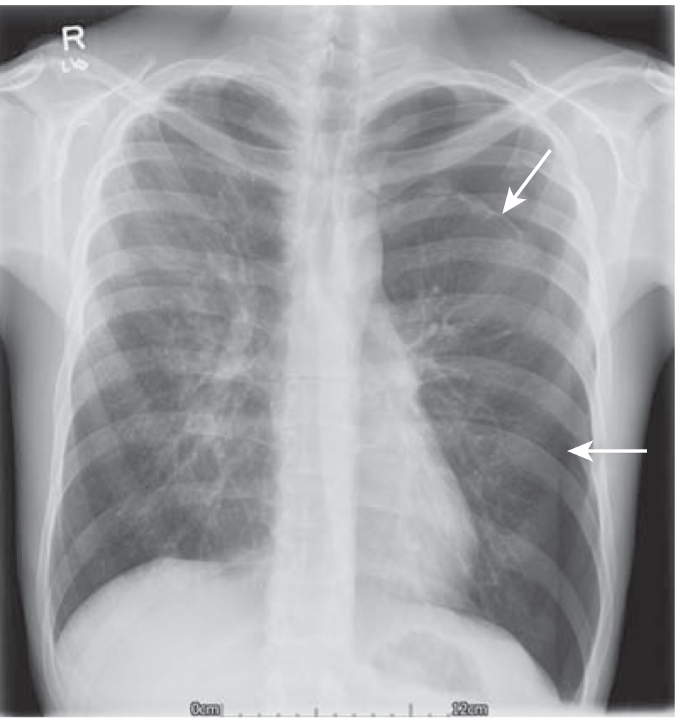
Figure 110.3 - Left-sided pneumothorax. White arrows mark the visceral pleural line. Note the absence of lung markings lateral to the line. No mediastinal shift = not under tension. (Murray & Nadel's, Ch. 110)
How to Distinguish PTX from a Skinfold (Common exam trap!)
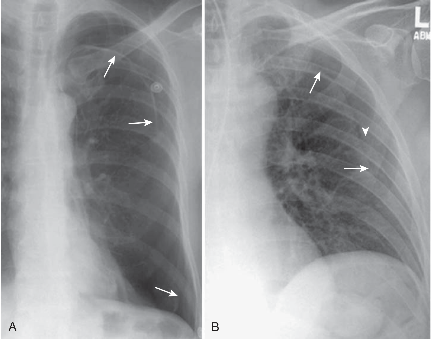
Figure 110.4 - Panel A: True pneumothorax - the line has air (lucency) on BOTH sides, no vessels beyond the line. Panel B: Skinfold - only lucency on ONE side of the edge; blood vessels can still be traced beyond the fold. (Murray & Nadel's, Ch. 110)
| Feature | Pneumothorax line | Skinfold |
|---|---|---|
| Lucency | Both sides of the line | One side only |
| Vessels beyond the line | None | Present |
| Line definition | Sharp, traceable | Poorly defined, fades |
The Deep Sulcus Sign (Supine Patient)
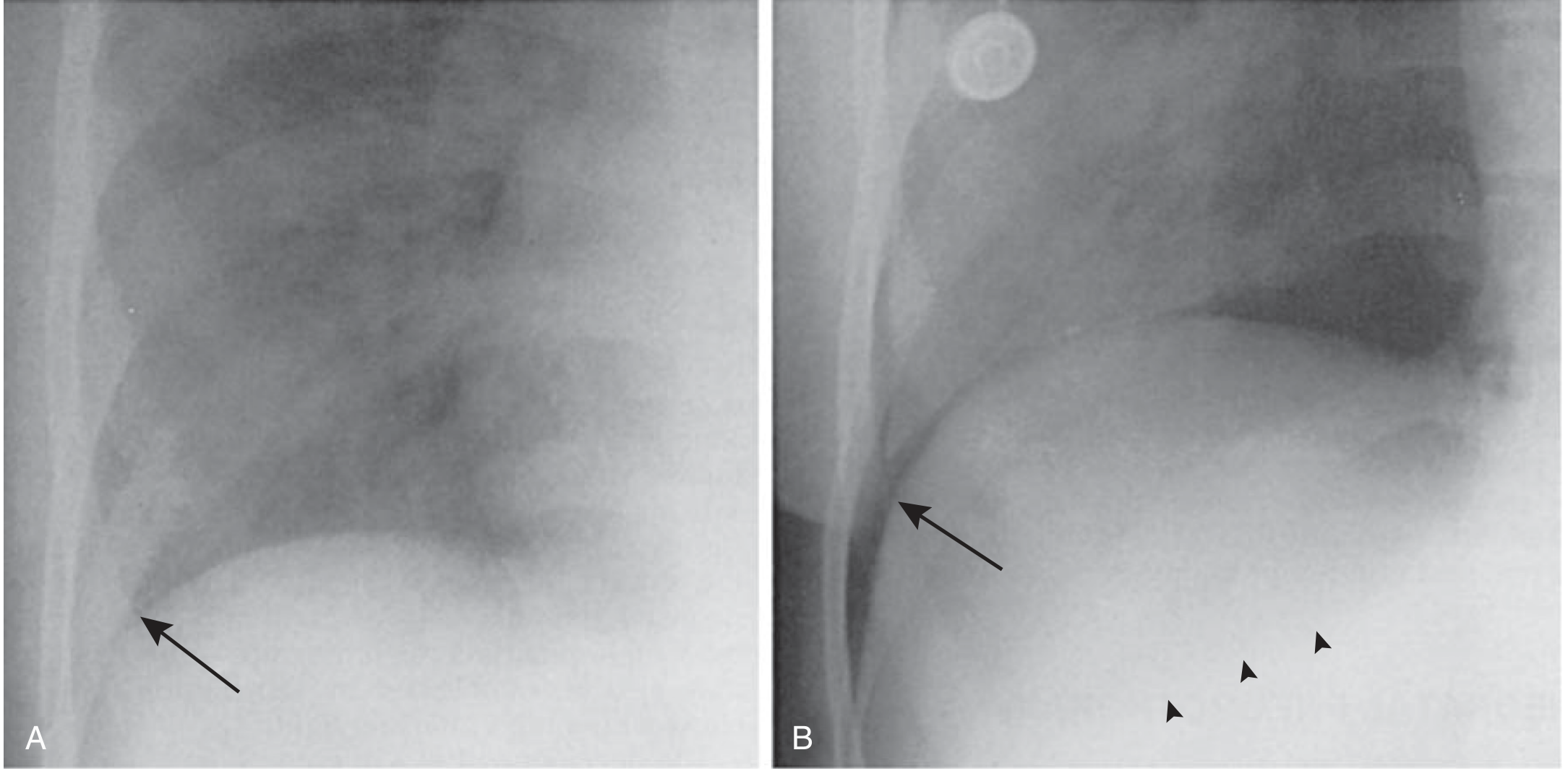
Figure 110.5 - In a supine patient, air floats anteriorly and inferiorly. Panel A: Normal costophrenic angle. Panel B: After developing a pneumothorax - the costophrenic angle deepens and sharpens (the deep sulcus sign, arrow). Always look for this in ICU/trauma CXRs. (Murray & Nadel's, Ch. 110)
Ultrasound (POCUS) - Increasingly Important
| Finding | Interpretation |
|---|---|
| Lung sliding present ("sea-shore sign" on M-mode) | Excludes PTX at that location |
| Absent sliding + A-lines ("bar code/stratosphere sign") | Suggests PTX (but not specific) |
| B-lines present | Excludes PTX (lung tissue is there) |
| Lung point (transition from sliding to no sliding) | Highly specific for PTX - marks the edge |
Ultrasound sensitivity is 78-90% vs CXR sensitivity 39-52% for PTX - especially in supine trauma patients.
CT Chest - The Gold Standard for Sizing and Differentiation
CT is mandatory when you need to distinguish bullous disease from pneumothorax - because inserting a chest tube into a giant bulla is dangerous (causes bronchopleural fistula).
BTS Size Classification (used clinically)
- Small: rim of air < 2 cm at the hilum level
- Large: rim of air ≥ 2 cm
LECTURE 7: The Life-Threatening Emergency - Tension Pneumothorax
This is the one you cannot miss. Here is the mechanism:
Visceral pleural tear acts as ONE-WAY VALVE
↓
Air enters pleural space with each breath
Air CANNOT escape (valve closes on expiration)
↓
Progressive rise in intrapleural pressure
↓
Compresses RIGHT VENTRICLE
Kinks / compresses SVC + IVC
↓
↓ Venous return → ↓ Cardiac output
↓
OBSTRUCTIVE SHOCK: Hypotension + Hypoxia
Important clinical fact: Hypoxia is present in 92% of ventilated patients but only 50% of spontaneously breathing patients with tension PTX (because spontaneous breathing generates negative pressure that partially compensates).
DO NOT wait for a chest X-ray if the clinical picture is clear (trauma + absent breath sounds + hypotension). Treat first, image later.
LECTURE 8: Management - A Structured Approach
Primary Spontaneous Pneumothorax (PSP)
PSP diagnosed on CXR
│
├─── Clinically stable + SMALL (< 2 cm)
│ │
│ CONSERVATIVE: Observation + High-flow O₂
│ (O₂ accelerates reabsorption ~4x)
│ Can be managed as OUTPATIENT
│
└─── Symptomatic OR LARGE (≥ 2 cm)
│
First-line: NEEDLE ASPIRATION
(or tube drainage)
│
Lung re-expands? → Discharge + follow up
│
Fails / Recurrence?
│
THORACOSCOPY + bleb stapling + PLEURODESIS
Secondary Spontaneous Pneumothorax (SSP)
Nearly all patients require tube drainage - they cannot tolerate even a small PTX due to limited reserve. Surgical pleurodesis is often needed if the patient can tolerate it. For non-surgical candidates: chemical pleurodesis.
Tension Pneumothorax
| Step | Action |
|---|---|
| 1. Immediate | Needle decompression - 5 cm angiocatheter |
| 2. Where? | 4th-5th ICS, midaxillary line (ATLS current recommendation) |
| 3. Why not 2nd ICS MCL? | Chest wall is thicker there; 38% failure rate vs 13% at midaxillary 4th-5th ICS |
| 4. Also avoids | Internal mammary artery, subclavian artery laceration |
| 5. Follow with | Formal chest tube thoracostomy |
Open Pneumothorax (Sucking Chest Wound)
When the chest wall defect approaches the size of the trachea, air preferentially enters through the wound rather than the trachea.
- Immediate management: Cover with a Heimlich (flutter) valve or 3-sided occlusive dressing (allows air out, not in)
- Definitive: Surgical repair
LECTURE 9: Preventing Recurrence - Pleurodesis
After a recurrence (or first episode in high-risk patients), the aim is to obliterate the pleural space so there is no room for air to collect.
| Method | Recurrence Rate | Notes |
|---|---|---|
| Observation alone | High (30-50%) | - |
| Chest tube only | ~30% | - |
| Thoracoscopic talc poudrage | ~5% | 2-4 g talc; no long-term sequelae; safe; preferred |
| VATS + bleb stapling + pleurodesis | ~5% | Risk: long-term post-op pain in >30%, lasting 3-18 months |
| Bulectomy alone | 27.5% at 10 yrs | Unacceptably high - MUST add pleurodesis |
| Open surgical pleurodesis | ~0% | Nearly 100% effective but rarely needed |
Talc safety: In long-term follow-up (22-35 years after talc poudrage), patients had near-normal lung function (TLC ~89% predicted) and no mesothelioma was observed. Talc is relatively contraindicated - not absolutely - in future lung transplant candidates.
LECTURE 10: Complications to Know
| Complication | Mechanism | Management |
|---|---|---|
| Tension PTX | One-way valve air trapping | Immediate decompression |
| Haemopneumothorax | Torn adhesions / vascularized bullae rupture | Drain: one tube superiorly (air), one inferiorly (blood) |
| Re-expansion pulmonary edema (RPE) | Rapid lung re-inflation → increased vascular permeability | Usually mild; supportive (O₂ + diuretics) |
| Prolonged air leak (PAL) | Visceral pleural tear not healing after >5-7 days | Wait up to 14 days (100% resolve by day 14 in PSP); consider VATS if persistent |
| Bronchopleural fistula | Failure to close, especially post-surgery | Surgical repair |
Quick Exam Summary
| Type | Who | Key Cause | Management |
|---|---|---|---|
| PSP | Young, tall, thin male smoker | Bleb/bulla rupture, pleural porosity | Observation vs aspiration; pleurodesis if recurrent |
| SSP | Any age, underlying lung disease | COPD (UK), TB (worldwide) | Tube drainage; pleurodesis |
| Traumatic | Trauma/procedures | Penetrating injury, iatrogenic | Tube drainage unless very small |
| Tension | ICU/ventilated, trauma | One-way valve mechanism | Immediate needle at 4th-5th ICS MAL → chest tube |
| Catamenial | Reproductive age women | Thoracic endometriosis | OCP/GnRH + surgery |
All textbook figures from: Murray & Nadel's Textbook of Respiratory Medicine, Chapter 110 (Pneumothorax)
Harrison's Principles of Internal Medicine, 22E (2025), p. 2313
This is a shared conversation. Sign in to Orris to start your own chat.