I now have comprehensive content from multiple authoritative textbooks. Let me compile the full, detailed response.
Difficult Airway Scores
Predicting a difficult airway before induction is one of the most important acts in perioperative and emergency medicine. No single test achieves 100% sensitivity, but combining multiple assessments substantially improves predictive accuracy. Below is a systematic review of all major scoring systems and tests.
1. Mallampati Classification
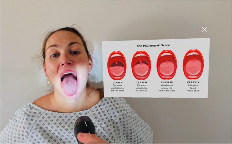
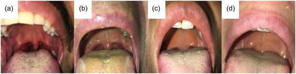
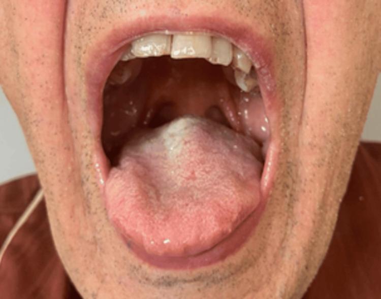
The most widely used single test. The patient sits upright, opens the mouth maximally, protrudes the tongue, and does not phonate. The visible oropharyngeal structures are classified into four classes.
| Class | Visible Structures |
|---|
| I | Entire palatal arch, bilateral faucial pillars visible down to their bases; uvula fully seen |
| II | Upper faucial pillars and most of the uvula visible |
| III | Only soft and hard palate visible |
| IV | Only the hard palate visible |
Interpretation: Classes III and IV predict a potentially difficult laryngoscopy. A meta-analysis confirmed the four-class score performs well as a predictor of difficult laryngoscopy (less reliably for difficult intubation). As a standalone test it is insufficient; it gains predictive power when combined with thyromental and sternomental distances.
Modified Mallampati - performed with the patient phonating "aah"; some evidence shows this slightly improves predictive value in obese patients.
- Morgan and Mikhail's Clinical Anesthesiology, 7e, p. 570-571
- Rosen's Emergency Medicine, Box 1.1
2. LEMON Assessment
The LEMON mnemonic is the standard tool in emergency airway management (Walls Manual). It has reasonable sensitivity and a high negative predictive value for difficult direct laryngoscopy.
L - Look Externally
Clinical gestalt - examine for external markers:
- Facial trauma, burns, hematoma
- Large incisors or prominent upper teeth
- Beard or mustache (impairs mask seal)
- Large tongue
- Morbid obesity
- Obvious masses, goiter, prior neck surgery
E - Evaluate the 3-3-2 Rule
Three geometric relationships predict glottic accessibility for direct laryngoscopy:
| Step | Measurement | Significance |
|---|
| 3 fingers | Incisor-to-incisor distance (mouth opening) | <3 fingerbreadths = restricted access |
| 3 fingers | Hyoid-mental (mentum to hyoid along mandibular floor) | <3 fingerbreadths = small submandibular space |
| 2 fingers | Thyroid notch to floor of chin | <2 fingerbreadths = high-riding larynx |
A receding mandible + high-riding larynx is exceptionally difficult for direct laryngoscopy because the operator cannot adequately displace the tongue to overcome the acute viewing angle.
M - Mallampati Scale
Classes III and IV predict moderate-to-high difficulty (see above).
O - Obstruction or Obesity
Conditions that can mechanically prevent glottic visualization or ETT passage:
- Epiglottitis, Ludwig angina, peritonsillar abscess
- Head and neck cancer, glottic polyps
- Neck hematoma, oropharyngeal edema
- Foreign body
- Obesity (contributes to rapid desaturation and difficult BMV)
N - Neck Mobility
Full flexion and extension is assessed. Severe restriction (ankylosing spondylitis, rheumatoid arthritis, cervical collar) may make direct laryngoscopy impossible.
LEMON Scoring Table (Quantitative Version)
| Criterion | Points |
|---|
| L: Facial trauma | 1 |
| L: Large incisors | 1 |
| L: Beard/mustache | 1 |
| L: Large tongue | 1 |
| E: Incisor distance <3 fingerbreadths | 1 |
| E: Hyoid-mental distance <3 fingerbreadths | 1 |
| E: Thyroid-to-mouth <2 fingerbreadths | 1 |
| M: Mallampati score >3 | 1 |
| O: Obstruction present | 1 |
| N: Limited neck mobility | 1 |
| Total | 10 |
Higher scores correlate with poor glottic visualization. Excluding the Mallampati component makes it more practical in emergency settings where the patient cannot cooperate.
A variant mnemonic LEMONS adds "S" for oxygen Saturation - not a direct predictor of difficult laryngoscopy, but a low SpO₂ shortens safe apnea time, constraining the intubation attempt. Another mnemonic, HEAVEN (Hypoxemia, Extremes of size, Anatomic challenges, Vomit/blood/fluid, Exsanguination, Neck immobility), has been proposed as a predictor covering both direct and video laryngoscopy difficulty, though its components are considered too broad for reliable clinical use.
- Rosen's Emergency Medicine, p. 24-29
- Roberts and Hedges' Clinical Procedures in Emergency Medicine, Fig. 6.4
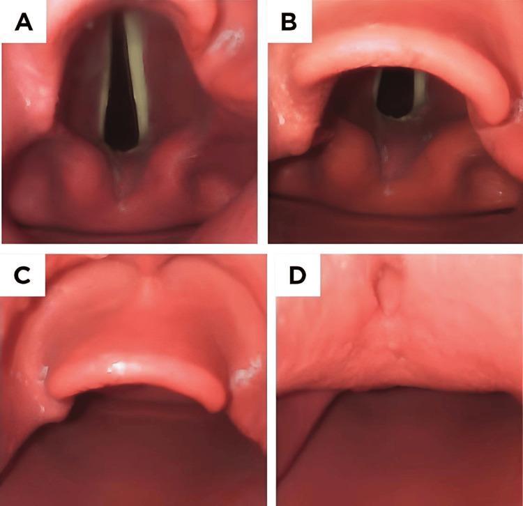
3. Cormack-Lehane Grading
Cormack and Lehane described this grading scale in 1984 to characterize the intraoperative laryngoscopic view rather than a preoperative predictor. It is used post-hoc to document and communicate airway difficulty.
| Grade | View | Intubation |
|---|
| I | Full glottis visible - vocal cords and anterior commissure seen | Easy |
| II | Partial glottis - posterior commissure or arytenoids seen; anterior commissure not visible | Usually possible |
| III | Only epiglottis seen; no glottic structures visible | Difficult; bougie often needed |
| IV | Neither epiglottis nor glottis visible | Failed airway; requires alternative technique |
Modified Yentis classification subdivides Grade II:
- IIA: partial view of glottis (anterior commissure not seen)
- IIB: only arytenoids or posterior vocal cords visible
Grades IIB and III are associated with a significantly higher incidence of failed intubation. Grade IV requires an alternate method.
A related scale - the POGO (Percentage of Glottic Opening) score - quantifies what percentage of the vocal cord length (anterior commissure to arytenoid notch) is visible. POGO has higher interobserver reliability than Cormack-Lehane and is more useful in research studies.
- Miller's Anesthesia, 10e, p. 5911-5912
4. Intubation Difficulty Scale (IDS)
Introduced by Adnet et al. (Anesthesiology 1997), the IDS is a post-intubation numeric score quantifying the overall complexity of an intubation. It is more useful for research documentation and intraoperative records than as a preoperative prediction tool.
Seven descriptors, each scored:
| Parameter | Score |
|---|
| N1 - Number of supplementary intubation attempts beyond the first | 1 per attempt |
| N2 - Number of supplementary operators beyond the first | 1 per operator |
| N3 - Number of alternative techniques used (video laryngoscope, bougie, fiberoptic, etc.) | 1 per technique |
| N4 - Cormack-Lehane grade minus 1 | 0-3 points |
| N5 - Lifting force used (normal = 0, increased = 1) | 0 or 1 |
| N6 - External laryngeal manipulation applied (no = 0, yes = 1) | 0 or 1 |
| N7 - Vocal cord position (abducted/not seen = 0; adducted = 1) | 0 or 1 |
IDS = N1 + N2 + N3 + N4 + N5 + N6 + N7
-
IDS = 0: no difficulty
-
IDS 1-5: minor difficulty
-
IDS >5: moderate-to-major difficulty
-
Miller's Anesthesia, 10e, Airway Management in Otolaryngology chapter
5. Wilson Risk Sum Score
Proposed by Wilson et al. (1988), this multivariate score assigns points to five preoperative physical characteristics:
| Factor | 0 | 1 | 2 |
|---|
| Weight | <90 kg | 90-110 kg | >110 kg |
| Head/neck movement | >90° | ~90° | <90° |
| Jaw movement (inter-incisor gap + mandibular subluxation) | IG>5 cm or SL+ve | IG<5 cm or SL=0 | IG<5 cm and SL-ve |
| Receding mandible | Normal | Moderate | Severe |
| Buck teeth | Normal | Moderate | Severe |
Maximum score = 10. A score ≥ 2 was associated with difficult intubation in the original study. Its sensitivity is limited when used alone.
6. El-Ganzouri Risk Index (EGRI)
A multivariate preoperative score incorporating seven variables. A score ≥ 4 identifies patients at high risk of difficult intubation.
| Variable | Score |
|---|
| Mouth opening (inter-incisor distance >4 cm = 0; 3-4 cm = 1; <3 cm = 2) | 0-2 |
| Thyromental distance (>6.5 cm = 0; ≤6.5 cm = 1) | 0-1 |
| Mallampati class (I = 0; II = 1; III/IV = 2) | 0-2 |
| Neck movement (>90° = 0; ~90° = 1; <90° = 2) | 0-2 |
| Ability to prognath (can advance lower teeth in front of upper = 0; edge-to-edge = 1; cannot = 2) | 0-2 |
| Body weight (<90 kg = 0; 90-110 kg = 1; >110 kg = 2) | 0-2 |
| History of difficult intubation (none = 0; questionable = 1; definite = 2) | 0-2 |
- Miller's Anesthesia, 10e - described as one of the models using several risk factors that "improve predictive value of airway assessment," p. 5851
7. Thyromental Distance (TMD) and Other Individual Measurements
| Test | Cut-off | Significance |
|---|
| Thyromental distance | <6.5 cm (or <3 fingerbreadths) | Reduced mandibular space; predicts anterior larynx |
| Sternomental distance | <12.5 cm | Limited neck extension |
| Mouth opening (interincisor gap) | <3 cm (or <2 fingerbreadths) | Restricted access |
| Neck circumference | >43 cm (17 inches) | Better predictor than BMI alone |
| Upper Lip Bite Test (ULBT) | Lower incisors cannot cover the upper lip | Predicts difficult laryngoscopy with higher specificity and less interobserver variability than Mallampati |
The ULBT has three classes:
- Class I: lower incisors bite above vermilion border of upper lip
- Class II: lower incisors bite below vermilion border
- Class III: lower incisors cannot bite the upper lip
8. Predictors of Difficult Mask Ventilation (DMV)
Predicting difficult mask ventilation is as important as predicting difficult intubation. The OBESE mnemonic covers key predictors:
| Factor |
|---|
| Obesity (BMI > 26 kg/m²) |
| Beak (beard) |
| Edentulous |
| Snoring / obstructive sleep apnea |
| Elderly (>55 years) |
Additional predictors: limited mandibular protrusion, Mallampati III/IV, presence of a neck mass.
9. ASA Difficult Airway Algorithm - 11-Point Assessment
The 2003 (and updated) ASA Difficult Airway Algorithm recommends an 11-point preoperative airway examination:
- Length of upper incisors
- Relationship of upper and lower incisors (prognathism/retrognathism)
- Inter-incisor distance on maximal mouth opening
- Visibility of uvula (Mallampati class)
- Shape of the palate (narrow/arched)
- Compliance of mandibular space
- Thyromental distance
- Length of the neck
- Thickness/circumference of the neck
- Range of motion of the head and neck
- Overall clinical gestalt
Summary: When to Use Each Score
| Score | Best Setting | When to Use |
|---|
| Mallampati | Elective/pre-op | Always; combine with other tests |
| LEMON | Emergency/ED | Rapid bedside assessment before RSI |
| ULBT | Elective/pre-op | Higher specificity; less observer variation |
| Wilson Score | Elective/pre-op | Multivariate preoperative screening |
| EGRI | Elective/pre-op | Comprehensive preoperative risk stratification |
| Cormack-Lehane | Intraoperative | Documenting and communicating laryngoscopic view |
| POGO | Intraoperative / research | More precise; better interobserver reliability |
| IDS | Post-intubation | Documenting intubation complexity; research |
Key principle: No single test has 100% sensitivity. Individual tests have limited sensitivity and positive predictive value, but a good negative test reliably predicts ease of intubation. Combining multiple tests (especially Mallampati + TMD + neck mobility + mouth opening) substantially improves prediction. A previous history of difficult intubation remains one of the most predictive single factors. - Miller's Anesthesia, 10e, p. 5845; Morgan and Mikhail's Clinical Anesthesiology, 7e, p. 571