
Knee osteoarthritis stages
Reading File
Knee osteoarthritis (OA) is classified using the Kellgren-Lawrence (KL) grading system (Grade 0-4) based on radiographic findings. Let me pull the full detail from the medical library.
Searching sources...
Finding Sources
Finding Sources
Searching PubMed
"knee osteoarthritis" AND "Kellgren-Lawrence" AND staging
Finding Sources
Finding Sources
Reading File
Reading File
Finding Sources
Reading File
Reading File
Finding Sources
Finding Sources
Reading File
Reading File
Loading Image
Here is a complete, textbook-grounded overview of knee osteoarthritis staging.
Knee Osteoarthritis - Staging
Kellgren-Lawrence (KL) Radiographic Grading System
The Kellgren-Lawrence (KL) scale is the most widely used system for grading OA severity on plain radiographs. It is a 5-point scale (Grade 0-4), sometimes described as a "five-point scale" in the literature. The key radiographic features assessed are: osteophytes, joint space narrowing (JSN), subchondral sclerosis, and bony deformity.
| Grade | Name | Radiographic Features |
|---|---|---|
| 0 | Normal | No radiographic features of OA |
| 1 | Doubtful | Doubtful narrowing of joint space; possible osteophytic lipping |
| 2 | Mild | Definite osteophytes; possible (doubtful) joint space narrowing |
| 3 | Moderate | Multiple moderate osteophytes; definite narrowing of joint space; some sclerosis; possible deformity of bone ends |
| 4 | Severe | Large osteophytes; marked narrowing of joint space; severe sclerosis; definite deformity of bone ends |
"Among the most well known is the Kellgren and Lawrence five-point scale."
- Firestein & Kelley's Textbook of Rheumatology, 2-Volume Set
Clinical Interpretation of Grades
-
KL 0-1 = Pre-radiographic / early OA. Draft classification criteria define "early OA" as KL grade 0 or 1, combined with either patient-reported symptoms (KOOS ≤85% in ≥2 categories) or clinical findings (joint line tenderness, crepitus). These criteria still require validation.
-
KL ≥2 = "Definite" radiographic OA - this threshold is used in most epidemiologic and research studies.
-
KL 4 = End-stage disease. A dedicated study (the MOST study) explored whether KL grade 4 truly represents end-stage knee OA from a structural standpoint.
-
Rheumatology, 2-Volume Set (2022, Elsevier), p. 1662 & 1738
Radiographic Examples (KL Grade 2 and 3 Knee)
The image below shows side-by-side knee radiographs demonstrating KL Grade 3 (left) and KL Grade 2 (right), with annotations for osteophytes and medial tibiofemoral joint space narrowing:
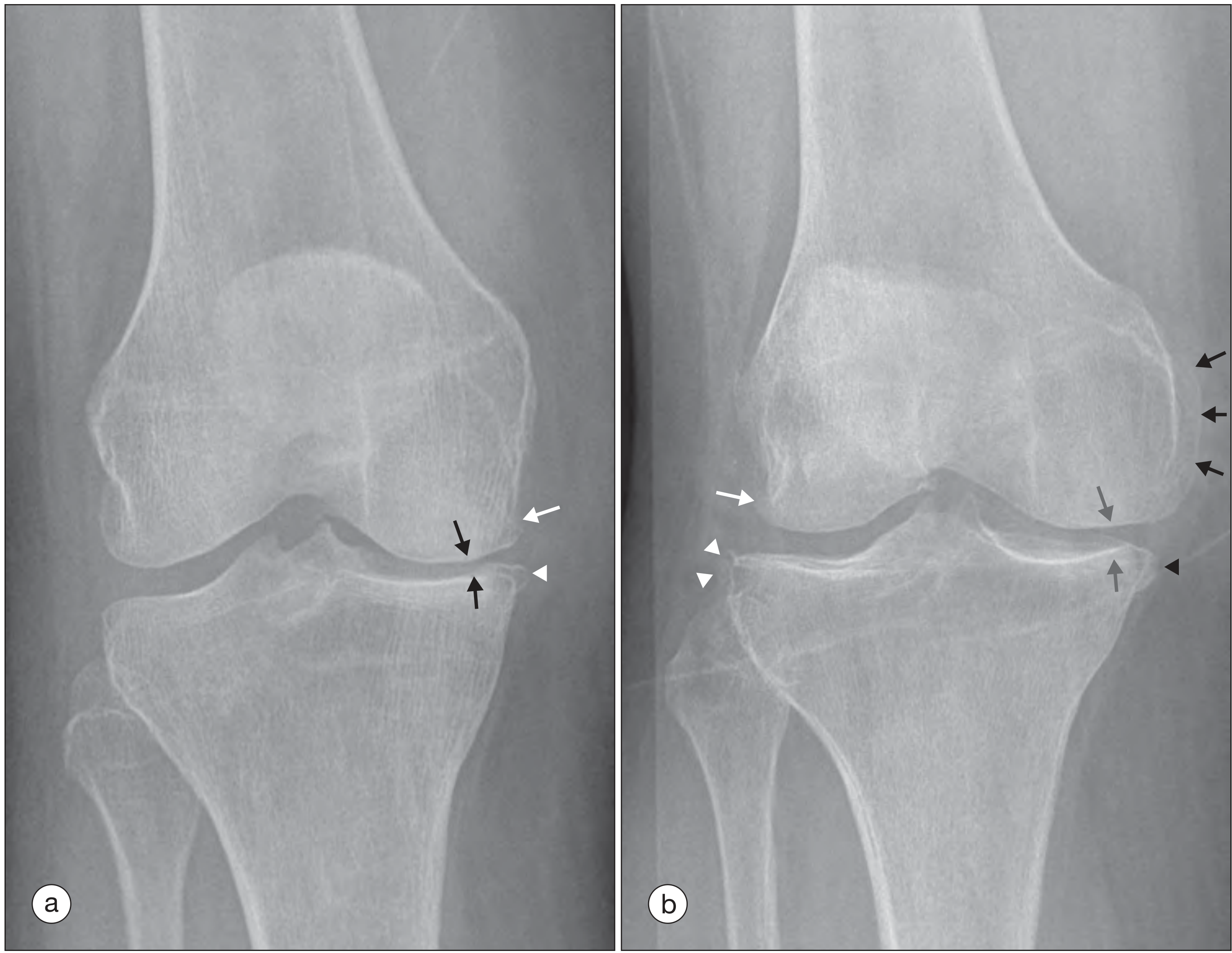
(a) KL Grade 3: Medial femoral osteophyte (OARSI grade 1), medial tibial osteophyte (OARSI grade 2), medial tibiofemoral joint space narrowing (OARSI grade 2).
(b) KL Grade 2: Lateral and medial osteophytes present at multiple sites; medial tibiofemoral joint space width reduced (OARSI grade 1).
- Rheumatology, 2-Volume Set (2022, Elsevier), Fig. 181.2
OARSI Grading (Supplementary System)
The Osteoarthritis Research Society International (OARSI) grading scheme provides finer granularity by scoring individual radiographic features (e.g., osteophytes at specific sites, joint space width per compartment) separately. It is primarily used in research and clinical trials for more accurate discrimination than the composite KL scale.
ACR Clinical Classification Criteria (Not Radiographic)
The American College of Rheumatology (ACR) criteria classify knee OA based on a combination of:
- Age >50 years
- Morning stiffness <30 minutes
- Crepitus on active motion
- Bony tenderness / enlargement
- No palpable warmth
- ± Radiographic osteophytes
These are used for diagnosis, not staging severity.
Outcome Measures by Stage
| Stage | Typical Management Approach |
|---|---|
| KL 1-2 (mild) | Physiotherapy, weight loss, NSAIDs, intra-articular injections (hyaluronic acid, corticosteroids) |
| KL 2-3 (moderate) | Above + bracing, activity modification, possibly arthroscopic procedures (limited evidence) |
| KL 3-4 (severe) | High tibial osteotomy (younger patients, unicompartmental disease), unicompartmental or total knee arthroplasty |
The AAOS recommends physical activity, low-impact aerobic exercise, and NSAIDs with strong evidence; weight loss (BMI >25) with moderate evidence, as the primary nonoperative measures regardless of KL grade. - Campbell's Operative Orthopaedics 15th Ed 2026, Table 10.4
Important Caveats
- Radiographic grade and symptom severity are poorly correlated - patients with KL grade 2-3 may have minimal symptoms, and those with KL 1 can have significant pain.
- Therapy decisions should not be based solely on radiographic appearance. - Firestein & Kelley's Textbook of Rheumatology
- MRI adds information beyond plain films (cartilage, subchondral bone marrow lesions, synovitis) and is increasingly used in clinical trials, but plain radiography with the KL scale remains the standard for clinical staging.
This is a shared conversation. Sign in to Orris to start your own chat.