
Applied anatomy of surgical limbus
surgical limbus anatomy corneoscleral junction diagram
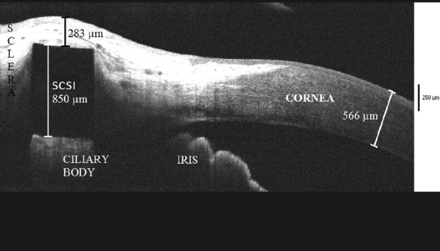
**Modality:** Anterior segment optical coherence tomography (AS-OCT). **Anatomical Region:** Anterior segment of the eye, specifically the corneoscleral junction (limbus), ciliary body, and iris. **Observed Findings:** The image displays a cross-sectional view of a supraciliary contraction segment implant (SCSI) positioned within the supraciliary space. The SCSI is visualized as a highly reflective, rectangular signal-void shadow measuring 850 µm in height. It is situated deep to the sclera and superficial to the ciliary body. **Characteristic Visual Features:** - **Scleral Thinning/Proximity:** The distance between the anterior edge of the SCSI and the external scleral surface is measured at 283 µm, indicating a superficial migration or shallow placement of the device. - **Corneal Morphometry:** Central/peripheral corneal thickness is annotated at 566 µm. - **Structural Displacement:** There is a visible anterior elevation of the overlying scleral tissue corresponding to the location of the implant. **Clinical Context:** This AS-OCT image assesses the anatomical positioning and potential migration of supraciliary implants used in the surgical treatment of presbyopia. The primary finding is the thinning of the overlying scleral cover, which may precede device exposure or extrusion.
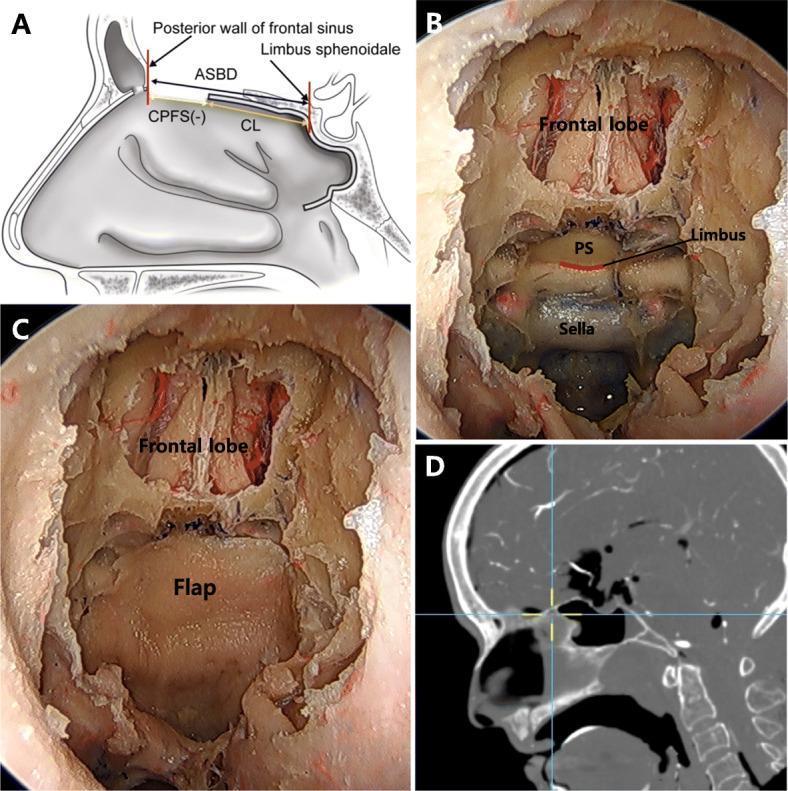
This composite image illustrates the surgical anatomy and reconstruction of the anterior skull base (ASB) following sphenoidotomy. Panel A is a sagittal anatomical diagram showing the relationship between the posterior wall of the frontal sinus and the limbus sphenoidale. It defines key surgical measurements: Anterior Skull Base Distance (ASBD), Coverage from the Limbus (CL), and Coverage at the Posterior wall of the Frontal Sinus (CPFS). Panels B and C are intraoperative 0-degree endoscopic views. Panel B shows the exposed surgical field with the frontal lobe, planum sphenoidale (PS), limbus, and sella turcica visible. Panel C demonstrates the subsequent placement of a pedicled nasoseptal flap (PNSF) over the ASB defect. Panel D is a sagittal CT-based navigation image used to confirm the spatial orientation and the endpoint of flap coverage relative to landmarks like the frontal and sphenoid sinuses. This educational material is designed for skull base surgeons to visualize the reach and limitations of nasoseptal flaps in endoscopic endonasal approaches.
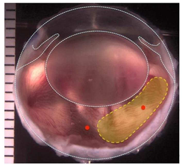
Clinical photograph and annotated diagram of a rabbit (Dutch belted) eye following a two-port partial pars plana vitrectomy. The image illustrates the surgical anatomy of the posterior segment, with the globe demarcated by a dashed white line. A yellow dotted outline highlights an elongated, horizontally oriented region in the temporal vitreous chamber where 0.3–0.4 mL of natural vitreous gel was surgically removed and replaced with a hydrogel-based vitreous substitute (1.5G_10Cop), silicone oil, or balanced salt solution (BSS). The surgical approach involved two 23-gauge trocars inserted 2 mm from the limbus for infusion and vitrector access. The surrounding tissue shows a pinkish-red hue with visible striations, while the replaced vitreous zone appears more opaque and yellowish. This visual documentation supports research into biocompatible vitreous substitutes, evaluating their effects on retinal health, intraocular pressure, and long-term functional stability.
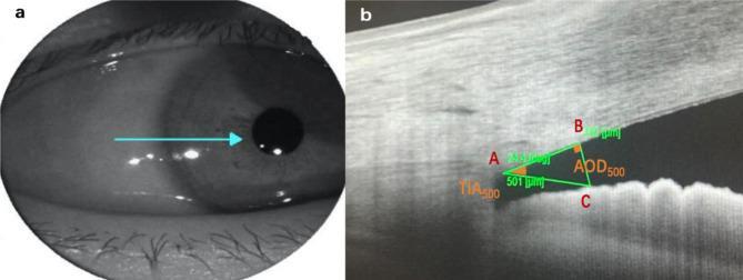
This dual-panel image demonstrates the methodology for quantifying anterior chamber angle (ACA) parameters using Swept-Source Anterior Segment Optical Coherence Tomography (AS-OCT). Panel A is an infrared clinical photograph of the eye, showing the horizontal scan line orientation directed toward the limbus. Panel B is a high-resolution AS-OCT cross-sectional scan of the iridocorneal angle. Key anatomical landmarks and measurements are annotated: Point A indicates the scleral spur, the primary reference point at the corneoscleral junction. The Anterior Opening Distance at 500 µm (AOD500) is illustrated by the vertical line segment B-C, measuring the distance from the corneal endothelium to the anterior iris surface. The Trabecular Iris Angle (TIA500) is shown as the vertex angle at point A, formed between the corneal and iris surfaces. These metrics are critical in ophthalmology for assessing glaucoma risk, characterizing angle closure, and evaluating physiological changes following surgical interventions like laser peripheral iridotomy.
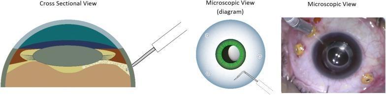
This composite educational graphic illustrates the surgical technique of pneumatic dissection of the anterior hyaloid membrane (AHM) during ophthalmic surgery. It consists of three panels: a cross-sectional diagram, a top-down microscopic diagram, and a real-time intraoperative surgical photograph. The cross-sectional view details the ocular anatomy, including the cornea, anterior chamber, iris, crystalline lens, and the posterior segment layers (sclera and choroid/retina). A 30-gauge needle is shown entering through the pars plana, with the tip positioned posterior to the lens. The top-down diagram illustrates the needle orientation relative to the iris and limbus. The clinical photograph demonstrates the actual surgical field, showing an eye with multiple sclerotomy cannulas (trocars) in place and a needle-mounted syringe performing the air injection near the pupillary margin. The primary educational focus is on the safe entry and positioning of the needle tip behind the lens within the vitreous cavity to facilitate AHM dissection while avoiding iatrogenic damage to the lens or retina.
applied anatomy surgical limbus ophthalmology incision trabecular Schlemm
limbus trabecular meshwork canal of Schlemm iridocorneal angle histology
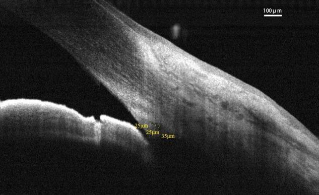
This diagnostic image is an Optical Coherence Tomography (OCT) cross-section of the human anterior segment of the eye, specifically focusing on the iridocorneal angle. The image demonstrates the anatomical relationship between the peripheral cornea, limbus, and iris. Centrally, the iridocorneal angle is visible, showing the trabecular meshwork and Schlemm’s canal (SC). Schlemm’s canal is identified as a low-reflectivity (hyporeflective) elliptical structure within the limbal sclera. Yellow quantitative annotations indicate measurements of 25µm and 35µm, representing the cross-sectional area (CSA) or dimensions of Schlemm’s canal and its proximity to the angle recess. A 100µm scale bar is provided in the upper right for spatial reference. This imaging modality is critical for evaluating aqueous humor outflow pathways in ophthalmology, particularly in the management of glaucoma and assessing surgical outcomes of micro-invasive glaucoma surgery (MIGS).
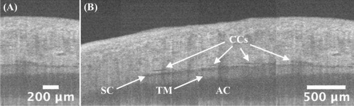
This diagnostic image displays high-resolution, circumferential spectral-domain optical coherence tomography (SD-OCT) of the human iridocorneal angle. Panel (A) provides a detailed cross-sectional view of the limbus region with a 200 µm scale bar. Panel (B) presents a larger 'panoramic' concatenated view (3.5 mm coverage) with a 500 µm scale bar, illustrating the aqueous outflow pathway. Key anatomical structures labeled include the Trabecular Meshwork (TM), visible as a band of intermediate reflectivity; Schlemm's Canal (SC), appearing as a low-intensity, horizontally oriented lumen; and multiple Collector Channels (CCs) branching from the SC. The Anterior Chamber (AC) is visualized at the bottom of the scans. These circumferential images demonstrate morphological variations and pattern changes in the outflow system, which is clinically significant for preoperative planning in glaucoma microinvasive surgeries. The imaging highlights the continuity and branching patterns of the ocular drainage structures necessary for understanding aqueous humor dynamics.
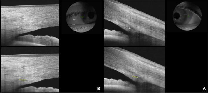
This diagnostic image set showcases Optical Coherence Tomography (OCT) of the anterior segment, specifically focusing on the iridocorneal angle and Schlemm’s canal (SC). The images compare a healthy control (Panel B) with a patient (Panel A). The cross-sectional OCT scans visualize the corneal stroma, the scleral spur, and the trabecular meshwork. Schlemm’s canal is identified as a thin, black, hypo-reflective lumen located external to the trabecular meshwork. In the healthy subject (B), the canal lumen is clearly visible and elongated, highlighted by a yellow outline in the lower-magnification view to demonstrate the Schlemm’s canal area (SCA). In the patient image (A), the SC lumen appears significantly smaller, narrower, and more compressed, indicating a reduction in both length and area. Accompanying clinical infrared photographs illustrate the probe positioning at the temporal limbus during the 3.0 mm line scan acquisition. This comparison is clinically relevant for ophthalmological studies involving aqueous humor outflow resistance and conditions such as glaucoma or Graves' ophthalmopathy.
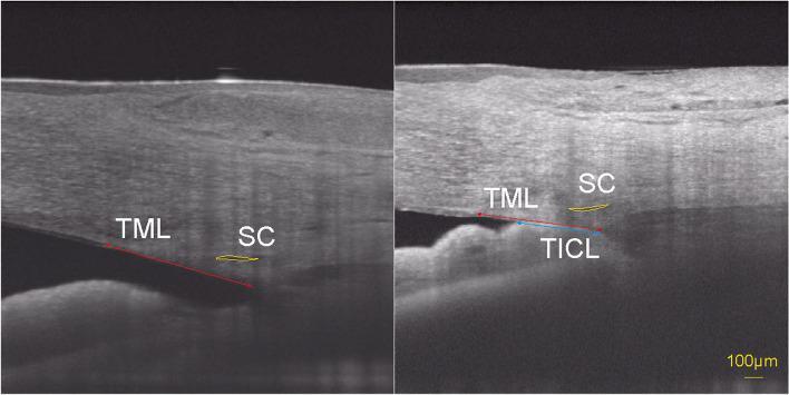
This diagnostic image consists of two side-by-side Swept-Source Optical Coherence Tomography (SS-OCT) B-scans of the human nasal iridocorneal angle. The images compare an open angle (left) with a closed angle demonstrating iridotrabecular contact (right). Key anatomical structures and measurements are annotated: Schlemm's canal (SC) is outlined with a yellow curve, showing a larger cross-sectional area in the open-angle image compared to the collapsed appearance in the closed-angle image. The trabecular meshwork length (TML) is marked by a red double-headed arrow extending from the scleral spur to Schwalbe's line. In the right image, the trabecular-iris contact length (TICL) is specifically highlighted with a blue double-headed arrow, quantifying the physical apposition between the iris and the trabecular meshwork. This comparison illustrates the morphological changes in SC parameters associated with primary angle-closure disease (PACD) and emphasizes how iridotrabecular contact correlates with reduced SC diameter and cross-sectional area, which is clinically relevant for understanding aqueous humor outflow obstruction.
https://www.ovid.com/jnls/kjop/fulltext/10.4103/kjo.kjo_26_2…
Applied Anatomy of the Surgical Limbus
Definition and Overview
Anatomical Boundaries
| Boundary | Landmark |
|---|---|
| Anterior (outer) | Corneolimbal junction - termination of Bowman's membrane |
| Posterior (outer) | Sclerolimbal junction |
| Anterior (inner) | Schwalbe's line - thickened termination of Descemet's membrane |
| Posterior (inner) | Scleral spur |
Macroscopic Appearance
The Surgical Limbus: Blue Zone and White Zone
1. Blue Zone (Anterior Zone)
- Corresponds to the area from the termination of Bowman's membrane to Schwalbe's line
- Appears bluish-grey clinically, overlying the peripheral corneal stroma
- The deep stroma here is avascular
- Surgical relevance: Incisions here are in clear corneal territory - the classic "clear corneal incision" (CCI) used in phacoemulsification cataract surgery
2. White Zone (Posterior Zone)
- Corresponds to the area from Schwalbe's line to the scleral spur
- Appears white/opaque, overlying scleral tissue
- The scleral spur is a wedge-shaped projection from the sclera that lodges the canal of Schlemm at its base
- Surgical relevance: Incisions here access the scleral tissue and the trabecular meshwork/Schlemm's canal - used in trabeculectomy, goniotomy, and MIGS (micro-invasive glaucoma surgery)
Histological Layers of the Limbus
| Layer | Structures |
|---|---|
| Outer limbus | Conjunctival epithelium, Bowman's membrane, bulbar conjunctiva, Tenon's capsule |
| Mid limbus | Transition zone - collagen changes from the regular lamellar pattern of corneal substantia propria to the irregular, interwoven bundles of scleral collagen |
| Inner limbus | Endothelium, canal of Schlemm, trabecular meshwork |
Key Histological Features
-
Epithelium: The 5-layer corneal epithelium thickens to 8-12 layers at the limbus, becoming continuous with bulbar conjunctiva. Contains the critically important limbal stem cells in the basal layer.
-
Stroma: Corneal collagen lamellae (regular, orthogonal) merge with scleral collagen (irregular, oblique bundles). This irregular, vascularized stroma is what gives the limbus its characteristic opacity.
-
Bowman's layer: Terminates at the anterior limbal margin. Its endpoint is a key surgical landmark.
-
Descemet's membrane: Terminates at Schwalbe's line (the internal anterior limbal boundary). The Descemet endothelium transitions to the trabecular endothelium here.
-
Trabecular meshwork (TM): A sponge-like network of collagenous channels lined by specialized endothelial cells that filter aqueous humor from the anterior chamber into the canal of Schlemm. - Junqueira's Basic Histology, 17e
-
Canal of Schlemm (Scleral Venous Sinus): A circumferential endothelium-lined sinus encircling the eye, embedded in the scleral stroma at the base of the scleral spur. Receives aqueous humor from the TM via bulk flow and vacuolar transport, then drains via aqueous veins of Ascher into episcleral and conjunctival veins. - Histology: Text and Atlas, Pawlina
Histology: Corneoscleral Junction (CSJ) and Trabecular Meshwork
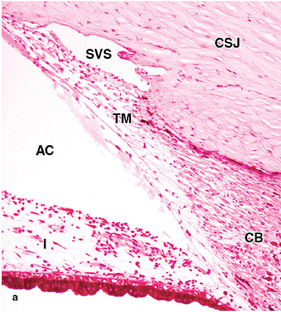
Aqueous Humor Outflow Pathway Through the Limbus

Limbal Stem Cells (LSCs): Applied Significance
- Continuously regenerate the entire corneal epithelium via centripetal migration
- Form a barrier preventing conjunctival epithelium from invading the cornea (a phenomenon called conjunctivalization)
- Their destruction (chemical burns, Stevens-Johnson syndrome, contact lens overwear) causes limbal stem cell deficiency (LSCD), manifesting as chronic photophobia, recurrent erosions, and corneal vascularization
Surgical Applications at the Limbus
1. Cataract Surgery - Site of Incision Choice
- Clear corneal incision (CCI): Placed in the blue zone, 1-1.5 mm anterior to the limbus. Avascular, rapid healing, no conjunctival disturbance.
- Scleral tunnel incision: Placed in the white zone or posterior to it. Gives a longer tunnel, more self-sealing, preferred in pediatric and complicated cases.
- Limbal incision (limbal section): At the exact corneoscleral junction. The traditional ECCE entry point.
2. Glaucoma Surgery
- Trabeculectomy: A scleral flap is raised just posterior to the limbus (white zone); a full-thickness block including TM is excised to create a fistula.
- Goniotomy/Trabeculotomy: Direct incision into the TM via the internal limbal angle, under gonioscopic control.
- Micro-invasive glaucoma surgery (MIGS): Devices (iStent, Trabectome, Kahook Dual Blade, OMNI system) access Schlemm's canal from the internal limbal angle via a temporal corneal incision.
- Viscocanalostomy/Deep sclerectomy: Ab externo (external) approach; a scleral flap at the limbus is deepened to expose and de-roof the canal of Schlemm without penetrating into the anterior chamber.
3. Vitreoretinal Surgery
- The pars plana entry (for vitrectomy and intravitreal injections) is placed 4 mm posterior to the limbus in phakic eyes and 3.5 mm posterior in pseudophakic eyes, to safely traverse the pars plana and enter the vitreous cavity without damaging the lens or retina. - Kanski's Clinical Ophthalmology, 10e
4. Trabeculectomy Bleb Management
- Late bleb leaks (post-trabeculectomy) may be revised by dissecting backwards from the limbus and advancing/resuturing conjunctiva. - Kanski's Clinical Ophthalmology, 10e
5. Corneal Transplantation
- Limbal allografts are combined with penetrating keratoplasty in severe LSCD.
- The limbus is the reference circle for trephine placement in keratoplasty.
6. Forced Duction Testing
- Fine-toothed (0.5) forceps are applied at the limbus to assess restriction of ocular movement in suspected entrapment versus paresis. - Lee's Essential Otolaryngology
Clinical Correlates
| Condition | Limbal Involvement |
|---|---|
| Glaucoma | Obstruction at trabecular meshwork or Schlemm's canal |
| LSCD | Destruction of palisades of Vogt / LSCs |
| Limbal dermoid | Congenital benign tumor at inferotemporal limbus |
| Pterygium | Fibrovascular growth from limbal conjunctiva onto cornea |
| Episcleritis/scleritis | Inflammation at the limbal/perilimbal vessels |
| Angle-closure glaucoma | Iris apposition to TM at internal limbus |
| Kayser-Fleischer rings | Copper deposition in peripheral Descemet's (at limbus) |
Summary of Key Surgical Landmarks
| Landmark | Location | Surgical Significance |
|---|---|---|
| Termination of Bowman's layer | Anterior external limbus | Anterior boundary of limbus; guides CCI placement |
| Schwalbe's line | Anterior internal limbus | Marks start of TM; goniosurgery landmark |
| Canal of Schlemm | Embedded in white zone | Target for MIGS, viscocanalostomy |
| Scleral spur | Posterior internal limbus | Marks posterior extent of TM; gonioscopy landmark |
| Palisades of Vogt | External surface | Limbal stem cell niche; avoid in peritomy |
| 3.5-4 mm from limbus | Pars plana | Vitrectomy/intravitreal injection entry |
- Junqueira's Basic Histology: Text and Atlas, 17e, pp. 1179-1181
- Histology: A Text and Atlas with Correlated Cell and Molecular Biology (Pawlina), pp. 2387-2389
- Kanski's Clinical Ophthalmology: A Systematic Approach, 10e
- Gopakumar G et al. "The Limbus and the Pupil: The Unsung Guardians of Vision." Kerala Journal of Ophthalmology 36(1):86-89, 2024