
Embryology of heart
heart embryology cardiac development stages
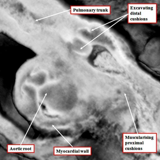
A high-magnification diagnostic image in grayscale showing the embryological development of the human cardiac outflow tract. The image identifies the early morphological stages of arterial valve formation within the intermediate component of the outflow tract. Key labeled structures include the pulmonary trunk and aortic root, illustrating the ongoing septation process. A significant educational feature is the 'excavating distal cushions,' which represent the primordial leaflets of the arterial valves undergoing tissue remodeling. The 'myocardial wall' is clearly visible, forming a myocardial turret that still encases the intermediate outflow tract at this stage. Additionally, the 'muscularising proximal cushions' are highlighted, demonstrating the transition of the cardiac jelly into muscular tissue that will eventually contribute to the ventricular infundibulum. This visual content serves as an educational tool for embryology and pediatric cardiology, focusing on the second heart field's contribution to heart development and the complex transition from simple cushions to structured semilunar valves.
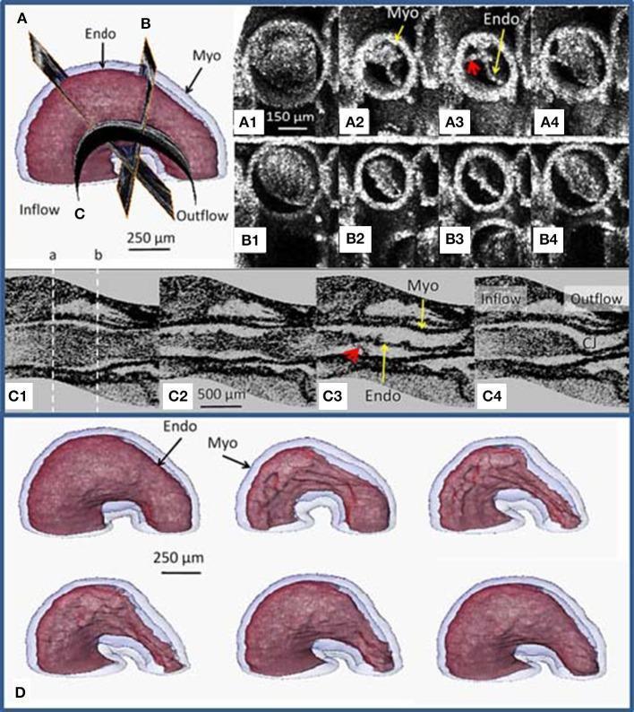
This composite image presents Optical Coherence Tomography (OCT) imaging and 3D surface renderings of a stage 13 embryonic avian heart (quail model), illustrating early cardiac looping and structural dynamics. The visual content is organized into four sections. Section A/B shows a 3D model with cross-sectional planes and corresponding time-sequence OCT slices (A1-A4 and B1-B4) that demonstrate the heart tube's eccentric deformation during the cardiac cycle; red arrows highlight tether-like connections between the myocardium (Myo) and endocardium (Endo). Section C1-C4 displays curved longitudinal OCT slices through the center of the heart tube, revealing non-uniform tissue morphology, scalloped endocardial cushions, and the spatial relationship between the inflow and outflow tracts. Section D features segmented 3D surface renderings of the myocardium (blue) and endocardium (red) at different stages of contraction, showing the endocardium folding into longitudinal ridges. This content is designed for developmental cardiology and embryology education, focusing on the mechanical relationship between tissue layers and cardiac jelly redistribution during early heart development.
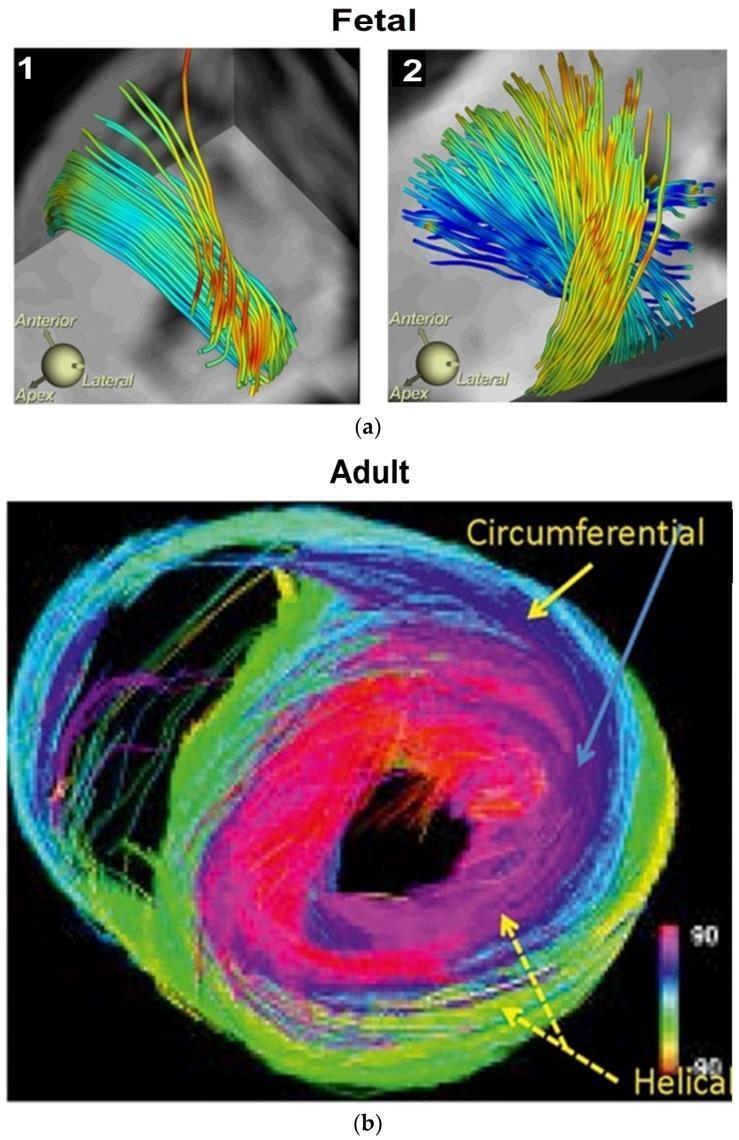
This medical visual consists of Diffusion Tensor MRI (DT-MRI) tractography images illustrating human myocardial fibrillogenesis and adult ventricular fiber architecture. Panel (a) displays fetal heart development at 10 weeks (1) and 14 weeks (2), showing the progression from simple, parallel fiber bundles to a more complex, interwoven helical arrangement of myocardial fibers. Panel (b) presents an adult heart in cross-section, utilizing a color-coded directional map to define spatial fiber orientation. The adult model highlights the distinct layers of the Helical Ventricular Myocardial Band (HVMB): right-handed helical fibers (red), left-handed helical fibers (green/yellow), and circumferential fibers (blue/purple) forming an outer wrap. A key anatomical feature demonstrated is the absence of circumferential fibers within the interventricular septum, where oblique helical fibers predominate. The progression from fetal to adult stages illustrates the development of the ventricular rope-like configuration necessary for efficient twisting and suction mechanics during the cardiac cycle. This diagnostic imaging serves as an educational tool for understanding cardiac embryology, anatomy, and functional pathophysiology.
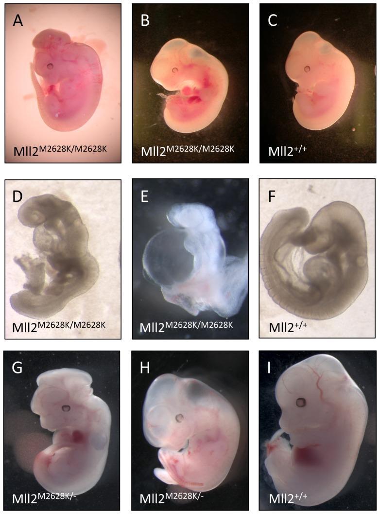
This composite of clinical photographs displays a side-by-side comparison of embryonic development in a mouse model, specifically examining the effects of the Mll2 M2628K mutation. The image is organized into three rows (A-C, D-F, G-I) comparing mutant (Mll2 M2628K/M2628K and Mll2 M2628K/-) embryos against wild-type (Mll2+/+) controls. Key developmental abnormalities in the mutants include exencephaly (protrusion of brain tissue due to neural tube defects), visible in panels A and G; severe pericardial oedema (fluid accumulation around the heart), prominent in B, E, and H; and abnormal heart looping and growth retardation shown in D and E. Panel E specifically illustrates anterior truncation defects at 9.5 dpc. In contrast, the wild-type embryos (C, F, I) demonstrate normal morphology, proper axial turning, and typical developmental progression for their respective stages (9.5 and 12.5 dpc). This figure serves as an educational tool for embryology and genetics, illustrating how specific genetic mutations disrupt midline development, cardiac morphogenesis, and neural tube closure.
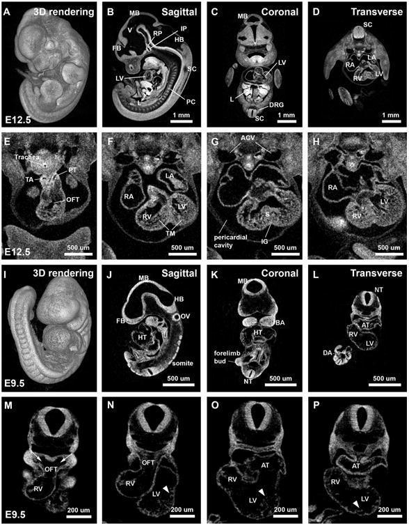
This composite diagnostic image features high-resolution 2D virtual sections and 3D reconstructions of mouse embryos at developmental stages E12.5 (A–H) and E9.5 (I–P), obtained via iodine-contrast micro-computed tomography (microCT). Panels A–D and I–L provide comparative 3D renderings and orthogonal sections (sagittal, coronal, and transverse) of the whole embryos. Key neurological structures labeled include the forebrain (FB), midbrain (MB), hindbrain (HB), ventricles (V), and neural tube (NT). In E12.5 sections, advanced organogenesis is visible, including the liver (L), primordial cartilage of vertebrae (PC), and dorsal root ganglion (DRG). High-magnification transverse cardiac views (E–H and M–P) illustrate the progression of heart development. At E12.5, the heart exhibits clear septation with distinct right and left atria (RA/LA) and ventricles (RV/LV), along with the trabeculated myocardium (TM), interventricular septum (S), thoracic aorta (TA), and pulmonary trunk (PT). Conversely, E9.5 images depict earlier stages featuring a primitive heart tube (HT), common atrium (AT), and early trabeculae formation (arrowheads). These images serve as an educational resource for embryology and high-resolution developmental imaging.
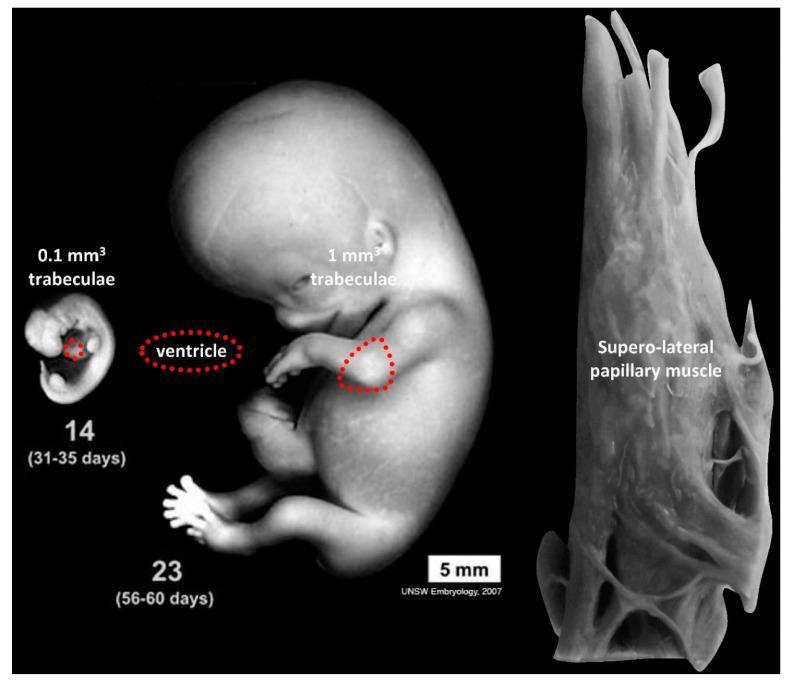
This educational image illustrates the morphological evolution of cardiac trabeculae and the ventricle across human embryonic development and into adulthood. The figure presents three distinct stages: a Carnegie stage 14 embryo (31-35 days), a Carnegie stage 23 embryo (56-60 days), and a high-resolution view of an adult supero-lateral papillary muscle. Dashed red lines on the embryos demarcate the cardiac ventricle, highlighting its growth. Quantitative annotations indicate that the volume of trabecular muscle increases by an order of magnitude from 0.1 mm3 at stage 14 to 1 mm3 at stage 23. The adult supero-lateral papillary muscle is shown as a complex, branching macroscopic structure, illustrating its developmental origin from the primitive embryonic trabecular network. The comparison emphasizes key concepts in embryology and cardiology, specifically the transition from a highly trabeculated embryonic ventricle to the specialized muscular structures of the mature heart, such as papillary muscles and the compacted ventricular wall.
cardiac septation foramen ovale atrial ventricular development
Embryology of the Heart
1. Origin of Cardiac Cells - The Heart Fields
- Primary (First) Heart Field (PHF): Pre-cardiac mesoderm migrates rostrally from the primitive streak to form a horseshoe-shaped region in the cranial part of the embryonic disc. These cells form the left ventricle and parts of the atria.
- Secondary (Second) Heart Field (SHF): A proliferative pool of mesodermal cells located in the dorsal wall of the pericardial cavity and the pharyngeal arches. SHF cells are added to both the cranial (outflow) and caudal (inflow) poles of the forming heart tube, contributing to the right ventricle, outflow tract, and portions of the atria. Disruption of SHF cell addition leads to outflow tract defects (DORV, Tetralogy of Fallot, VSD, pulmonary atresia).
- Neural crest cells migrate from the cranial neural folds into the outflow tract region (conotruncal region), where they populate the endocardial cushions and are essential for outflow tract septation.
2. Formation of the Heart Tube (~Day 18-22)
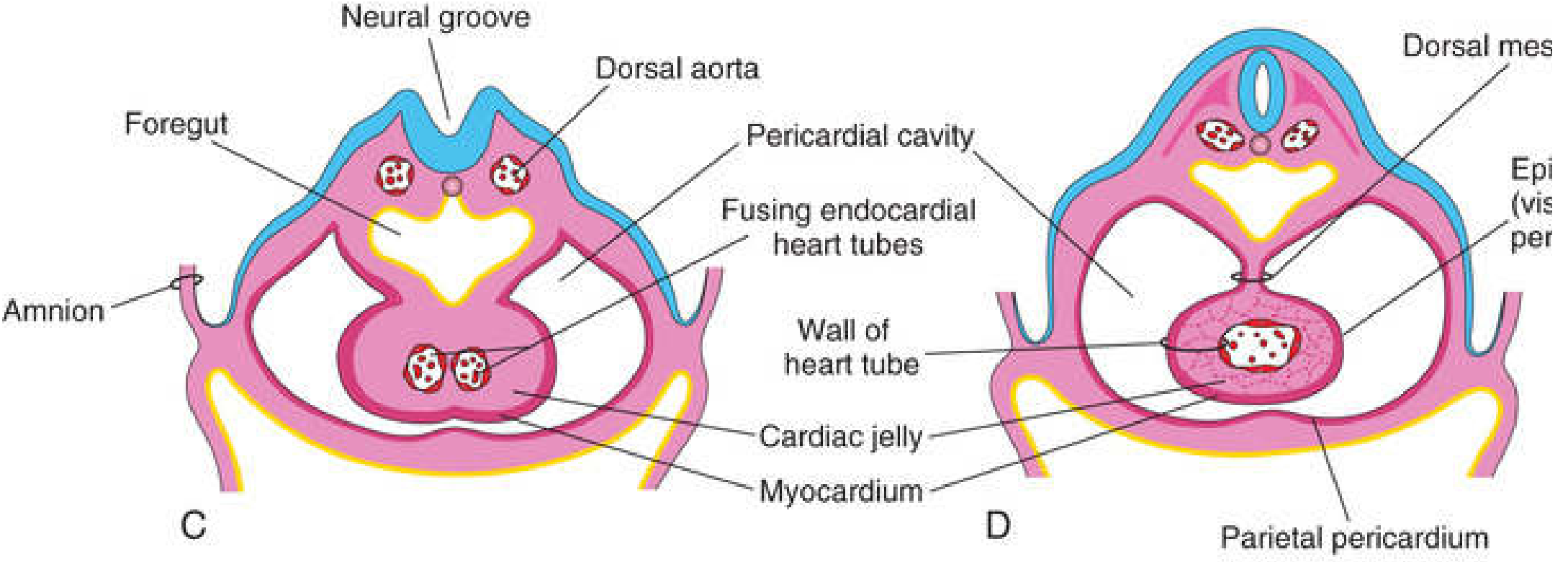
- Initially two bilateral endocardial tubes form in the cardiogenic area on either side of the midline.
- Lateral folding of the embryo brings these tubes together in the midline, where they fuse into a single tubular heart (around day 21-22).
- Simultaneously, the myocardium thickens and secretes a layer of extracellular matrix rich in hyaluronic acid called cardiac jelly, which separates it from the endothelium.
- The three-layered heart tube is thus formed:
- Endocardium - inner endothelial lining
- Myocardium - muscular middle wall
- Epicardium (visceral pericardium) - derived from the proepicardial organ, spreads over the myocardium from the external surface of the sinus venosus. The epicardium is also responsible for forming the coronary arteries (including their endothelial lining and smooth muscle).
- The heart tube is initially suspended in the pericardial cavity by the dorsal mesocardium. The central part of the dorsal mesocardium later degenerates, creating the transverse pericardial sinus.
3. Cardiac Looping (~Day 23-28)
- The straight heart tube elongates by addition of cells from the SHF.
- On day 23, the cephalic portion bends ventrally, caudally, and to the right (D-loop = dextral/rightward looping), while the atrial (caudal) portion shifts dorsocranially and to the left.
- Looping is complete by day 28.
- The looped tube has identifiable segments from cranial to caudal:
- Truncus arteriosus - leads to the aortic sac
- Bulbus cordis (= truncus arteriosus + conus arteriosus + conus cordis)
- Primitive ventricle
- Primitive atrium
- Sinus venosus
- The atrioventricular (AV) canal is the narrow junction between the common atrium and the early embryonic ventricle.
- Ballooning outgrowth of myocytes at specified points around the outer curvature of the looped tube creates the chambers (~5th week, Carnegie Stage 13).
4. Atrial Septation (~Week 5-7)
| Event | Detail |
|---|---|
| Septum primum grows | Thin crescent-shaped muscular septum grows from roof of atrium toward AV endocardial cushions |
| Foramen primum | Large gap between free edge of septum primum and AV cushions; allows R→L oxygenated blood shunt |
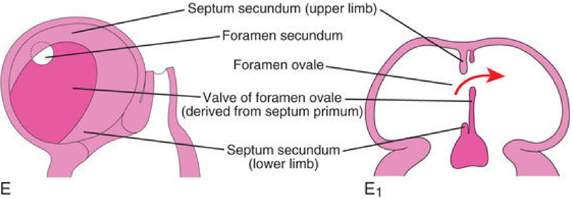
| Foramen secundum forms | Apoptosis in the upper septum primum creates perforations that coalesce → foramen secundum forms before foramen primum closes |
| Septum secundum grows | A thick, muscular second septum grows from the anterosuperior atrial wall to the right of septum primum; it is crescentic and never completely closes |
| Foramen ovale | The gap between septum secundum's free edge and the posterior atrial wall = foramen ovale. Septum primum serves as a flap valve |
| At birth | With first breath, left atrial pressure rises, pressing septum primum against septum secundum → functional closure; anatomical closure (fusion) follows |
5. AV Canal and Endocardial Cushion Development
- Endocardial cushions form in the AV canal (superior and inferior) and in the outflow tract as expansions of the cardiac jelly (ECM).
- Cells from the endocardium undergo epithelial-to-mesenchymal transition (EMT) and migrate into the AV cushions.
- Cells from neural crest populate the conotruncal cushions.
- Fusion of the AV cushions divides the single AV canal into right (tricuspid) and left (mitral) orifices.
- The cushions also give rise to the leaflets of the AV valves.
6. Ventricular Septation (~Week 5-7)
- A muscular interventricular septum begins forming as a ridge growing from the floor of the common ventricle toward the fused AV endocardial cushions.
- An interventricular foramen persists at the top between the muscular septum and the AV cushions.
- The foramen closes by contributions from three sources:
- Right bulbar ridge
- Left bulbar ridge
- Endocardial cushion tissue (+ neural crest cells)
- Together they form the membranous part of the interventricular septum (derived from endocardial cushion extension to the right side, merging with the aorticopulmonary septum and the muscular septum).
- After closure, the pulmonary trunk communicates with the right ventricle and the aorta communicates with the left ventricle.
7. Outflow Tract Septation - Bulbus Cordis Partitioning
-
The bulbus cordis has three parts:
- Truncus arteriosus - forms roots and proximal aorta and pulmonary artery
- Conus arteriosus (conus cordis) - forms outflow tracts of both ventricles
- Trabeculated part - becomes the right ventricle
-
Two conotruncal/bulbar ridges (populated by neural crest cells) grow spirally and fuse to form the aorticopulmonary septum.
-
Because the ridges grow in a spiral (helical) fashion, the resulting aorta and pulmonary trunk are spirally twisted around each other.
-
The bulbus cordis is incorporated into the right ventricle (conus arteriosus becomes the infundibulum/outflow tract of RV).
-
Failure of neural crest cell migration into the conotruncal region causes transposition of the great arteries, persistent truncus arteriosus, tetralogy of Fallot, etc.
8. Sinus Venosus and Venous Development
- The sinus venosus initially receives three paired veins: vitelline (omphalomesenteric), umbilical, and common cardinal veins.
- Due to left-to-right shunting:
- The right sinus horn enlarges and is incorporated into the right atrium as the sinus venarum (smooth-walled posterior part of RA).
- The left sinus horn regresses to become the coronary sinus and the oblique vein of the left atrium.
- The right and left venous valves guard the entrance of the sinus venosus into the atrium; the right valve contributes to the crista terminalis, valve of inferior vena cava (Eustachian valve), and valve of coronary sinus (Thebesian valve).
9. Development of the Conducting System
- The SA node develops from cells of the right sinus horn where it joins the right atrium.
- The AV node and bundle of His develop from cells of the AV canal myocardium.
- The conduction system is established early, consistent with the heart's function from day 22.
10. Clinical Correlates
| Defect | Embryological Basis |
|---|---|
| Atrial Septal Defect (ASD) - Secundum type | Excessive resorption of septum primum OR inadequate development of septum secundum; associated with NKX2.5 mutations |
| Patent Foramen Ovale (PFO) | Failure of septum primum and secundum to fuse postnatally (~25% of population) |
| VSD | Failure of muscular or membranous interventricular septum to close (most common congenital heart defect) |
| Transposition of Great Arteries | Failure of aorticopulmonary septum to spiral; associated with SHF or neural crest cell disruption |
| Tetralogy of Fallot | Unequal division of outflow tract by displaced conotruncal septum; linked to SHF elongation failure |
| Persistent Truncus Arteriosus | Failure of aorticopulmonary septum to form; neural crest cell defect |
| Dextrocardia / Situs Inversus | Failure of normal D-looping (L-loop instead); disruption of laterality pathway |
| Ebstein Anomaly | Abnormal tricuspid valve development from AV endocardial cushions |
| Holt-Oram Syndrome | TBX5 mutation - ASD + radial limb defects (autosomal dominant, 1/100,000) |
| Down Syndrome (Trisomy 21) | 50% have CHD, most commonly complete AV canal defect |
Summary Timeline
| Week | Key Events |
|---|---|
| Week 3 | Cardiogenic mesoderm forms; bilateral heart tubes appear |
| Week 3-4 (Day 22) | Heart tubes fuse; tubular heart begins beating |
| Week 4 (Day 23-28) | Cardiac looping (D-loop); chamber bulging begins |
| Week 5-6 | Atrial septation begins (septum primum); endocardial cushions fuse |
| Week 5-7 | Ventricular septation; outflow tract septation |
| Week 7-8 | Membranous IVS closes; heart structurally complete |
| Birth | Functional closure of foramen ovale; ductus arteriosus closes |
- The Developing Human: Clinically Oriented Embryology (Moore & Persaud), Ch. 13
- Langman's Medical Embryology, Ch. 13
- Braunwald's Heart Disease, Ch. 82 (Congenital Heart Disease)

Explain
Your Notes Explained
Topic: Embryology of the Pericardium
1. The Central Diagram - Lateral Plate Mesoderm (LPM) Splitting
- Ectoderm = outer layer of the embryo
- Endoderm = inner layer lining the gut
- The green-shaded mesoderm in the middle = Lateral Plate Mesoderm (LPM)
| Layer | Full Name | What it Forms |
|---|---|---|
| Somatic / Somatopleuric LPM (upper layer) | Somatic lateral plate mesoderm | Lines the body wall (parietal layer) |
| Splanchnic / Splanchnopleuric LPM (lower layer, with heart symbol) | Splanchnic lateral plate mesoderm | Surrounds the gut and viscera (visceral layer) |
2. Left Side - Septum Transversum → Fibrous Pericardium
Septum Transversum → Rises to become → Fibrous Pericardium
- Part of it descends to form the central tendon of the diaphragm (shown in the right diagram with the arrow labeled "Central tendon of diaphragm")
- Part of it migrates upward (cranially) to surround the heart and forms the fibrous pericardium
3. Boxed Note (Bottom Left) - Somatopleuric LPM
Somatopleuric LPM → Parietal layer of serous pericardium
4. Right Diagram - The Heart and its Pericardial Layers
- Derived from the Splanchnopleuric LPM (same layer that gives rise to the heart itself)
- Covers the outer surface of the heart directly
- Derived from Somatopleuric LPM
- Lines the inside of the fibrous sac
"Two layers continuous with each other at root of great blood vessels"
Summary in One Table
| Structure | Embryological Origin |
|---|---|
| Myocardium (heart muscle) | Splanchnopleuric LPM (cardiogenic mesoderm) |
| Epicardium (visceral pericardium) | Splanchnopleuric LPM (proepicardial organ) |
| Parietal serous pericardium | Somatopleuric LPM |
| Fibrous pericardium | Septum transversum mesoderm |
| Central tendon of diaphragm | Septum transversum |