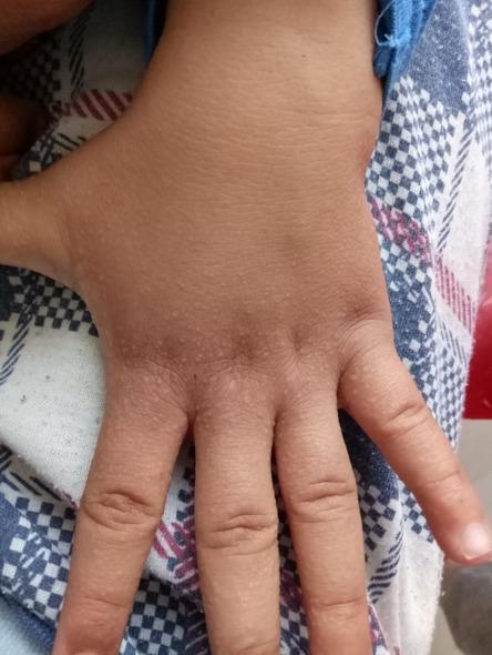
small micronodules 1 mm in size on fingers of 35 year old male. provide diagnosis
Reading File
Finding Sources
Finding Sources
Searching the Web
1mm micronodules fingers diagnosis dermatology differential
Finding Sources
Finding Sources
Finding Sources
Finding Sources
Finding Sources
Searching Images
small 1mm papules micronodules fingers skin
Reading File
Loading Image
Loading Image
Reading File
I now have sufficient information to provide a comprehensive differential diagnosis. Here is a thorough clinical answer:
Differential Diagnosis: Small 1mm Micronodules on Fingers — 35-Year-Old Male
The size (1 mm), location (fingers), and patient demographics narrow the differential considerably. Below are the most likely diagnoses in order of probability, followed by less common but important considerations.
🥇 Top Diagnosis: Lichen Nitidus
This is the classic condition presenting as multiple 1–2 mm, discrete, smooth, round, skin-colored (flesh-toned) papules on the fingers and dorsal hands in children and young adults.
Key features:
- Papules are typically 1–2 mm — exactly matching your description
- Individual lesions have a glistening, shiny surface; may be umbilicated
- Skin-colored to pinkish/red-brown; no Wickham striae (distinguishes from lichen planus)
- Predilection sites: trunk, genitalia, dorsal hands/fingers, forearms
- Usually asymptomatic (pruritus is uncommon — unlike lichen planus)
- Shows Koebner phenomenon in ~12% of cases
- No association with systemic disease
- More common in children and young adults; no sex predilection
Histopathology: Circumscribed infiltrate of lymphocytes, histiocytes, and giant cells in papillary dermis beneath a thinned epidermis — described as a "ball-in-claw" pattern
— Fitzpatrick's Dermatology, 8e, Chapter 33
🥈 Verruca Plana (Flat Warts / HPV)
- 1–4 mm, smooth, flesh-colored to slightly brownish flat-topped papules
- Caused by HPV-3, -10, -28
- Dorsal hands and fingers are classic locations
- Often appear in linear arrays (Koebner effect from scratching)
- Young adults are a common demographic
- Slightly rough surface texture on palpation
— Dermatology 2-Volume Set 5e
🥉 Granuloma Annulare (Perforating or Subcutaneous type)
- Papules/nodules on dorsal hands, fingers, and extensor extremities
- Usually larger annular rings, but perforating GA presents with small (~1–3 mm) umbilicated papules on the dorsal fingers
- Skin-colored to erythematous
- Necrobiotic collagen surrounded by palisading histiocytes on biopsy
- Associated with diabetes in disseminated forms (less relevant for localized)
— Textbook of Family Medicine 9e; Fitzpatrick's Dermatology 8e
Other Important Differentials
| Condition | Key distinguishing features |
|---|---|
| Molluscum contagiosum | 2–5 mm umbilicated pearly papules; central dimple with caseous plug; caused by poxvirus |
| Intradermal tophaceous gout | Whitish-yellowish firm papules on finger pads/joints; elevated uric acid; may be tender |
| Acquired digital fibrokeratoma | Single firm fibrous papule with collarette; usually periungual |
| Lichen planus (acral) | Violaceous polygonal papules; Wickham striae; usually pruritic |
| Rheumatoid nodules | Firm subcutaneous nodules, extensor surfaces; associated with RF-positive RA |
| Mpox (monkeypox) | ~1–2 mm pale papules on knuckles/fingers in early stage; systemic symptoms |
| Frictional lichenoid dermatitis | Small papules on knuckles; seen in atopic individuals, especially children |

Recommended Workup
| Step | Purpose |
|---|---|
| Dermoscopy | Flat warts show mosaic pattern; lichen nitidus shows homogeneous structureless pattern |
| Skin biopsy (punch) | Definitive — differentiates lichen nitidus, GA, LP, tophaceous gout |
| Serum uric acid | Rule out gout if tophi suspected |
| RF / anti-CCP | Rule out rheumatoid arthritis if nodules are periarticular |
| HPV typing (if needed) | Usually clinical for flat warts |
Summary
In a 35-year-old male with 1 mm micronodules on fingers, the most likely diagnosis is lichen nitidus given the precise size match, location, and age group. Flat warts (verruca plana) are the leading infectious differential. Perforating granuloma annulare is a close third. A skin biopsy will confirm the diagnosis if clinical features are ambiguous.
Sources: Fitzpatrick's Dermatology 8e; Andrews' Diseases of the Skin; Dermatology 2-Volume Set 5e
This is a shared conversation. Sign in to Orris to start your own chat.