
Grading of hepatic encephalopathy Signs of splenomegaly,arees of portocaval anastomosis, methods of percussion of spleen
Reading File
Now let me search the medical library for all three topics in parallel.
Finding Sources
Finding Sources
Finding Sources
Good results. Now let me read the detailed content from these sources in parallel.
Reading File
Reading File
Finding Sources
The Harrison's content on percussion was truncated. Let me read more of it, and also search for portocaval anastomosis.
Reading File
Finding Sources
Now let me search for portocaval anastomosis more specifically, and also get the Harrison's HE grading:
Finding Sources
Finding Sources
Reading File
Finding Sources
Excellent - found an image of portosystemic collateral sites. Let me read that section and fetch the image in parallel.
Reading File
Reading File
Loading Image
I now have all the information needed. Let me also search for signs of splenomegaly more specifically:
Finding Sources
I have all the necessary content. Let me now compile a comprehensive answer.
1. Grading of Hepatic Encephalopathy (HE)
West Haven Criteria (WHC) - the most widely used system
| Grade | Intellectual Function | Neuromuscular Function | Classification |
|---|---|---|---|
| 0 | Normal | Normal | Unimpaired |
| Minimal (MHE) | Normal clinical exam; subtle changes in work or driving | Minor abnormalities on psychometric/visual perception tests | Covert HE |
| Grade 1 | Personality changes, attention deficits, irritability, depressed state, shortened attention span | Tremor, incoordination, altered sleep rhythm | Covert HE |
| Grade 2 | Sleep-wake cycle changes, lethargy, apathy, disorientation to time, mood/behavioral changes | Asterixis, ataxic gait, slow/slurred speech | Overt HE |
| Grade 3 | Somnolence, confusion, gross disorientation, amnesia | Muscular rigidity, nystagmus, clonus, Babinski sign | Overt HE |
| Grade 4 | Stupor and coma | Oculocephalic reflex, unresponsiveness to noxious stimuli | Overt HE |
- Grades 0 and Minimal = Covert HE (CHE) - no clinical evidence of mental change but abnormal special tests
- Grades 2-4 = Overt HE (OHE) - clinically obvious
SONIC Classification (Spectrum of Neurocognitive Impairment in Cirrhosis)
A newer framework that divides patients into:
- Unimpaired - no clinical, neurophysiologic, or neuropsychometric abnormalities
- Covert HE - clinically normal but abnormal cognitive/neurophysiologic tests (= MHE + Grade 1)
- Overt HE - Grade 2 and above; the key distinguishing feature is disorientation to time
Types by Underlying Disease:
- Type A - associated with acute liver failure (ALF)
- Type B - associated with portosystemic bypass without intrinsic liver disease
- Type C - associated with cirrhosis and portal hypertension (most common)
Key Clinical Features:
- Asterixis (flapping tremor, elicited by wrist hyperextension) - hallmark of overt HE; present in grades 2-3, absent in coma
- EEG changes: triphasic wave patterns appear at grade 2-3, disappear in coma
- Pathophysiology: hyperammonemia → impaired blood-brain barrier, glutamine-mediated astrocyte swelling, cerebral edema
(Sources: Sleisenger and Fordtran's Gastrointestinal and Liver Disease; Current Surgical Therapy 14e; Miller's Anesthesia 10e)
2. Signs of Splenomegaly
Symptoms
- Pain and heaviness in the left upper quadrant (LUQ)
- Massive splenomegaly causes early satiety (compresses stomach)
- LUQ pain from capsule stretch, infarction, or capsule inflammation
- Visible fullness in the LUQ that descends on inspiration (massive splenomegaly)
Physical Signs
| Sign | Detail |
|---|---|
| Palpable spleen | The most important sign; normally not palpable (lies within the rib cage); recorded as cm below the left costal margin |
| Splenic notch | Medial notch palpable on the enlarged spleen - distinguishes spleen from other LUQ masses |
| Moves with respiration | Descends on inspiration - follows the diaphragm |
| Cannot get above it | Cannot insinuate fingers above the upper border (comes from under the left costal margin) |
| Dull to percussion | Percussion of Traube's space is dull |
| Auscultation | May reveal venous hum or friction rub over the spleen |
Bimanual Palpation Technique:
- Patient supine with flexed knees; examiner's left hand on lower rib cage pulling skin toward costal margin; right hand starts in left lower quadrant and moves toward left costal margin, feeling the spleen tip as the patient inspires deeply
- Bimanual palpation in right lateral decubitus adds nothing to the supine examination
Differentiating enlarged spleen from other masses:
- Has a medial notch
- Moves with respiration
- Dull to percussion (no resonant colon between spleen and anterior abdominal wall)
- Cannot get above it
(Source: Harrison's Principles of Internal Medicine 22E)
3. Areas of Portocaval (Portosystemic) Anastomosis
When portal pressure rises above ~10-12 mmHg, collateral portosystemic channels open. The classic sites are:
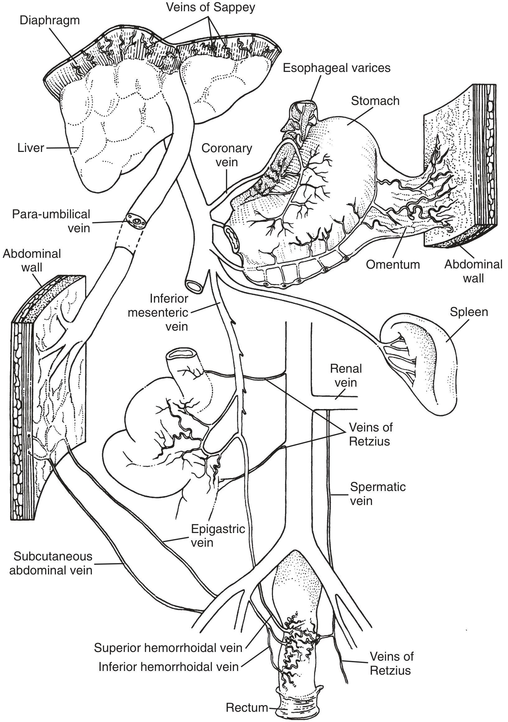
| Site | Portal Tributary | Systemic Tributary | Clinical Manifestation |
|---|---|---|---|
| Lower esophagus | Left gastric (coronary) vein | Azygos/hemiazygos veins | Esophageal varices (most dangerous) |
| Gastric fundus | Short gastric veins | Azygos system | Gastric varices |
| Umbilical region | Para-umbilical veins (along ligamentum teres) | Superficial epigastric/subcutaneous abdominal veins | Caput medusae - dilated periumbilical veins |
| Distal rectum/anus | Superior hemorrhoidal (rectal) vein (IMV) | Middle and inferior hemorrhoidal veins | Anorectal varices / hemorrhoids |
| Retroperitoneum | Veins of Retzius | Renal veins, inferior vena cava | Retroperitoneal collaterals |
| Bare area of liver/diaphragm | Veins of Sappey | Phrenic/diaphragmatic veins | Diaphragmatic varices |
| Splenorenal | Splenic vein | Left renal vein | Spontaneous splenorenal shunt |
(Sources: Maingot's Abdominal Operations; Tietz Textbook of Laboratory Medicine 7e)
4. Methods of Percussion of the Spleen
Three classic methods are described (Harrison's 22E):
1. Nixon's Method
- Patient positioned on right lateral decubitus (right side down) so spleen lies above the colon and stomach
- Percussion starts at the lower level of pulmonary resonance in the posterior axillary line, proceeding diagonally toward the lower midanterior costal margin
- Normal: upper border of dullness is 6-8 cm above the costal margin
- Splenomegaly: dullness >8 cm in an adult
2. Castell's Method
- Patient supine
- Percuss the lowest intercostal space in the anterior axillary line (8th or 9th space)
- Normal: resonant note during both expiration and full inspiration
- Splenomegaly: dull note on full inspiration (spleen comes down to occupy this space)
3. Traube's Semilunar Space Percussion
- Patient supine with left arm slightly abducted
- Borders of Traube's space: 6th rib superiorly, left midaxillary line laterally, left costal margin inferiorly
- Percuss from medial to lateral margins during normal breathing
- Normal: resonant sound throughout
- Splenomegaly: dull percussion note in Traube's space
Accuracy Notes (Harrison's):
- Sensitivity: 56-71% for palpation, 59-82% for percussion
- Reproducibility is better for palpation than percussion
- Both are less reliable in obese patients or patients who just ate (full stomach gives dullness)
- Recommended approach: percuss first; if positive, proceed to palpation - if spleen is palpable, splenomegaly is likely
- Ultrasonography is the current gold standard (normal spleen = max cephalocaudad diameter of 13 cm)
This is a shared conversation. Sign in to Orris to start your own chat.