
mastectomy with lymph node clearance and its surgical anatomy
Reading File
Mastectomy with axillary lymph node clearance is the surgical removal of the entire breast along with the axillary lymph nodes. The procedure requires precise knowledge of breast and axillary anatomy - including fascial layers, neurovascular structures, and lymphatic drainage - to achieve oncological clearance while avoiding complications like brachial plexus injury or lymphedema.
Searching sources...
Finding Sources
Finding Sources
Finding Sources
Finding Sources
Reading File
Reading File
Reading File
Reading File
Reading File
Reading File
Reading File
Reading File
Loading Image
Loading Image
I now have comprehensive information from multiple authoritative surgical textbooks. Let me compile a thorough response.
Mastectomy with Axillary Lymph Node Clearance: Surgical Anatomy and Technique
1. Surgical Anatomy of the Breast
Boundaries
The mature breast extends from the 2nd rib superiorly to the 6th rib inferiorly, from the sternal edge medially to the midaxillary line laterally. Breast tissue also extends into the axilla as the axillary tail of Spence. Posteriorly, the breast overlies portions of:
- Pectoralis major (upper medial)
- Serratus anterior (inferolateral)
- External abdominal oblique and upper rectus sheath (inferior)
For the mastectomy dissection, glandular tissue extends to the clavicle superiorly, the lateral sternum medially, the inframammary fold/rectus sheath inferiorly, and the anterior border of latissimus dorsi laterally.
- Mulholland and Greenfield's Surgery, 7e, p. 3866
Fascial Layers
- The breast parenchyma sits within the superficial fascia of the anterior thoracic wall
- Posteriorly, the deep investing fascia of pectoralis major (pectoralis fascia) forms the deep boundary - this is excised en bloc with the breast during mastectomy
- The clavipectoral fascia (deep to pectoralis minor) must be incised to enter the axilla during nodal clearance
Cooper's Ligaments
Fibrous connective bands extending from the deep fascia to the dermis. The mastectomy flap dissection is carried in the avascular plane defined by these ligaments - between breast parenchyma and subcutaneous fat. The thickness of flaps varies with individual subcutaneous tissue volume.
Breast Parenchyma
Consists of 15-20 lobes of glandular tissue, separated by fibrous stroma and adipose tissue. Each lobe is composed of 20-40 lobules, which open via lactiferous ducts that drain into lactiferous sinuses, then through the nipple. The upper outer quadrant contains the most parenchyma and is the most frequent site of breast cancer.
2. Blood Supply
The breast receives arterial supply from three main sources:
| Source | Branch | Territory |
|---|---|---|
| Internal thoracic (mammary) artery | Medial perforators (intercostal spaces II-IV) | Medial breast; most dominant supply |
| Axillary artery | Superior thoracic a., lateral thoracic a., pectoral branches of thoracoacromial a. | Lateral and upper breast |
| Posterior intercostal arteries | Lateral branches | Lateral breast |
During mastectomy, the internal mammary perforators are identified at the lateral sternal edge and controlled with clips or ties at their origin. These are the main bleeding vessels during the medial dissection.
The venous system parallels arterial supply, draining into the internal thoracic, axillary, and intercostal veins.
- Mulholland and Greenfield's Surgery, 7e, p. 3867
3. Innervation
- Sensory: Lateral and anterior cutaneous branches of intercostal nerves T2-T6 supply the breast parenchyma and skin. The upper breast is also supplied by anterior branches of the supraclavicular nerve (cervical plexus, C3-C4).
- The intercostobrachial nerve (lateral cutaneous branch of T2) crosses the axilla medially to laterally to reach the medial upper arm. It is often sacrificed during axillary dissection, causing sensory loss/numbness over the medial arm; preservation should be attempted when possible.
4. Lymphatic Drainage of the Breast
More than 75% of breast lymph drains to the axillary nodes; the remainder drains to parasternal (internal mammary) nodes. This occurs regardless of the quadrant of origin - it is not anatomically quadrant-dependent.
Three lymphatic plexuses:
- Intrinsic (primary) - interlobular and along lactiferous ducts within the gland
- Subareolar plexus - drains skin, nipple/areola, and central gland
- Deep (posterior) plexus - on the deep surface, communicates with fascia of pectoralis
- Mulholland and Greenfield's Surgery, 7e, p. 3869
5. Axillary Anatomy
The axilla is a pyramidal compartment with:
- Anterior wall: Pectoralis major, pectoralis minor, clavipectoral fascia
- Posterior wall: Subscapularis, teres major, latissimus dorsi
- Medial wall: Serratus anterior + upper 4-5 ribs
- Lateral wall: Humerus / bicipital groove
- Apex: Cervicoaxillary canal (bounded by clavicle anteriorly, scapula posteriorly, 1st rib medially) - communicates with posterior triangle of neck
- Base: Axillary fascia and skin
Axillary Lymph Node Levels (defined relative to pectoralis minor)
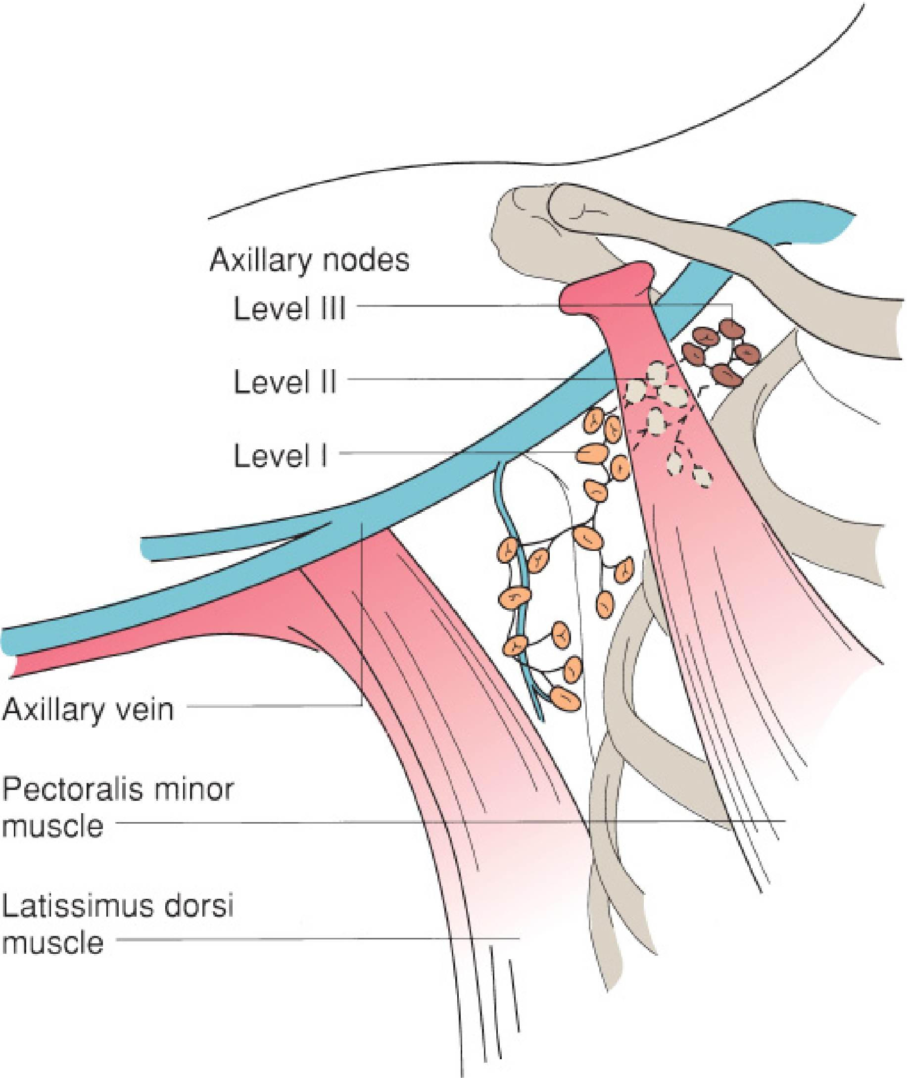
| Level | Location | Node Groups |
|---|---|---|
| Level I | Inferior and lateral to pectoralis minor | External mammary (anterior/pectoral), axillary vein (lateral), scapular (posterior/subscapular) |
| Level II | Posterior to (behind) pectoralis minor | Central nodes; some subclavicular nodes |
| Level III | Medial to pectoralis minor | Subclavicular (infraclavicular/apical) nodes |
| Rotter's nodes (interpectoral) | Between pectoralis major and minor, along lateral pectoral nerve | Sampled based on involvement |
Standard axillary lymph node dissection (ALND) for breast cancer clears levels I and II, removing at least 10 lymph nodes. Level III is cleared when there is gross involvement at level II.
- Mulholland and Greenfield's Surgery, 7e, p. 3870; Current Surgical Therapy, 14e, p. 807
6. Key Structures at Risk During ALND - Critical Surgical Anatomy
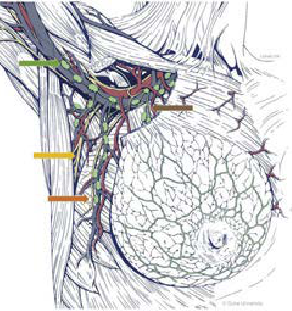
Thoracodorsal Neurovascular Bundle
- Nerve: branch of posterior cord of brachial plexus (C6, C7, C8); artery: branch of subscapular artery
- Enters the axilla in a deep posterior position, approximately halfway between the chest wall and latissimus muscle - typically in the center of the specimen
- Identified and must be preserved - injury causes weakness of shoulder adduction/internal rotation (latissimus dorsi paralysis), limiting arm movements and affecting reconstructive options (TRAM/LD flap)
- The thoracodorsal vein is a major tributary of the axillary vein; care needed to distinguish it from the anterior thoracoepigastric vein (which must be ligated)
Long Thoracic Nerve (Nerve of Bell)
- Arises from roots C5, C6, C7
- Runs vertically along the medial chest wall (serratus anterior surface), entering the muscle inferiorly
- Carefully protected against the chest wall throughout the dissection; can be pulled laterally by retractors and must be specifically sought if not visible
- Injury causes winged scapula (serratus anterior paralysis)
Intercostobrachial Nerve
- Lateral cutaneous branch of T2 intercostal nerve
- Runs horizontally (medial to lateral) across the axillary field - easily distinguished from the vertically running thoracodorsal and long thoracic nerves
- Sacrifice causes numbness/dysesthesia over the medial upper arm; preservation is attempted but often not possible
Axillary Vein
- Forms the superior boundary of the ALND
- Identified laterally first (safest approach - no critical structures laterally) by following the anterior surface of latissimus dorsi superiorly where the vein crosses its tendinous portion
- Dissection must not go superior to the axillary vein - this dramatically worsens lymphedema risk
- Small venous tributaries on the inferior aspect are clipped and divided as dissection proceeds medially
Brachial Plexus
- Located superior to the axillary vein; not typically encountered in standard levels I-II dissection
- At risk with level III dissection or aggressive superior dissection
7. Types of Mastectomy
| Type | What is Removed | Axillary Surgery |
|---|---|---|
| Simple (total) mastectomy | All breast glandular tissue + NAC | None |
| Modified radical mastectomy (MRM) | Entire breast + NAC + pectoralis fascia | Levels I-II ALND |
| Skin-sparing mastectomy | Glandular tissue + NAC; skin envelope preserved | As indicated |
| Nipple-sparing mastectomy | Glandular tissue only; entire skin envelope + NAC preserved | As indicated |
MRM is performed for biopsy-proven positive axillary nodes. It preserves both pectoralis major and minor (unlike the historical Halsted radical mastectomy which sacrificed both).
- Current Surgical Therapy, 14e, p. 778-779
8. Operative Steps: Modified Radical Mastectomy
Patient Setup
- Supine; ipsilateral arm extended and widely prepped
- General anaesthesia (regional block e.g. thoracic paravertebral block is a useful adjunct)
- Prophylactic antibiotics, sequential compression devices
Mastectomy Phase
- Incision: Broad transverse-oblique ellipse encompassing the nipple-areolar complex; orientation facilitates subsequent axillary dissection
- Flap elevation: Raise mastectomy flaps in the avascular plane between breast parenchyma and subcutaneous fat, extending to:
- Clavicle superiorly
- Lateral sternal edge medially
- Rectus sheath/inframammary fold inferiorly
- Anterior border of latissimus dorsi laterally
- Include the axillary tail of Spence during the superolateral dissection
- Chest wall dissection: Breast and pectoralis fascia are excised off the pectoralis major muscle (from superior to inferior). Small perforating vessels from pectoralis are controlled with electrocautery/clips.
- Medial hemostasis: Internal mammary perforators are identified in the intercostal spaces and ligated at the sternal border
- The breast may be left in continuity with axillary tissue for en bloc ALND
- Current Surgical Therapy, 14e, p. 807
Axillary Lymph Node Dissection Phase
- Enter the axilla: Incise the clavipectoral fascia at the lateral border of pectoralis major, progressing superiorly toward the apex
- Identify the axillary vein: Follow latissimus dorsi anteriorly/superiorly until the vein crosses its tendinous band (lateral approach - safest)
- Vein dissection: Dissect fatty and nodal tissue off the inferior aspect of the axillary vein from lateral to medial, clipping all small tributaries. Do not go superior to the vein.
- Identify thoracodorsal bundle: Located centrally/posteriorly in the axilla. Identify and protect throughout.
- Identify long thoracic nerve: Along medial chest wall, running vertically over serratus anterior. Preserve against the chest wall; ease retractors if not seen.
- Intercostobrachial nerve: Running horizontally - attempt preservation but may sacrifice for exposure
- Rotter's space: Palpate and inspect for involved Rotter's nodes between the two pectoralis muscles; the medial pectoral neurovascular bundle is identified here and may be preserved or ligated depending on nodal involvement
- Apex dissection: Connect cleared axillary vein to the chest wall below pectoralis minor. Mobilize the nodal specimen inferiorly, divide tissue between thoracodorsal and long thoracic nerves (included in specimen)
- Final sweep: Blunt sweep clears specimen from the latissimus/subscapularis floor. Anterior branches of thoracodorsal bundle are clipped and divided.
- Verification: Visually confirm or stimulate thoracodorsal and long thoracic nerves for integrity; palpate for residual adenopathy in Rotter's space and level III
- Current Surgical Therapy, 14e, p. 807-808
9. Closure and Drainage
- Two closed-suction (Jackson-Pratt) drains: one looped into the apex of the axilla, one under the mastectomy flap
- Wound closed in layers (deep dermal interrupted + subcuticular skin closure)
- Compression binder/wrap applied
- Drains removed when output falls below 30 mL per 24-hour period (sequentially, not same day)
10. Complications and Their Anatomical Basis
| Complication | Anatomy / Mechanism |
|---|---|
| Lymphedema | Disruption of axillary lymphatics; worsened by dissection above axillary vein |
| Winged scapula | Long thoracic nerve injury (serratus anterior paralysis) |
| Shoulder weakness | Thoracodorsal nerve injury (latissimus dorsi paralysis) |
| Arm numbness | Intercostobrachial nerve sacrifice (medial upper arm) |
| Seroma | Dead space in axilla post-dissection; commonest complication |
| Hematoma | Failure to control internal mammary perforators or thoracoepigastric vein |
| Skin necrosis | Thin flaps or excessive tension, especially with nipple-sparing technique |
| Chest wall numbness | Intercostal nerve branches divided during flap elevation |
Sources:
- Mulholland and Greenfield's Surgery: Scientific Principles and Practice, 7e - Surgical Anatomy of the Breast, pp. 3866-3870
- Current Surgical Therapy, 14e - Mastectomy and Axillary Lymph Node Dissection, pp. 778-809
This is a shared conversation. Sign in to Orris to start your own chat.