
CLL PBS picture
CLL chronic lymphocytic leukemia peripheral blood smear smudge cells
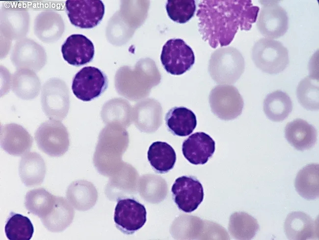
Peripheral blood smear from a patient with suspected chronic lymphocytic leukemia (CLL) shows a predominance of small, mature-appearing lymphocytes with scant cytoplasm and clumped nuclear chromatin, accompanied by fragile cells that readily disrupt during smear preparation, producing numerous smudge or basket cells visible on the slide. The image depicts a single smudge cell in the upper right, with numerous small darkly staining lymphocytes occupying the field. The smear was prepared with Wright-Giemsa stain and examined under light microscopy at high magnification (approximately 1000x total magnification with oil immersion). Clinically, CLL is defined by absolute mature lymphocytosis of ≥5.0 × 10^9/L sustained for ≥3 months, often asymptomatic and detected incidentally on complete blood count. Morphology alone is insufficient for diagnosis; immunophenotyping showing co-expression of CD5, CD19, CD23 with weak surface immunoglobulin and light chain restriction supports CLL diagnosis. Smudge cells result from fragile leukemic cells and reflect underlying cytoskeletal fragility, though they are not specific. The image illustrates typical features: monomorphic lymphocytes, scant cytoplasm, condensed chromatin, and absent prominent nucleoli. This finding has diagnostic significance, guiding further workup, including flow cytometry, cytogenetics, and molecular studies, to distinguish CLL from other chronic lymphoproliferative disorders such as small lymphocytic lymphoma (SLL), mantle cell lymphoma, prolymphocytic leukemia, and reactive lymphocytosis.
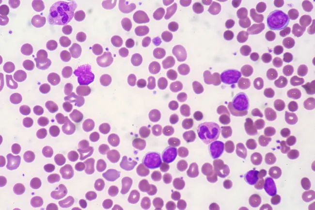
Peripheral blood smear from a patient with known chronic lymphocytic leukemia (CLL) showing autoimmune hemolytic anemia (AIHA). Brightfield microscopy of Wright-Giemsa stained smear at 1000x (oil immersion) reveals two dominant cell populations: numerous small mature lymphocytes typical of CLL and red blood cells showing spherocytic deformation. A smudge cell is evident just left of center, a classical clue for CLL. Red cells display reduced central pallor consistent with spherocytes, indicating intravascular or extravascular hemolysis driven by autoantibodies. The background shows mild polychromasia and rare nucleated RBCs, consistent with a robust reticulocyte response; the corrected reticulocyte count in this case is markedly elevated (>7%). Platelets are not significantly abnormal. These features collectively support autoimmune destruction of erythrocytes in the setting of CLL. Diagnostic significance lies in recognizing AIHA as a common autoimmune complication in CLL, with a clinical picture of sudden Hb decline and compensatory erythropoiesis. Differential considerations include drug-induced hemolysis, other causes of hemolytic anemia, or marrow infiltration by CLL. Clinical correlation includes therapy implications (steroids, rituximab) and close monitoring of hemolysis markers. This image is a valuable teaching resource for hematology morphology, AIHA in CLL, and differential diagnosis in anemia.
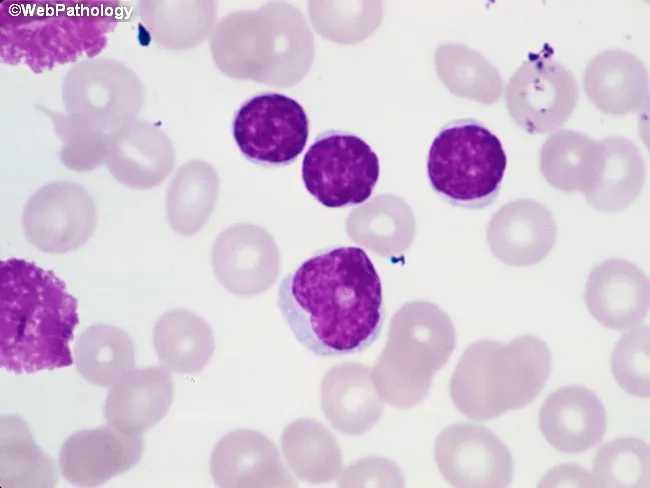
Imaging Modality: Brightfield light microscopy of a Wright-Giemsa stained peripheral blood smear. The primary subject is leukocytes from circulating blood, with focus on small mature-appearing lymphocytes and rare prolymphocytes. At center-right, several lymphocytes exhibit condensed chromatin and scant cytoplasm, consistent with chronic lymphocytic leukemia (CLL) morphology. Along the left edge, characteristic smudge cells are visible, reflecting fragile lymphocytes commonly seen in CLL. A prolymphocyte located just below center presents with slightly irregular nuclear contours, more dispersed chromatin, a prominent nucleolus, and modestly increased cytoplasm. In CLL, prolymphocytes usually comprise less than 2% of neoplastic cells; when 10–15%, the term atypical CLL is used and carries implications of aberrant immunophenotype, cytogenetic abnormalities, cytopenias, refractoriness to therapy, and worse prognosis. If prolymphocytes predominate, consideration should be given to B-cell prolymphocytic leukemia. This image demonstrates key diagnostic features including lymphoid morphology, prolymphocytic variant, and smear artifacts. Clinically, these findings correlate with lymphocytosis and potential anemia or thrombocytopenia in affected patients. Definitive characterization requires ancillary testing such as flow cytometry, immunophenotyping, and cytogenetics. The morphology supports a differential diagnosis that includes CLL with prolymphocytic transformation, atypical CLL, and B-PLL, guiding prognosis and treatment planning.
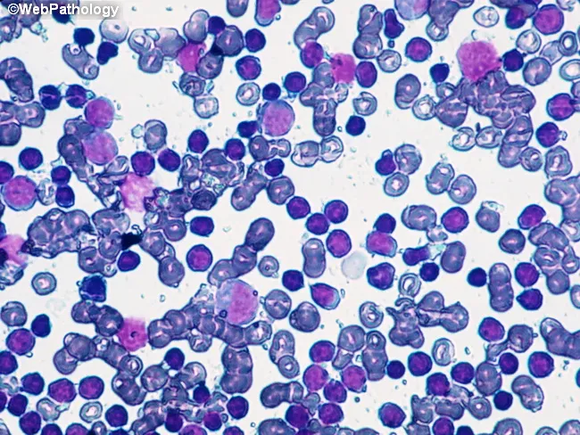
Imaging modality: Brightfield light microscopy of a Wright-Giemsa stained peripheral blood smear. The slide shows a predominant population of small to medium-sized lymphoid cells with scant pale cytoplasm and dense, clumped chromatin. Numerous cells exhibit mature-appearing B lymphocyte morphology. A fine background of erythrocytes and occasional smudge cells is present, with no overt blasts or granulocytic abnormalities. The overall pattern is lymphocytosis with a monomorphic lymphocytic population, compatible with monoclonal B-cell lymphocytosis (MBL) or the chronic lymphocytic leukemia (CLL) spectrum. Immunophenotypic confirmation (CD5+, CD23+, CD19+, surface Ig) is typically required for definitive classification, but is outside the scope of this image. The cluster appears relatively uniform, suggesting clonality rather than reactive lymphocytosis. Genetic associations commonly reported with CLL/MBL include 13q14 deletion and trisomy 12, though such findings require molecular testing. Clinically, CLL is defined by an absolute lymphocyte count ≥5.0 x 10^9/L in peripheral blood for at least 3 months; values below this threshold meet criteria for MBL. The image illustrates morphologic correlates of indolent clonal B-cell expansions and underscores the need to integrate flow cytometry and cytogenetics for diagnosis, prognosis, and management decisions. This image is educational for hematology, pathology, and cytology reference libraries.
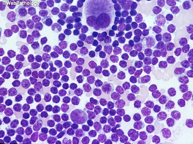
Bone marrow aspirate smear analyzed by light microscopy with high magnification reveals marrow involvement by chronic lymphocytic leukemia (CLL) cells. The predominant population consists of small, dark lymphocytes with scant cytoplasm and condensed, clumped nuclear chromatin, producing high nuclear-to-cytoplasmic ratios. Occasional larger forms and prolymphocytes may be present but are uncommon. A consistent finding is the relative scarcity of cytoplasm and uniform round to slightly irregular nuclei among the bulk of lymphoid cells. The smear may show scattered megakaryocytes, reflecting concurrent hematopoietic activity within the aspirate. Morphology mirrors circulating leukemic cells, consistent with systemic disease. Clinically, such marrow infiltration is associated with cytopenias and advanced disease stage, especially when diffuse, solid infiltration predominates in core biopsy patterns; these patterns correlate with adverse prognosis and with expression of prognostic markers such as ZAP-70. The image corresponds to a high-magnification field intended to document cellular detail rather than architectural marrow patterns. Diagnostic significance lies in confirming lymphoid marrow involvement by CLL, quantifying blast equivalents is not applicable, and differentiating from other small B-cell neoplasms relies on adjunct flow cytometry, cytogenetics, and immunophenotyping. This representation is typical for educational, diagnostic, and research contexts describing CLL marrow morphology.
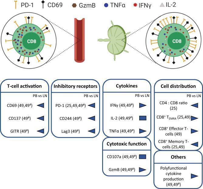
This medical infographic illustrates the comparative T-cell exhaustion phenotype of CD8+ T-cells in Chronic Lymphocytic Leukemia (CLL), contrasting peripheral blood (PB) versus lymph nodes (LN). The top section features anatomical diagrams of a blood vessel and a lymph node, each linked to a CD8+ T-cell illustration. Visual symbols indicate that CD8+ T-cells in the LN exhibit a higher density of surface markers PD-1 and CD69 and altered intracellular molecules (GzmB, TNFα, IFNγ, IL-2) compared to PB. The bottom section provides data summary boxes for 'T-cell activation' (CD69, CD137, GITR), 'Inhibitory receptors' (PD-1, CD244, Lag3), 'Cytokines' (IFNγ, IL-2, TNFα), 'Cytotoxic function' (CD107a, GzmB), and 'Cell distribution'. Blue arrow markers indicate a trend towards higher expression or prevalence in the lymph nodes for most markers, including exhausted cell populations (CD8+ TEMRA, Effector, and Memory T-cells). This diagram highlights that T-cell exhaustion in CLL is more severe in the lymphoid tissue microenvironment than in systemic circulation.
CLL - Peripheral Blood Smear (PBS) Picture
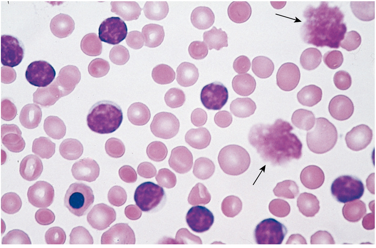
Key PBS Features of CLL
- Small cells (6-12 µm), similar in size to normal lymphocytes
- Round to slightly irregular nuclei
- Condensed/clumped "soccer ball" chromatin - the chromatin pattern is densely packed, not fine or blastic
- Scant cytoplasm - very thin rim, high N:C ratio
- Nucleoli are inconspicuous or absent
- Fragile leukemic lymphocytes rupture when the smear is being spread
- Appear as amorphous, smeared nuclear material with no identifiable cell membrane
- Two smudge cells are visible (arrowed) in the Robbins image above
- Their presence strongly suggests CLL but is not pathognomonic
- Spherocytes (hyperchromatic, round RBCs without central pallor) - indicating coexistent autoimmune hemolytic anemia (AIHA), a known complication of CLL
- Nucleated RBC (bottom left, blue cell) - from premature release due to severe anemia or marrow infiltration (leukoerythroblastosis)
Additional CLL PBS Images

Diagnostic Criteria Summary
| Feature | CLL PBS Finding |
|---|---|
| Lymphocyte morphology | Small, round, mature-appearing |
| Chromatin | Condensed/clumped ("cracked mud") |
| Cytoplasm | Scant |
| Nucleolus | Inconspicuous |
| Pathognomonic artifact | Smudge cells (Gumprecht shadows) |
| ALC threshold | ≥5.0 × 10⁹/L for ≥3 months |
Differential Diagnosis on PBS
- Reactive lymphocytosis - more pleomorphic, mixed cell sizes, no smudge cells
- Mantle cell lymphoma - irregular/cleaved nuclei, CD5+ but CD23-
- Follicular lymphoma - cleaved "buttock cells"
- B-PLL (Prolymphocytic Leukemia) - >55% prolymphocytes (large cells with prominent nucleolus)
- Hairy cell leukemia - "hairy" cytoplasmic projections, TRAP+
Robbins, Cotran & Kumar - Pathologic Basis of Disease, p. 560 Goldman-Cecil Medicine, Diagnosis section
Smudge cells
smudge cells Gumprecht shadows CLL peripheral blood smear
Peripheral blood smear from a patient with suspected chronic lymphocytic leukemia (CLL) shows a predominance of small, mature-appearing lymphocytes with scant cytoplasm and clumped nuclear chromatin, accompanied by fragile cells that readily disrupt during smear preparation, producing numerous smudge or basket cells visible on the slide. The image depicts a single smudge cell in the upper right, with numerous small darkly staining lymphocytes occupying the field. The smear was prepared with Wright-Giemsa stain and examined under light microscopy at high magnification (approximately 1000x total magnification with oil immersion). Clinically, CLL is defined by absolute mature lymphocytosis of ≥5.0 × 10^9/L sustained for ≥3 months, often asymptomatic and detected incidentally on complete blood count. Morphology alone is insufficient for diagnosis; immunophenotyping showing co-expression of CD5, CD19, CD23 with weak surface immunoglobulin and light chain restriction supports CLL diagnosis. Smudge cells result from fragile leukemic cells and reflect underlying cytoskeletal fragility, though they are not specific. The image illustrates typical features: monomorphic lymphocytes, scant cytoplasm, condensed chromatin, and absent prominent nucleoli. This finding has diagnostic significance, guiding further workup, including flow cytometry, cytogenetics, and molecular studies, to distinguish CLL from other chronic lymphoproliferative disorders such as small lymphocytic lymphoma (SLL), mantle cell lymphoma, prolymphocytic leukemia, and reactive lymphocytosis.
Peripheral blood smear from a patient with known chronic lymphocytic leukemia (CLL) showing autoimmune hemolytic anemia (AIHA). Brightfield microscopy of Wright-Giemsa stained smear at 1000x (oil immersion) reveals two dominant cell populations: numerous small mature lymphocytes typical of CLL and red blood cells showing spherocytic deformation. A smudge cell is evident just left of center, a classical clue for CLL. Red cells display reduced central pallor consistent with spherocytes, indicating intravascular or extravascular hemolysis driven by autoantibodies. The background shows mild polychromasia and rare nucleated RBCs, consistent with a robust reticulocyte response; the corrected reticulocyte count in this case is markedly elevated (>7%). Platelets are not significantly abnormal. These features collectively support autoimmune destruction of erythrocytes in the setting of CLL. Diagnostic significance lies in recognizing AIHA as a common autoimmune complication in CLL, with a clinical picture of sudden Hb decline and compensatory erythropoiesis. Differential considerations include drug-induced hemolysis, other causes of hemolytic anemia, or marrow infiltration by CLL. Clinical correlation includes therapy implications (steroids, rituximab) and close monitoring of hemolysis markers. This image is a valuable teaching resource for hematology morphology, AIHA in CLL, and differential diagnosis in anemia.
Imaging Modality: Brightfield light microscopy of a Wright-Giemsa stained peripheral blood smear. The primary subject is leukocytes from circulating blood, with focus on small mature-appearing lymphocytes and rare prolymphocytes. At center-right, several lymphocytes exhibit condensed chromatin and scant cytoplasm, consistent with chronic lymphocytic leukemia (CLL) morphology. Along the left edge, characteristic smudge cells are visible, reflecting fragile lymphocytes commonly seen in CLL. A prolymphocyte located just below center presents with slightly irregular nuclear contours, more dispersed chromatin, a prominent nucleolus, and modestly increased cytoplasm. In CLL, prolymphocytes usually comprise less than 2% of neoplastic cells; when 10–15%, the term atypical CLL is used and carries implications of aberrant immunophenotype, cytogenetic abnormalities, cytopenias, refractoriness to therapy, and worse prognosis. If prolymphocytes predominate, consideration should be given to B-cell prolymphocytic leukemia. This image demonstrates key diagnostic features including lymphoid morphology, prolymphocytic variant, and smear artifacts. Clinically, these findings correlate with lymphocytosis and potential anemia or thrombocytopenia in affected patients. Definitive characterization requires ancillary testing such as flow cytometry, immunophenotyping, and cytogenetics. The morphology supports a differential diagnosis that includes CLL with prolymphocytic transformation, atypical CLL, and B-PLL, guiding prognosis and treatment planning.
Imaging modality: Brightfield light microscopy of a Wright-Giemsa stained peripheral blood smear. The slide shows a predominant population of small to medium-sized lymphoid cells with scant pale cytoplasm and dense, clumped chromatin. Numerous cells exhibit mature-appearing B lymphocyte morphology. A fine background of erythrocytes and occasional smudge cells is present, with no overt blasts or granulocytic abnormalities. The overall pattern is lymphocytosis with a monomorphic lymphocytic population, compatible with monoclonal B-cell lymphocytosis (MBL) or the chronic lymphocytic leukemia (CLL) spectrum. Immunophenotypic confirmation (CD5+, CD23+, CD19+, surface Ig) is typically required for definitive classification, but is outside the scope of this image. The cluster appears relatively uniform, suggesting clonality rather than reactive lymphocytosis. Genetic associations commonly reported with CLL/MBL include 13q14 deletion and trisomy 12, though such findings require molecular testing. Clinically, CLL is defined by an absolute lymphocyte count ≥5.0 x 10^9/L in peripheral blood for at least 3 months; values below this threshold meet criteria for MBL. The image illustrates morphologic correlates of indolent clonal B-cell expansions and underscores the need to integrate flow cytometry and cytogenetics for diagnosis, prognosis, and management decisions. This image is educational for hematology, pathology, and cytology reference libraries.
Smudge Cells (Gumprecht Shadows)
What are Smudge Cells?
- No cell membrane
- No identifiable nuclear outline
- Smeared, wispy purple chromatin
- Gumprecht shadows (eponym)
- Basket cells (basket-like chromatin appearance)
Why Do They Form in CLL?
- Decreased expression of vimentin (an intermediate filament that normally protects cells during smear preparation)
- The cells are inherently fragile - they lyse easily under mechanical shear stress when the smear is spread
Clinical Significance
| Point | Detail |
|---|---|
| Classic association | CLL - strongly suggestive |
| Not pathognomonic | Can occur in other conditions with lymphocytosis |
| Quantity correlates | Higher smudge cell count may correlate with more fragile cells |
| Diagnostic utility | Presence on routine PBS prompts flow cytometry workup |
Other Conditions with Smudge Cells
- Other lymphoproliferative disorders (lymphoma in leukemic phase)
- Infectious mononucleosis (reactive lymphocytosis)
- Normal smears in very small numbers
When to say that marrow smear is diluted
bone marrow aspirate diluted smear peripheral blood contamination
This is a Wright–Giemsa stained bone marrow aspirate smear imaged by bright-field light microscopy at high magnification, showing a cytology-level field of hematopoietic cells. Nuclei are round to irregular with coarse chromatin; cells range from mature myeloid/erythroid elements to immature precursor forms. Clusters of cells and occasional smearing artefacts reflect aspirate preparation. Staining emphasizes nuclear detail and cytoplasmic basophilia, enabling assessment of lineage commitment, maturation, cellularity, and potential neoplastic involvement. The image is clinically relevant for evaluating marrow cellularity, dysplasia, and infiltration, and for distinguishing reactive hematopoiesis from malignant processes. Diagnostic significance lies in guiding ancillary studies such as flow cytometry, cytogenetics, and molecular testing. Potential educational and diagnostic applications include hematopathology training, differential diagnosis of acute leukemia and myelodysplastic syndrome, aplastic anemia evaluation, and assessment of marrow involvement by lymphoma. Clinically, correlate with CBC, differential counts, peripheral smear, and immunophenotyping to determine prognosis and treatment planning. Interpretation should consider sample quality and ancillary data to ensure accurate diagnosis.
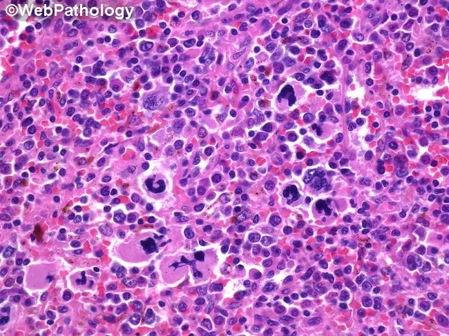
High-magnification brightfield micrograph of a human bone marrow aspirate smear stained with Hematoxylin and Eosin, demonstrating dysplastic megakaryocytes in a hematopoietic background. The megakaryocytes exhibit atypical nuclear morphology, including multinucleation, irregular lobulation, and nuclear fragmentation, with variable cytoplasmic basophilia. Background cellularity shows concurrent dyserythropoiesis and dysplastic myeloid precursors consistent with a myelodysplastic spectrum. Overall architecture may be hypercellular or variably cellular, with scattered normal hematopoietic elements interspersed among abnormal cells. Features are most prominent in the megakaryocytic lineage, where dysplasia is a salient hallmark. The image illustrates morphologic criteria that support a diagnosis in the context of possible progression toward acute leukemia, and should be correlated with peripheral blood counts, marrow blast percentage, cytogenetic and molecular studies. Clinically, such dysplastic megakaryocytes are associated with cytopenias and splenomegaly, and approximately a fifth of patients with related myelodysplastic/myeloproliferative conditions progress to acute myeloid leukemia. This representation is valuable for educational, diagnostic, and research purposes, particularly in studies of megakaryocytic dysplasia, MDS/AML continuum, prognostication, and therapeutic planning focused on palliation and cytopenia management. Careful documentation of features and context enhances reproducibility, supports standardized reporting, and facilitates cross-institutional data sharing for outcome studies and biomarker-driven clinical trials.
Bone marrow aspirate smear analyzed by light microscopy with high magnification reveals marrow involvement by chronic lymphocytic leukemia (CLL) cells. The predominant population consists of small, dark lymphocytes with scant cytoplasm and condensed, clumped nuclear chromatin, producing high nuclear-to-cytoplasmic ratios. Occasional larger forms and prolymphocytes may be present but are uncommon. A consistent finding is the relative scarcity of cytoplasm and uniform round to slightly irregular nuclei among the bulk of lymphoid cells. The smear may show scattered megakaryocytes, reflecting concurrent hematopoietic activity within the aspirate. Morphology mirrors circulating leukemic cells, consistent with systemic disease. Clinically, such marrow infiltration is associated with cytopenias and advanced disease stage, especially when diffuse, solid infiltration predominates in core biopsy patterns; these patterns correlate with adverse prognosis and with expression of prognostic markers such as ZAP-70. The image corresponds to a high-magnification field intended to document cellular detail rather than architectural marrow patterns. Diagnostic significance lies in confirming lymphoid marrow involvement by CLL, quantifying blast equivalents is not applicable, and differentiating from other small B-cell neoplasms relies on adjunct flow cytometry, cytogenetics, and immunophenotyping. This representation is typical for educational, diagnostic, and research contexts describing CLL marrow morphology.
bone marrow aspirate smear diluted haemodiluted criteria diagnosis
https://www.ncbi.nlm.nih.gov/books/NBK603716
Diluted (Haemodiluted) Bone Marrow Aspirate Smear
The Key Criterion - Particles (Spicules)
| Finding | Interpretation |
|---|---|
| No particles + No megakaryocytes + No precursor cells | "Blood tap" - essentially peripheral blood only, not marrow at all |
| No particles + Megakaryocytes or other precursor cells present | "Dilute BM sample" - haemodiluted but some marrow elements present; qualitative evaluation only possible |
| Particles present + absent/very reduced cellularity | Hypocellular marrow - qualitative description only |
| Particles present + adequate cellularity | Adequate aspirate - full assessment can be performed |
Why Dilution Happens
- Too large a volume aspirated - progressive dilution with peripheral blood occurs with increasing syringe volumes; the first 0.5 ml drawn is the most cellular and least diluted
- Aspiration needle placed too superficially in cortical bone rather than marrow cavity
- Aspiration through a trephine needle - the ICSH specifically warns this technique commonly leads to haemodilution
- Re-aspiration at same site - previously aspirated site fills with peripheral blood
- Technically difficult aspirations - fibrotic marrows (myelofibrosis), hypocellular marrows
How to Recognise on the Smear Itself
- Marrow particles/spicules - small pink/grey-purple gritty fragments visible to the naked eye on the slide; on microscopy, islands of haemopoietic cells surrounded by fat spaces and stromal cells
- Megakaryocytes - large multi-lobed cells; their presence confirms genuine marrow material was obtained
- Haemopoietic precursors - myeloid/erythroid precursors (normoblasts, myelocytes, metamyelocytes) that are not normally seen in peripheral blood
- M:E ratio assessable - only meaningful if particles are present
Practical Reporting
| Smear Adequacy | Report Wording |
|---|---|
| Good | "Particulate BM aspirate, adequate for assessment" |
| Diluted but some marrow | "Aparticulate, dilute BM sample - qualitative evaluation only" |
| No marrow at all | "Blood tap / peripheral blood only - no marrow elements identified" |
| Fibrotic / no material | "Dry tap - trephine biopsy required for assessment" |
Clinical Impact
- MRD (Minimal Residual Disease) assessment in myeloma - a haemodiluted sample can give a falsely low or undetectable MRD result, which should be interpreted with caution
- Blast percentage in AML/MDS - dilution underestimates the true blast count
- Plasma cell % in myeloma diagnosis - falsely low percentages
Brief about Bethesda SYSTEM OF reporting Thyroid
Bethesda system thyroid cytopathology categories FNA table
The Bethesda System for Reporting Thyroid Cytopathology (TBSRTC)
The 6 Categories
| Category | Diagnosis | Risk of Malignancy | Management |
|---|---|---|---|
| I | Nondiagnostic / Unsatisfactory | 1-4% (but actual surgical risk ~20%) | Repeat FNA with ultrasound guidance |
| II | Benign | <3% | Clinical follow-up / serial US |
| III | Atypia of Undetermined Significance (AUS) / Follicular Lesion of Undetermined Significance (FLUS) | ~10-30% | Repeat FNA, molecular testing, or lobectomy |
| IV | Follicular Neoplasm / Suspicious for Follicular Neoplasm | 25-40% | Diagnostic lobectomy / molecular testing |
| V | Suspicious for Malignancy | 50-75% | Surgery in most cases |
| VI | Malignant | 97-99% | Definitive surgical management |
Category-by-Category Detail
- Cyst fluid only, virtually acellular specimen, or obscured by clotting/blood
- Adequacy criterion (not met): at least 6 groups of ≥10 well-preserved follicular cells
- Action: repeat US-guided FNA; if persistently nondiagnostic, consider diagnostic lobectomy
- Benign follicular nodule (adenomatoid/colloid nodule)
- Lymphocytic (Hashimoto) thyroiditis
- Granulomatous (subacute) thyroiditis
- Lowest risk category - observe with serial US; repeat FNA only if significant change in nodule characteristics
- Cytologic atypia that is more than benign but insufficient for Bethesda IV/V
- A "wastebasket" category that should be used sparingly (<7% of all FNAs ideally)
- Molecular testing (e.g. ThyroSeq v3, Afirma) is increasingly used to risk-stratify before surgery
- Follicular or Hürthle cell (oncocytic) pattern; cannot distinguish adenoma from carcinoma on cytology alone (requires capsular/vascular invasion on histology)
- Specify if Hürthle cell (oncocytic) type
- Architectural atypia more than cytologic atypia - FNA has intrinsic limitation here
- Suspicious for: papillary carcinoma, medullary carcinoma, metastatic carcinoma, lymphoma
- Most cases proceed to surgery; molecular testing may further guide extent
- Includes:
- Papillary thyroid carcinoma (PTC) - most common
- Medullary thyroid carcinoma (MTC)
- Poorly differentiated / anaplastic carcinoma
- Squamous cell carcinoma
- Non-Hodgkin lymphoma
- Metastatic carcinoma
Key Points to Remember
- Categories V and VI → surgery in most cases
- Categories III and IV → "indeterminate" zone - account for ~20% of FNAs; molecular testing (ThyroSeq, Afirma) helps avoid unnecessary surgery
- Category II → safe to observe (false-negative rate 1-6% at high-volume centers)
- Nodule size caveat: nodules >3-4 cm with benign cytology carry a higher false-negative risk (malignancy found in 11-13% at surgery due to sampling error)
- The system was updated in 2023 with refined malignancy risk estimates incorporating NIFTP (Non-Invasive Follicular Thyroid neoplasm with Papillary-like nuclear features), which is no longer counted as malignant, lowering the malignancy risk in some categories
Fischer's Mastery of Surgery 8e, Table 34.1; Mulholland & Greenfield's Surgery 7e, p. 4043-4044
Micro follicle vs Macro folliclr
thyroid FNA microfollicle macrofollicle cytology smear comparison
Microfollicle vs Macrofollicle in Thyroid FNA Cytology
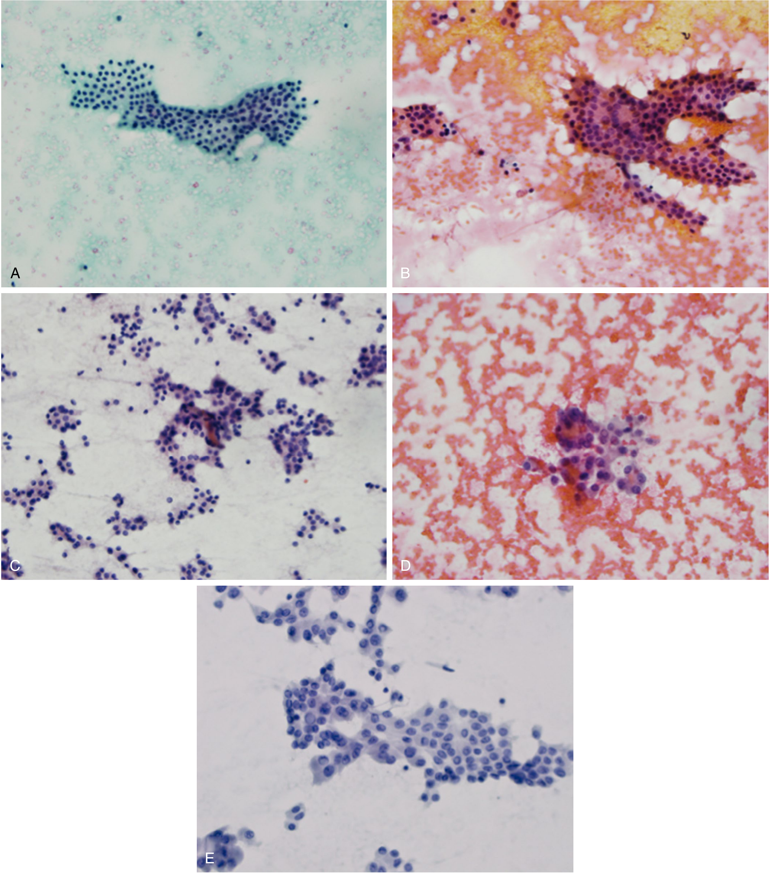
Core Comparison
| Feature | Macrofollicle | Microfollicle |
|---|---|---|
| Size | Large (>15-20 cells lining a follicle) | Small rings of <15 cells (often 6-12 cells) |
| Colloid | Abundant, watery/thin "cracked" colloid background | Scant or absent colloid |
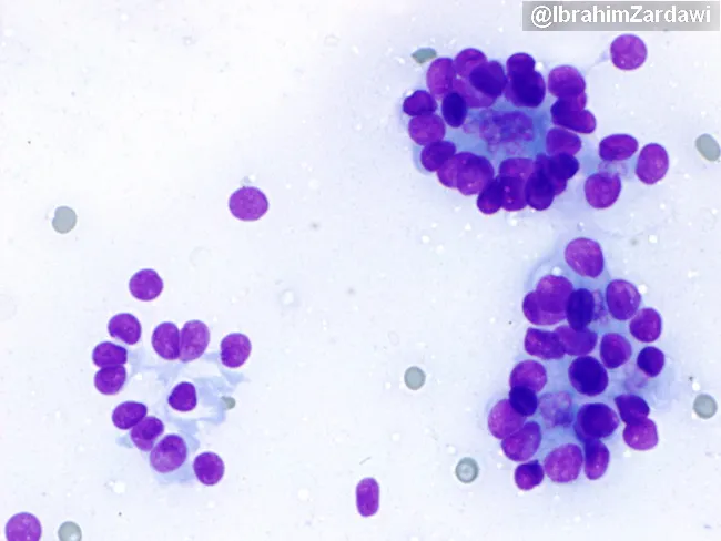
| Arrangement | Flat monolayered sheets, honeycomb pattern | Small tight 3D clusters, rosette-like rings |
| Cell crowding | Minimal, evenly spaced | Crowded, overlapping nuclei |
| Bethesda category | Category II - Benign | Category IV - Follicular Neoplasm |
| Clinical significance | Hyperplastic/colloid nodule, multinodular goitre | Raises suspicion for follicular adenoma or carcinoma |
| Malignancy risk | <3% | 17-28% (NIFTP ≠ cancer) |
Macrofollicular Pattern - Benign Features
- Dispersed, thin watery colloid in background ("cracked mud" or "chicken wire" colloid)
- Monolayered flat sheets of bland follicular cells
- Large ring-like follicular structures (can see the lumen)
- Cells are evenly spaced, uniform, no crowding
- Nuclei are small, round, basally oriented
- Low cellularity overall
Microfollicular Pattern - Suspicious Features
- High cellularity - the aspirate is flooded with cells
- Small ring-like structures (rosettes) of 6-12 follicular cells arranged around a tiny central lumen
- Paucity of colloid or dense colloid globules (inspissated)
- Cell crowding, 3-dimensional (acervate) groups
- Nuclear overlapping
Key rule: Focus on the predominant pattern - a few microfollicles in an otherwise macrofollicular smear does NOT constitute Bethesda IV.
Why FNA Cannot Distinguish Adenoma from Carcinoma
- Microfollicular FNA → Bethesda IV → Diagnostic lobectomy (or molecular testing)
- Final diagnosis only made on surgical pathology
Quick Memory Aid
| Pattern | Think of... | Action |
|---|---|---|
| Macrofollicle + abundant colloid | Macro = big and benign | Observe |
| Microfollicle + scant colloid + hypercellular | Micro = might be malignant | Operate (lobectomy) |
Scott-Brown's Otorhinolaryngology, p. 9163-9166; Sabiston Surgery, Fig. 73.17 & Table 73.3