Ecg
Reading File
Asking for Preferences
Finding Sources
Finding Sources
Finding Sources
Finding Sources
Finding Sources
Reading File
Finding Sources
Finding Sources
Reading File
Finding Sources
Reading File
Finding Sources
Loading Image
Reading File
Loading Image
Here is a complete guide to ECG basics and how to read one, drawn from Medical Physiology (Boron & Boulpaep) and Braunwald's Heart Disease.
What is an ECG?
An ECG (electrocardiogram) records the time-dependent electrical activity of the heart from electrodes on the body surface. It gives direct information about:
- Rate - how fast the heart is beating
- Rhythm - where the impulse originates and how it conducts
- Axis - the direction of the electrical vector
- Waveform morphology - signs of ischemia, hypertrophy, or conduction problems
The ECG Paper
- Vertical axis: 0.1 mV per small box (1 mm)
- Horizontal axis: 0.04 sec per small box (1 mm); 0.2 sec per large box (5 mm)
- 5 large boxes = 1.0 second
The 12 Leads
A standard ECG has 12 leads - 6 limb leads and 6 chest (precordial) leads:
| Group | Leads | View |
|---|---|---|
| Limb leads | I, II, III | Frontal plane |
| Augmented limb leads | aVR, aVL, aVF | Frontal plane |
| Precordial leads | V1 - V6 | Horizontal plane |
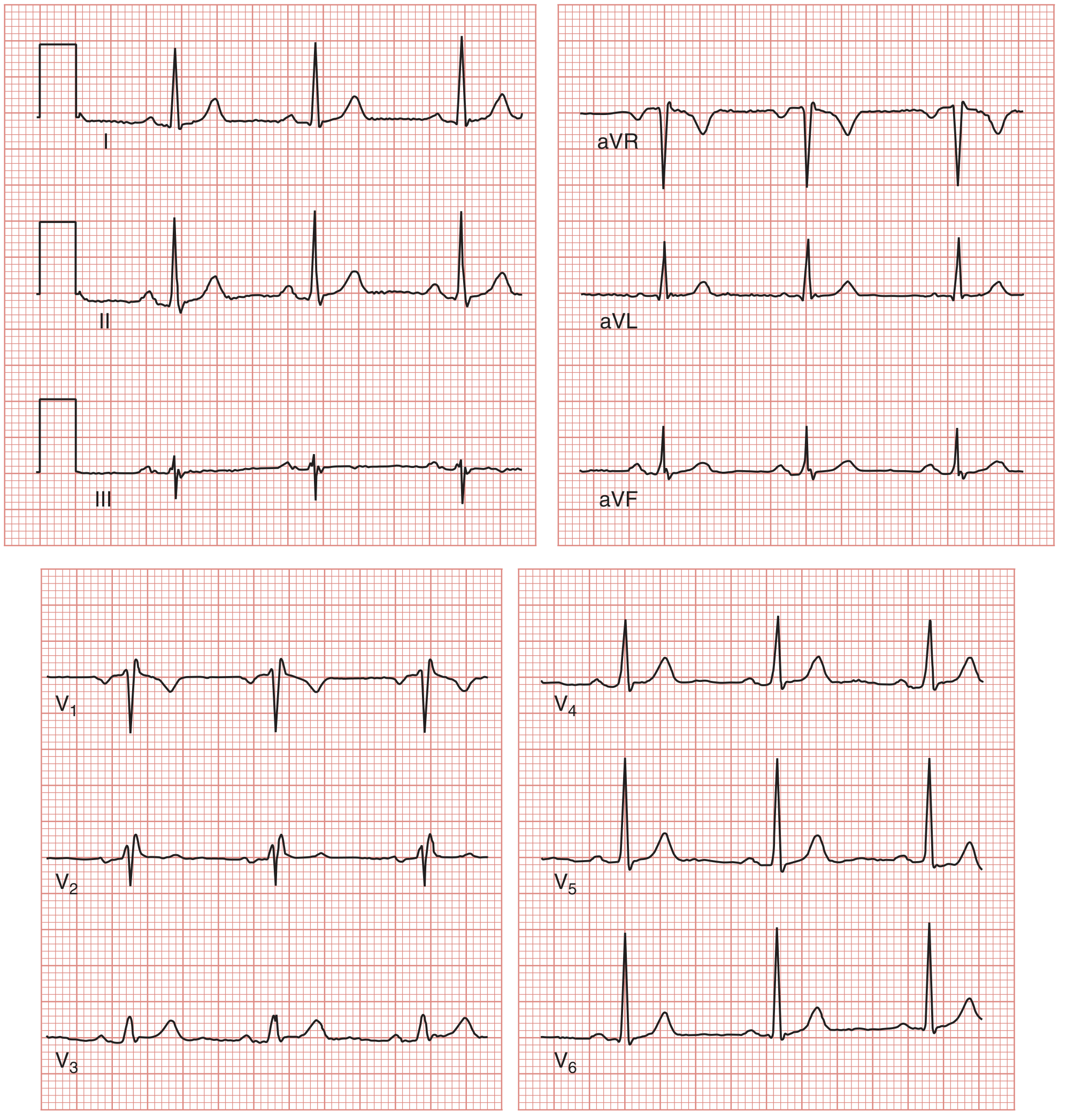
Here is what a normal 12-lead ECG looks like:
The ECG Waveforms
Each heartbeat produces a characteristic sequence of waves:
| Wave / Interval | What it represents |
|---|---|
| P wave | Atrial depolarization |
| PR interval | Time from atrial to ventricular activation (through AV node) |
| QRS complex | Ventricular depolarization |
| ST segment | Plateau phase of ventricular action potential (between QRS and T) |
| T wave | Ventricular repolarization |
| QT interval | Total ventricular electrical activity (depolarization + repolarization) |
| U wave | Small wave after T wave (cause debated; seen in hypokalemia) |
QRS Wave Naming Convention
- Q = first negative deflection before an R wave
- R = first positive deflection
- S = negative deflection after an R wave
- R' = second positive deflection (e.g., in RBBB)
- Capital letters (Q, R, S) = large amplitude; lowercase (q, r, s) = small amplitude
Normal Interval Values
| Interval | Normal Duration |
|---|---|
| PR interval | 0.12 - 0.20 sec (3-5 small boxes) |
| QRS duration | < 0.12 sec (< 3 small boxes) |
| QT interval | Rate-dependent; corrected QTc < 0.44 sec (men), < 0.46 sec (women) |
How to Measure Heart Rate
Direct method: Rate = 60 / (R-R interval in seconds)
Quick method (count large boxes between two R waves):
- 1 large box = 300 bpm
- 2 boxes = 150
- 3 boxes = 100
- 4 boxes = 75
- 5 boxes = 60
- 6 boxes = 50
Memorize: 300 - 150 - 100 - 75 - 60 - 50
Systematic Approach to Reading an ECG
Use this 8-step method every time:
- Rate - Calculate using R-R interval (normal: 60-100 bpm)
- Rhythm - Regular or irregular? Is every QRS preceded by a P wave?
- P waves - Present? Upright in II? One per QRS?
- PR interval - Normal (0.12-0.20 sec)? Short (WPW)? Prolonged (heart block)?
- QRS duration - Narrow (< 0.12 sec) or wide (bundle branch block)?
- Axis - Normal axis is -30° to +90° in the frontal plane
- ST segment & T waves - Elevation (STEMI, pericarditis)? Depression (ischemia)?
- QT interval - Prolonged (risk of torsades)?
Normal Sinus Rhythm Criteria
- Rate 60-100 bpm
- Regular rhythm
- Upright P wave in lead II (negative in aVR)
- Each P wave followed by a QRS
- Normal PR interval (0.12-0.20 sec)
- Narrow QRS (< 0.12 sec)
Electrical Axis
The normal QRS axis is between -30° and +90° in the frontal plane.
- Left axis deviation (< -30°): left anterior fascicular block, inferior MI, LVH
- Right axis deviation (> +90°): right ventricular hypertrophy, left posterior fascicular block, PE
Quick axis check: Look at leads I and aVF:
- Both positive = normal axis
- I positive, aVF negative = possible left axis deviation
- I negative, aVF positive = right axis deviation
This is the foundation. Once you are comfortable with these basics, the next steps are recognizing specific patterns: bundle branch blocks, ST changes, hypertrophy criteria, and arrhythmias. Let me know which area you'd like to go into next.
- Medical Physiology (Boron & Boulpaep), Box 21-2, pp. 731-734
- Braunwald's Heart Disease, 12th Ed.
This is a shared conversation. Sign in to Orris to start your own chat.