
Detailed Notes On Cranial Nerve
Reading File
The cranial nerves are 12 pairs of nerves arising directly from the brain/brainstem, each with distinct origins, functions, and clinical significance.
Searching sources...
Finding Sources
Searching Images
cranial nerves overview diagram brainstem
Reading File
Finding Sources
Reading File
Finding Sources
Reading File
Finding Sources
Reading File
Reading File
Loading Image
Reading File
Finding Sources
Reading File
Loading Image
Reading File
I now have comprehensive information from Gray's Anatomy for Students to compile detailed notes. Let me compile the full, well-structured notes.
Detailed Notes on the Cranial Nerves
Source: Gray's Anatomy for Students, Rosen's Emergency Medicine, Localization in Clinical Neurology
Overview
There are 12 pairs of cranial nerves (CN I-XII), part of the peripheral nervous system (PNS). Their defining feature is that they exit the cranial cavity through foramina or fissures. All cranial nerves innervate structures in the head or neck. The exception is the vagus nerve [X], which descends into the thorax and abdomen to innervate viscera.
All cranial nerves except one (the accessory nerve [XI]) originate from the brain itself.
Cranial Nerves on the Base of the Brain
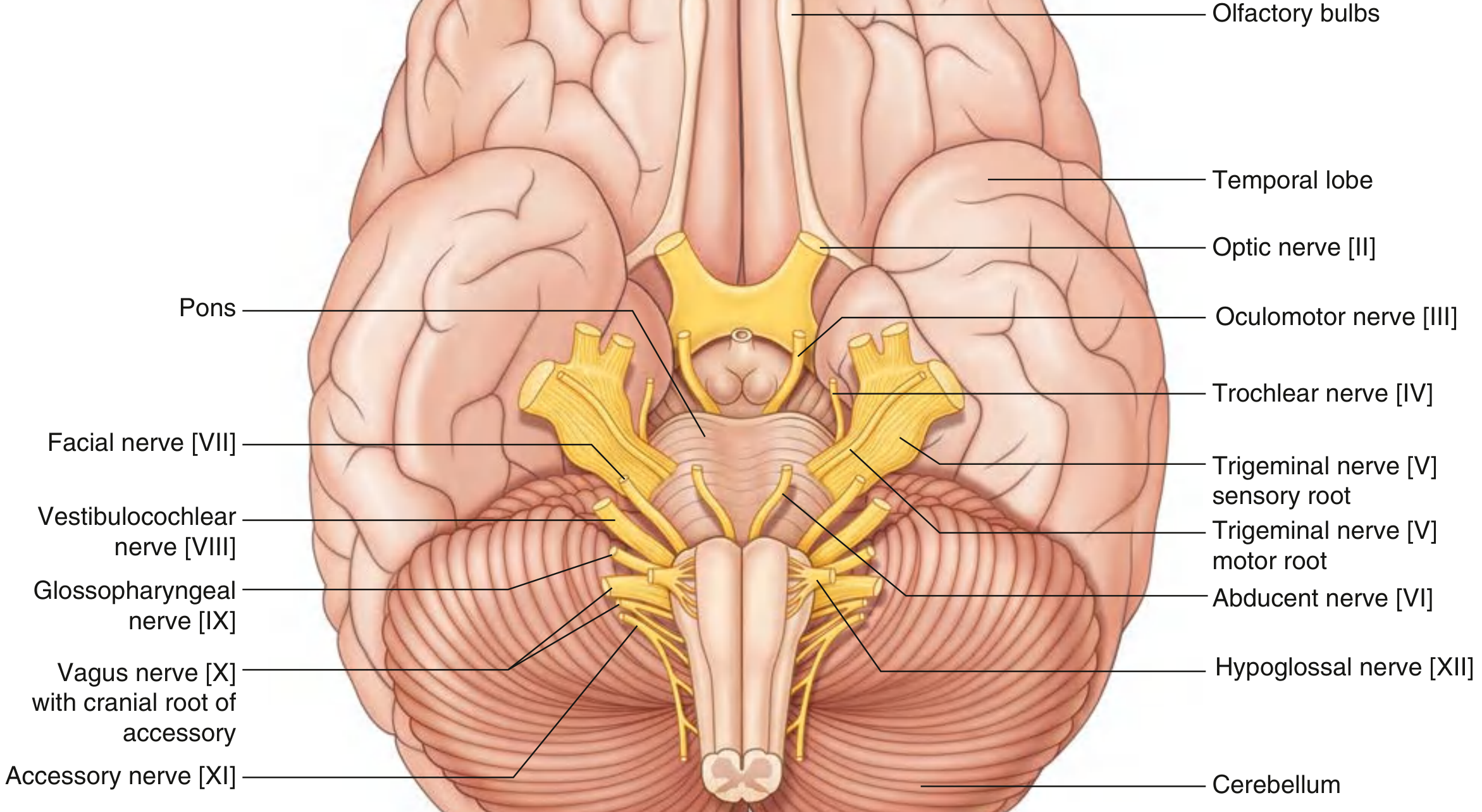
Gray's Anatomy for Students - Cranial Nerves at the Base of the Brain
Fiber Component Classification
Cranial nerves carry a mix of somatic, visceral, and special fiber types. The table below summarizes all functional components:
| Functional Component | Abbreviation | General Function | Cranial Nerves |
|---|---|---|---|
| General Somatic Afferent | GSA | Touch, pain, temperature | V, VII, IX, X |
| General Visceral Afferent | GVA | Sensory input from viscera | IX, X |
| Special Afferent | SA | Smell, taste, vision, hearing, balance | I, II, VII, VIII, IX, X |
| General Somatic Efferent | GSE | Motor to skeletal (voluntary) muscles | III, IV, VI, XII |
| General Visceral Efferent | GVE | Motor to smooth muscle, heart, glands (parasympathetic) | III, VII, IX, X |
| Branchial Efferent | BE | Motor to pharyngeal arch-derived muscles | V, VII, IX, X, XI |
Memory Mnemonics
Names (I-XII): Oh Oh Oh To Touch And Feel Very Good Velvet And Hair
(Olfactory, Optic, Oculomotor, Trochlear, Trigeminal, Abducens, Facial, Vestibulocochlear, Glossopharyngeal, Vagus, Accessory, Hypoglossal)
Fiber type (S=Sensory, M=Motor, B=Both): Some Say Marry Money But My Brother Says Big Brains Matter Most
Individual Cranial Nerves
CN I - Olfactory Nerve
| Feature | Detail |
|---|---|
| Fiber type | Special Afferent (SA) - smell |
| Origin | Nasal mucosa (roof + upper nasal cavity) |
| Course | Axon bundles pass through cribriform plate of ethmoid bone → synapse in olfactory bulbs |
| Foramen | Cribriform plate (ethmoid bone) |
| Function | Sense of smell (olfaction) |
| Lesion | Anosmia (loss of smell) - caused by cribriform plate fractures, closed head injury |
CN II - Optic Nerve
| Feature | Detail |
|---|---|
| Fiber type | Special Afferent (SA) - vision |
| Origin | Photoreceptors of the retina |
| Course | Retinal axons → optic nerve → optic canal → optic chiasm → optic tract → lateral geniculate nucleus |
| Foramen | Optic canal |
| Function | Vision; pupillary light reflex (afferent limb) |
| Lesion | Blindness, visual field defects, loss of pupillary constriction |
Note: The optic nerve is technically a CNS tract (myelinated by oligodendrocytes, not Schwann cells).
CN III - Oculomotor Nerve
| Feature | Detail |
|---|---|
| Fiber types | GSE + GVE (parasympathetic) |
| Origin | Midbrain (anterior surface, between midbrain and pons) |
| Course | Passes through lateral wall of cavernous sinus → exits via superior orbital fissure |
| GSE targets | Levator palpebrae superioris, superior rectus, inferior rectus, medial rectus, inferior oblique |
| GVE targets | Preganglionic parasympathetics → ciliary ganglion → sphincter pupillae (pupil constriction) + ciliary muscle (accommodation) |
| Foramen | Superior orbital fissure |
| Lesion | Dilated pupil, ptosis, eye turns "down and out" (due to unopposed CN IV + VI), loss of pupillary reflex |
CN IV - Trochlear Nerve
| Feature | Detail |
|---|---|
| Fiber type | GSE |
| Origin | Midbrain |
| Unique feature | Only CN to exit from the POSTERIOR surface of the brainstem; also the smallest cranial nerve |
| Course | Curves around midbrain → cavernous sinus lateral wall → superior orbital fissure |
| Target | Superior oblique muscle (depresses adducted eye; intorts the eye) |
| Foramen | Superior orbital fissure |
| Lesion | Vertical diplopia, head tilt (to compensate), inability to look down-and-in |
CN V - Trigeminal Nerve
The largest cranial nerve; major general sensory nerve of the face and motor nerve for mastication.
| Feature | Detail |
|---|---|
| Fiber types | GSA + Branchial Efferent (BE) |
| Origin | Anterolateral surface of pons (large sensory root + small motor root) |
| Ganglion | Trigeminal ganglion (Gasserian ganglion) in the trigeminal cave on the petrous bone |
| Foramen | V1 - Superior orbital fissure; V2 - Foramen rotundum; V3 - Foramen ovale |
Three divisions:
V1 - Ophthalmic nerve:
- Purely sensory (GSA)
- Sensation from forehead, anterior scalp, upper eyelid, cornea, dorsum of nose, nasal mucosa (upper), paranasal sinuses, dura of anterior cranial fossa
- Passes through cavernous sinus lateral wall → superior orbital fissure
V2 - Maxillary nerve:
- Purely sensory (GSA)
- Sensation from lower eyelid, cheek, upper lip, upper teeth and gums, hard palate, nasal mucosa (lower), maxillary sinus
- Exits via foramen rotundum → pterygopalatine fossa
V3 - Mandibular nerve:
- Mixed: sensory (GSA) + motor (BE)
- Sensation from lower lip, chin, lower teeth and gums, anterior two-thirds tongue (general sensation, NOT taste), ear, temporal scalp
- Motor to: muscles of mastication (masseter, temporalis, medial and lateral pterygoids), tensor tympani, tensor veli palatini, mylohyoid, anterior belly of digastric
- Exits via foramen ovale
Lesion: Loss of facial sensation, loss of corneal reflex (afferent limb V1), jaw deviation toward side of lesion (weakness of pterygoids), loss of mastication
CN VI - Abducens Nerve
| Feature | Detail |
|---|---|
| Fiber type | GSE |
| Origin | Junction of pons and medulla (pontomedullary junction) |
| Course | Long intracranial course → through cavernous sinus (most medial CN in sinus) → superior orbital fissure |
| Target | Lateral rectus muscle (abduction of eye) |
| Foramen | Superior orbital fissure |
| Lesion | Medial deviation of the eye (failure to abduct); most common cause of isolated diplopia; vulnerable in raised intracranial pressure (false localizing sign) |
CN VII - Facial Nerve
| Feature | Detail |
|---|---|
| Fiber types | GSA, SA (taste), GVE (parasympathetic), BE (motor) |
| Origin | Lateral brainstem (pontomedullary junction) - two roots: large motor root + smaller intermediate nerve |
| Course | Internal acoustic meatus → facial canal in petrous temporal bone → stylomastoid foramen |
| Ganglion | Geniculate ganglion (sensory cell bodies, in petrous bone) |
Key branches:
- Greater petrosal nerve (from geniculate ganglion): preganglionic parasympathetics → pterygopalatine ganglion → lacrimal gland, nasal/palatal glands
- Nerve to stapedius: motor to stapedius muscle (dampens loud sounds)
- Chorda tympani: taste (anterior 2/3 tongue) + parasympathetics → submandibular ganglion → submandibular and sublingual glands
Functions:
- BE: All muscles of facial expression, scalp muscles, posterior belly of digastric, stylohyoid, stapedius
- SA: Taste from anterior 2/3 of tongue (via chorda tympani)
- GVE: Lacrimal, submandibular, sublingual glands + nasal/palatal mucous glands
- GSA: Skin of external acoustic meatus and auricle
Lesion:
- LMN (Bell's palsy): Complete ipsilateral facial paralysis (forehead involved - forehead has bilateral cortical representation)
- UMN (stroke): Contralateral lower face weakness only (forehead spared)
- Additional features: loss of taste anterior 2/3 tongue, hyperacusis (stapedius paralysis), dry eye/mouth
CN VIII - Vestibulocochlear Nerve
| Feature | Detail |
|---|---|
| Fiber type | Special Afferent (SA) - hearing + balance |
| Origin | Lateral brainstem between pons and medulla |
| Course | Internal acoustic meatus → through petrous temporal bone (divides within bone) |
| Two divisions | Vestibular (balance) + Cochlear (hearing) |
| Foramen | Internal acoustic meatus |
| Lesion | Sensorineural hearing loss, tinnitus, vertigo, nystagmus |
CN IX - Glossopharyngeal Nerve
| Feature | Detail |
|---|---|
| Fiber types | GVA, GSA, SA (taste), GVE (parasympathetic), BE |
| Origin | Anterolateral surface of upper medulla oblongata (rootlets) |
| Course | Jugular foramen (2 ganglia: superior + inferior) |
| Foramen | Jugular foramen |
Functions:
- SA: Taste from posterior 1/3 of tongue
- GSA: Sensation from posterior 1/3 tongue, palatine tonsils, oropharynx, middle ear mucosa, pharyngotympanic tube, mastoid air cells
- GVA: Sensory from carotid body (chemoreceptors) and carotid sinus (baroreceptors)
- GVE: Preganglionic parasympathetics → otic ganglion → parotid gland (via lesser petrosal nerve)
- BE: Stylopharyngeus muscle (3rd pharyngeal arch)
Key branch: Tympanic nerve → tympanic plexus → lesser petrosal nerve → parotid gland
Lesion: Loss of gag reflex (afferent limb), loss of taste posterior 1/3 tongue, loss of carotid sinus reflex
CN X - Vagus Nerve
The most widely distributed cranial nerve.
| Feature | Detail |
|---|---|
| Fiber types | GSA, GVA, SA (taste), GVE (parasympathetic), BE |
| Origin | Anterolateral medulla (just below CN IX rootlets) |
| Course | Jugular foramen → carotid sheath → thorax → abdomen |
| Foramen | Jugular foramen |
Functions:
- GSA: Larynx, laryngopharynx, external acoustic meatus, auricle, posterior cranial fossa dura
- GVA: Aortic body/arch baroreceptors, esophagus, bronchi, lungs, heart, abdominal viscera (foregut + midgut)
- SA: Taste around the epiglottis
- GVE: Parasympathetics to pharynx, larynx, thoracic viscera (heart, lungs), abdominal viscera (foregut + midgut up to splenic flexure)
- BE: Palatoglossus (tongue), soft palate muscles (except tensor veli palatini), pharyngeal muscles (except stylopharyngeus), laryngeal muscles
Key branches:
- Pharyngeal branch: pharyngeal plexus (with CN IX)
- Superior laryngeal nerve: internal (sensory above vocal folds) + external (motor to cricothyroid)
- Recurrent laryngeal nerve: all intrinsic laryngeal muscles (except cricothyroid) + sensory below vocal folds
Lesion: Hoarseness (RLN palsy), dysphagia, loss of gag reflex (efferent limb), uvula deviation away from lesion, loss of cough reflex
CN XI - Accessory Nerve
| Feature | Detail |
|---|---|
| Fiber type | Branchial Efferent (BE) |
| Unique feature | Only CN that does NOT originate from the brain - arises from upper cervical spinal cord (C1-C5) and a small cranial root |
| Course | Cranial root joins vagus; spinal root ascends through foramen magnum → exits via jugular foramen |
| Foramen | Jugular foramen |
| Targets | Sternocleidomastoid (SCM) and trapezius muscles |
| Lesion | Weakness turning head to opposite side (SCM), shoulder drop, weakness elevating shoulder (trapezius) |
CN XII - Hypoglossal Nerve
| Feature | Detail |
|---|---|
| Fiber type | GSE |
| Origin | Anterior surface of medulla oblongata (between olive and pyramid) |
| Course | Hypoglossal canal → loops around occipital artery → enters tongue |
| Foramen | Hypoglossal canal (anterior condylar canal) |
| Target | All muscles of the tongue EXCEPT palatoglossus (which is CN X) |
| Lesion | Tongue deviates toward side of lesion on protrusion (LMN); ipsilateral tongue wasting and fasciculations |
Parasympathetic Ganglia of the Head
Four cranial nerves carry parasympathetic fibers: CN III, VII, IX, X. They relay in four ganglia:
| Ganglion | Preganglionic CN | Branch | Function |
|---|---|---|---|
| Ciliary | CN III | Branch to ciliary ganglion | Pupil constriction (sphincter pupillae) + lens accommodation (ciliary muscle) |
| Pterygopalatine | CN VII | Greater petrosal nerve | Lacrimal gland; nasal cavity, maxillary sinus, palate mucous glands |
| Otic | CN IX | Lesser petrosal nerve | Parotid gland |
| Submandibular | CN VII | Chorda tympani → lingual nerve | Submandibular + sublingual glands |
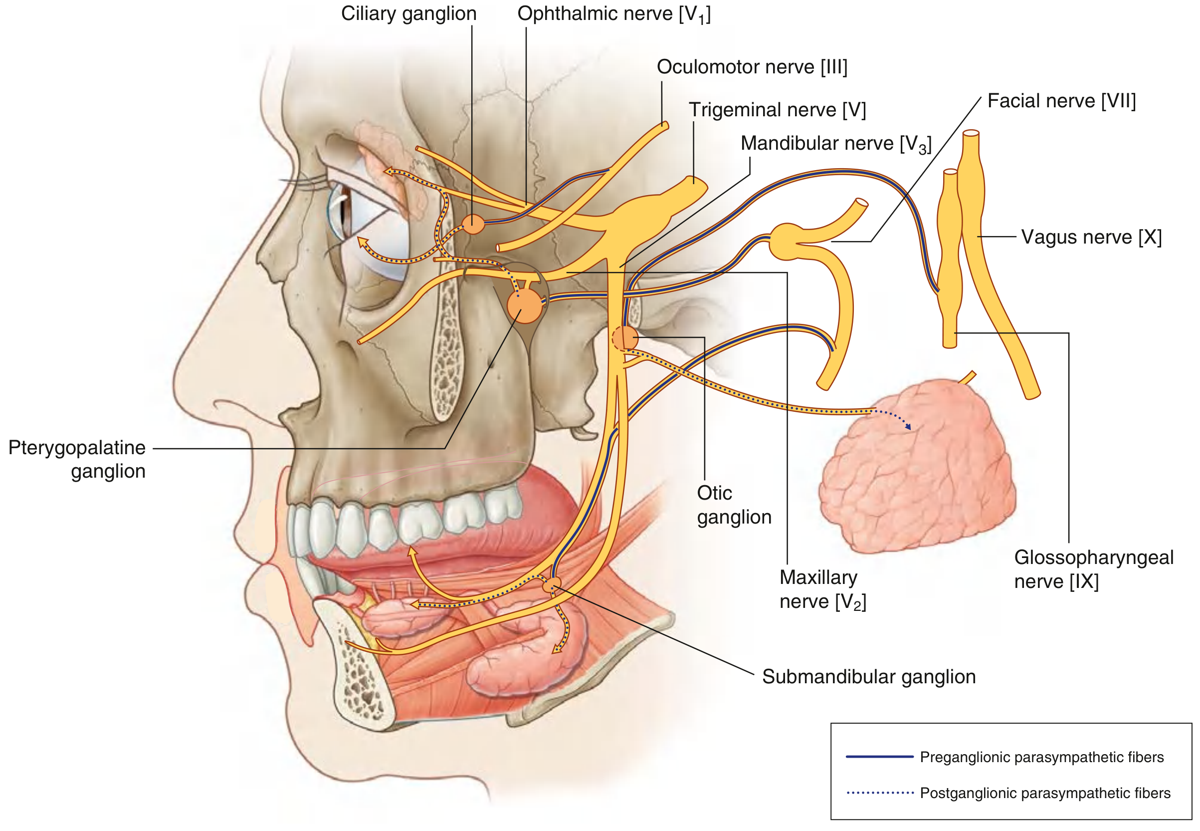
Gray's Anatomy for Students - Cranial Nerves and Parasympathetic Innervation
Foramina and Cranial Nerve Exit Points
| Cranial Nerve | Foramen/Opening |
|---|---|
| I - Olfactory | Cribriform plate (ethmoid) |
| II - Optic | Optic canal |
| III - Oculomotor | Superior orbital fissure |
| IV - Trochlear | Superior orbital fissure |
| V1 - Ophthalmic | Superior orbital fissure |
| V2 - Maxillary | Foramen rotundum |
| V3 - Mandibular | Foramen ovale |
| VI - Abducens | Superior orbital fissure |
| VII - Facial | Internal acoustic meatus → facial canal → stylomastoid foramen |
| VIII - Vestibulocochlear | Internal acoustic meatus |
| IX - Glossopharyngeal | Jugular foramen |
| X - Vagus | Jugular foramen |
| XI - Accessory | Jugular foramen |
| XII - Hypoglossal | Hypoglossal canal |
Brainstem Levels of Cranial Nerve Nuclei
| Level | Cranial Nerves |
|---|---|
| Midbrain | III (oculomotor), IV (trochlear) |
| Pons | V (trigeminal), VI (abducens), VII (facial), VIII (vestibulocochlear) |
| Medulla | IX (glossopharyngeal), X (vagus), XI (accessory - spinal cord C1-C5), XII (hypoglossal) |
| Cerebrum (not brainstem) | I (olfactory - olfactory bulb), II (optic - diencephalon/thalamus/LGN) |
Clinical Lesion Summary
| Cranial Nerve | Clinical Findings | Example Lesion |
|---|---|---|
| I (Olfactory) | Anosmia (loss of smell) | Cribriform plate fracture; closed head injury |
| II (Optic) | Blindness, visual field loss, absent pupil response | Orbital trauma; optic neuritis (MS) |
| III (Oculomotor) | Dilated pupil, ptosis, eye "down and out" | Posterior communicating artery aneurysm; uncal herniation |
| IV (Trochlear) | Vertical diplopia, head tilt | Head trauma (most common cause) |
| V (Trigeminal) | Facial numbness, jaw deviation, absent corneal reflex | Trigeminal neuralgia; acoustic neuroma |
| VI (Abducens) | Eye medially deviated, cannot abduct | Raised ICP (false localizing); pontine lesion |
| VII (Facial) | LMN: complete ipsilateral facial palsy; UMN: lower face only | Bell's palsy; stroke; parotid tumour |
| VIII (Vestibulocochlear) | Deafness, tinnitus, vertigo | Acoustic neuroma; Meniere's disease; ototoxic drugs |
| IX (Glossopharyngeal) | Loss of gag reflex (afferent), absent carotid sinus reflex | Jugular foramen tumours |
| X (Vagus) | Hoarseness, dysphagia, uvula away from lesion, absent gag (efferent) | Aortic aneurysm (left RLN); lung cancer |
| XI (Accessory) | Weak head turning + shoulder shrug | Neck surgery; jugular foramen syndrome |
| XII (Hypoglossal) | Tongue deviates toward lesion; tongue wasting | Skull base tumours; neck dissection |
High-Yield Clinical Points
-
CN III palsy with dilated pupil = compressive lesion (posterior communicating artery aneurysm, uncal herniation). Pupil-sparing CN III palsy = diabetic microvascular disease (vasa nervorum ischemia - pupillomotor fibers on the periphery are spared).
-
Bell's palsy vs. UMN facial palsy: Bell's palsy involves the forehead (complete ipsilateral paralysis). UMN lesion (stroke) spares the forehead because the frontalis muscle has bilateral cortical representation.
-
Jugular foramen syndrome (CN IX, X, XI) - caused by jugular foramen tumours (e.g. glomus jugulare, metastases).
-
CN VI palsy and raised ICP: CN VI has the longest intracranial course; it can be compressed by raised ICP independent of the site of lesion - hence a "false localizing sign."
-
The four parasympathetic cranial nerves: III, VII, IX, X (mnemonic: 3, 7, 9, 10). Remember: parasympathetic fibers in CN III, VII, and IX are distributed with branches of the trigeminal nerve to reach their target organs.
-
Chorda tympani (branch of CN VII): crosses the middle ear, exits via the petrotympanic fissure, joins the lingual nerve (CN V3). Carries taste (anterior 2/3 tongue) and presympathetic fibers to submandibular ganglion.
-
Tongue innervation summary:
- General sensation: CN V3 (lingual nerve)
- Taste anterior 2/3: CN VII (chorda tympani)
- Taste posterior 1/3: CN IX
- Motor (all except palatoglossus): CN XII
- Motor palatoglossus: CN X
-
Laryngeal innervation (CN X):
- All intrinsic laryngeal muscles innervated by the recurrent laryngeal nerve EXCEPT the cricothyroid (external branch of superior laryngeal nerve)
- Sensory above vocal folds: internal branch of superior laryngeal nerve
- Sensory below vocal folds: recurrent laryngeal nerve
Sources: Gray's Anatomy for Students (ISBN 9780323934237) | Localization in Clinical Neurology, 8e (ISBN 9781975160241) | Rosen's Emergency Medicine (ISBN 9780323757898)
This is a shared conversation. Sign in to Orris to start your own chat.