
Myocardial infarction
Myocardial infarction (MI) is death of heart muscle due to prolonged ischemia, most commonly from rupture of an atherosclerotic plaque causing coronary artery thrombosis. It presents with chest pain, ST changes on ECG, and elevated cardiac biomarkers (troponin). Management centers on rapid reperfusion - either thrombolysis or percutaneous coronary intervention (PCI).
Searching sources...
Reading File
The query is broad - MI covers pathology, clinical features, ECG, management, complications, etc. Let me clarify what aspect you need.
Asking for Preferences
What aspect of myocardial infarction are you looking for?
Researching further...
Running parallel searches across pathology, clinical features, management, and complications.
Finding Sources
Searching PubMed
"myocardial infarction"[MeSH Terms] AND management
Good - I have multiple textbook sources. Now let me pull the key sections from Braunwald's (cardiology), Robbins (pathology), and Ganong's (physiology) in parallel for a comprehensive overview.
Reading File
Reading File
Reading File
Good content from Robbins and Ganong's. Now let me pull deeper content - morphology/timeline from Robbins, clinical features from Goldman-Cecil, and management from Sabiston.
Reading File
Reading File
Reading File
Excellent content from Robbins. Let me now get the morphological timeline and complications from Robbins (the most detailed pathology source), plus the Guyton physiology section.
Reading File
Reading File
I have rich content from multiple authoritative textbooks. Let me also fetch the key pathology image showing MI morphology progression, which will be very useful visually.
Finding Sources
Reading File
Excellent - I have the full morphology table. Let me get the rest of the timeline table and complications from Robbins, and also fetch the key pathology image.
Reading File
Loading Image
Loading Image
I now have comprehensive content from multiple authoritative textbooks plus two excellent images. I have everything needed for a complete overview. Let me compile the full answer.
Myocardial Infarction - Complete Overview
Definition
Myocardial infarction (MI), commonly called a "heart attack," is necrosis of heart muscle resulting from ischemia. The 2018 joint U.S./European Cardiology task force defines it as "acute myocardial injury detected by abnormal cardiac biomarkers in the setting of evidence of acute myocardial ischemia."
Epidemiology & Risk Factors
- ~10% of MIs occur before age 40; ~45% before age 65
- Men are at greater risk than women, but the gap narrows with age
- Women are relatively protected during reproductive years; risk rises sharply after menopause (declining estrogen)
- Major risk factors mirror those for atherosclerosis: hypertension, dyslipidemia, diabetes, smoking, obesity, family history
Types of MI
As described in the Sabiston Textbook of Surgery:
- Type 1 MI - atherosclerotic plaque rupture causing occlusion and ischemic infarction (the classic "heart attack")
- Type 2 MI - demand ischemia; mismatch between oxygen supply and demand without plaque rupture (e.g., tachyarrhythmia, severe anemia, hypotension)
Type 1 MI is further classified as:
- STEMI - ST elevation + biomarker elevation (complete occlusion)
- NSTEMI - biomarker elevation without ST elevation (partial/transient occlusion)
- Unstable angina - ischemic symptoms without biomarker elevation
Pathogenesis
Robbins & Kumar Basic Pathology describes the typical sequence:
- An atheromatous plaque is eroded or disrupted by endothelial injury, intraplaque hemorrhage, or mechanical forces, exposing subendothelial collagen and necrotic contents
- Platelets adhere, aggregate, and activate, releasing thromboxane A2, ADP, and serotonin - causing further aggregation and vasospasm
- Coagulation is activated by exposure of tissue factor, adding to the thrombus
- Within minutes, the enlarging thrombus fully occludes the coronary lumen
Angiography within 4 hours of MI onset shows coronary thrombosis in ~90% of cases. When performed at 12-24 hours, only 60% still show thrombus - indicating spontaneous thrombolysis occurs.
In 10% of cases, transmural MI occurs without occlusive atherosclerosis - due to vasospasm, embolism from mural thrombi (e.g., atrial fibrillation), or valve vegetations.
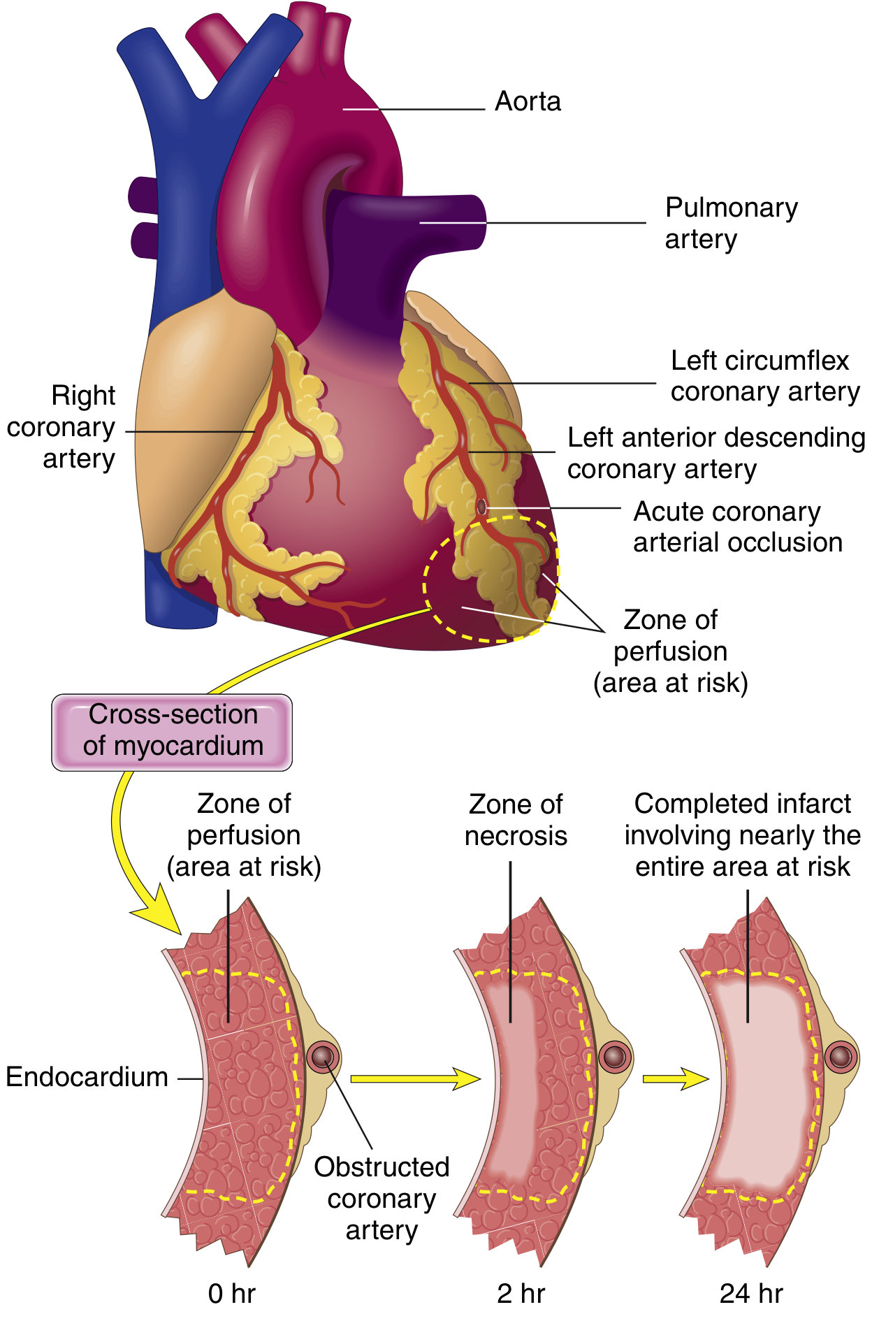
Myocardial Response to Ischemia
From Robbins & Kumar:
- Within seconds: aerobic metabolism ceases → ATP drops → lactic acid accumulates
- Within minutes: loss of contractility (reversible at this stage)
- 20-40 minutes of persistent ischemia: irreversible coagulative necrosis
- The subendocardial zone is most susceptible - it receives blood last from epicardial vessels and is compressed by high intramural pressures
Per Guyton and Hall Physiology: cardiac muscle requires ~1.3 mL O2/100g/min just to stay alive. Even 15-30% of normal resting coronary flow can prevent death of most cells - but the central core of a large infarct, with near-zero flow, dies.
A full-thickness infarct achieves its complete extent in 3 to 6 hours - the critical therapeutic window.
Coronary Artery Territories
| Artery | Territory | % of MIs |
|---|---|---|
| Left Anterior Descending (LAD) | Anterior LV wall, anterior 2/3 of septum, apex | 40-50% |
| Right Coronary Artery (RCA) | Most of RV, posterior LV (right-dominant) | 30-40% |
| Left Circumflex (LCX) | Lateral LV wall | 15-20% |
Morphological Evolution
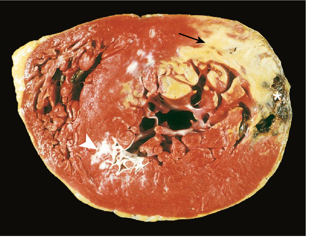
The table below is from Robbins & Kumar Basic Pathology:
| Time Frame | Gross Appearance | Light Microscopy |
|---|---|---|
| 0-½ hour | None | None |
| ½-4 hours | None | Wavy fibers at border; sarcolemmal disruption |
| 4-12 hours | Occasional dark mottling | Coagulation necrosis begins; edema; hemorrhage |
| 12-24 hours | Dark mottling | Coagulation necrosis; pyknotic nuclei; hypereosinophilic myocytes; early neutrophil infiltrate |
| 1-3 days | Mottling with yellow-tan center | Coagulation necrosis; increased neutrophils |
| 3-7 days | Hyperemic border; central yellow-tan softening | Neutrophil death; macrophages begin phagocytosis |
| 7-10 days | Maximally yellow-tan and soft; depressed red-tan margins | Well-developed macrophage phagocytosis; early granulation tissue |
| 10-14 days | Red-gray depressed borders | Established granulation tissue; new vessels; collagen deposition |
| 2-8 weeks | Gray-white scar (border to core) | Increasing collagen; decreasing cellularity |
| >2 months | Scarring complete | Dense collagenous scar |
Key point: Myocardial necrosis proceeds invariably to scar without regeneration.
ECG Changes
From Ganong's Review of Medical Physiology, three electrical defects occur in infarcted cells:
| Defect | Current Flow | ECG Change (leads over infarct) |
|---|---|---|
| Rapid repolarization | Out of infarct | ST elevation |
| Decreased resting membrane potential | Into infarct | TQ depression (recorded as ST elevation) |
| Delayed depolarization | Out of infarct | ST elevation |
Hallmark of acute MI: ST segment elevation in leads overlying the infarct; ST depression in reciprocal leads.
After days to weeks, the ST changes resolve. Dead tissue becomes electrically silent. The characteristic late change is the development of Q waves in leads overlying the infarct.
Localizing MI by ECG leads:
- Anterior (LAD): V1-V4
- Lateral (LCX): I, aVL, V5-V6
- Inferior (RCA): II, III, aVF
- Posterior (RCA/LCX): tall R in V1-V2 (reciprocal)
Cardiac Biomarkers
- Troponin I and T - most sensitive and specific; rise 3-6 hours post-MI, peak at 24-48h, remain elevated 7-10 days (troponin T) or 5-7 days (troponin I)
- CK-MB - rises in 4-6h, peaks at 24h, returns to normal in 48-72h; used to detect re-infarction
- Myoglobin - earliest to rise (1-3h) but not cardiac-specific
Clinical Presentation
Typical:
- Severe, crushing/pressure chest pain - often radiating to the left arm, jaw, or back
- Duration >20 minutes (differentiates from stable angina)
- Diaphoresis, nausea, vomiting
- Dyspnea
Atypical (especially women, elderly, diabetics):
- Epigastric pain, fatigue, jaw pain, or no pain at all ("silent MI")
Management
Acute STEMI - "Time is muscle"
-
Immediate reperfusion (within 90 min "door-to-balloon"):
- Primary PCI (preferred if available within 90 min)
- Thrombolysis (if PCI unavailable within 120 min)
-
Antiplatelet therapy:
- Aspirin 300 mg loading
- P2Y12 inhibitor (ticagrelor or clopidogrel)
-
Anticoagulation: heparin (UFH or LMWH)
-
Supplemental O2 if SpO2 <94%
-
Nitrates: sublingual/IV for chest pain and hypertension (avoid in RV infarction - can cause catastrophic preload reduction)
-
Beta-blockers: cardioprotective; start if no cardiogenic shock, bradycardia, or low EF
-
Morphine: for pain unrelieved by nitrates
NSTEMI / Unstable Angina
- Risk-stratify (TIMI/GRACE score)
- High-risk: early invasive strategy (angiography within 24-72h)
- Medical therapy: antiplatelet + anticoagulation + statin
Secondary Prevention
- Aspirin lifelong
- Statin (high-intensity, e.g., atorvastatin 40-80 mg)
- ACE inhibitor / ARB - especially if EF reduced or anterior MI; prevents remodeling
- Beta-blocker - reduces sudden cardiac death
- Aldosterone antagonist (eplerenone) if EF <40% and no renal failure
Recent meta-analysis (Chi et al., 2025) found beta-blockers for secondary prevention post-MI may not reduce mortality in patients without reduced ejection fraction or heart failure - an evolving evidence area.
Complications
From Guyton and Hall and Robbins:
Early (within days)
- Arrhythmias - most common cause of death; ventricular fibrillation accounts for 80-90% of ischemic cardiac deaths
- Cardiogenic shock - pump failure; mortality ~80% without intervention
- Acute heart failure - pulmonary edema from LV dysfunction
- Systolic stretch - non-contractile infarcted segment bulges outward during systole, worsening effective output
Mechanical complications (days 1-14, highest risk 3-7 days)
- Free wall rupture - leads to hemopericardium and tamponade; usually fatal
- Interventricular septal rupture - new harsh pansystolic murmur; requires surgical repair
- Papillary muscle rupture - acute severe mitral regurgitation; cardiogenic shock
Late complications
- Ventricular aneurysm - fibrotic bulge of infarcted wall; risk of mural thrombus, arrhythmia, and persistent heart failure
- Dressler syndrome (post-MI pericarditis) - autoimmune pericarditis 2-10 weeks post-MI; fever, pleuritic chest pain, elevated ESR; treated with NSAIDs/aspirin
- Chronic heart failure - progressive LV remodeling
- Sudden cardiac death - Peek et al., 2024 found in a meta-analysis of pooled cohorts that sudden cardiac death risk remains significantly elevated for years after MI
Key Pearls
- Irreversible injury begins at 20-40 minutes of ischemia
- Full infarct develops in 3-6 hours - the reperfusion window
- LAD occlusion = most common (40-50%); often fatal if proximal
- Subendocardium is always more vulnerable than epicardium
- ECG: ST elevation = acute injury; Q waves = established necrosis
- Right-sided MI: avoid nitrates and diuretics; maintain preload
- Cardiac troponin is the gold standard biomarker
- After MI, healing produces only scar - no myocardial regeneration
Sources: Robbins & Kumar Basic Pathology; Ganong's Review of Medical Physiology, 26th Ed; Guyton and Hall Textbook of Medical Physiology; Sabiston Textbook of Surgery; Braunwald's Heart Disease
This is a shared conversation. Sign in to Orris to start your own chat.