I now have comprehensive material with excellent diagrams. Let me compile a thorough, well-organized response on Animal Andrology.
Animal Andrology - Core Detail
Animal andrology is the science of male reproductive biology - covering anatomy, physiology, endocrinology, spermatogenesis, semen evaluation, and pathology of the male reproductive tract. While rooted in comparative biology, the core cellular and hormonal mechanisms are conserved across mammals. Below is a systematic, detailed overview.
1. Anatomy of the Male Reproductive Tract
The male reproductive system comprises:
- Testes - paired gonads performing spermatogenesis and steroidogenesis
- Excurrent duct system - rete testis, efferent ductules, epididymis, ductus deferens, urethra
- Accessory sex glands - seminal vesicles, prostate, bulbourethral glands (species-variable)
- External genitalia - penis, scrotum, prepuce
Testis Structure
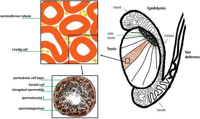
Each testis is enclosed in a fibrous capsule (tunica albuginea) and divided into lobules, each containing 1-4 coiled seminiferous tubules. The tubules are where spermatogenesis occurs. Between tubules in the interstitium lie Leydig cells (interstitial cells) - the primary site of androgen synthesis.
Tubule walls contain two cell types:
- Sertoli cells (sustentacular cells) - support and nourish germ cells
- Germ cells - at various stages of division
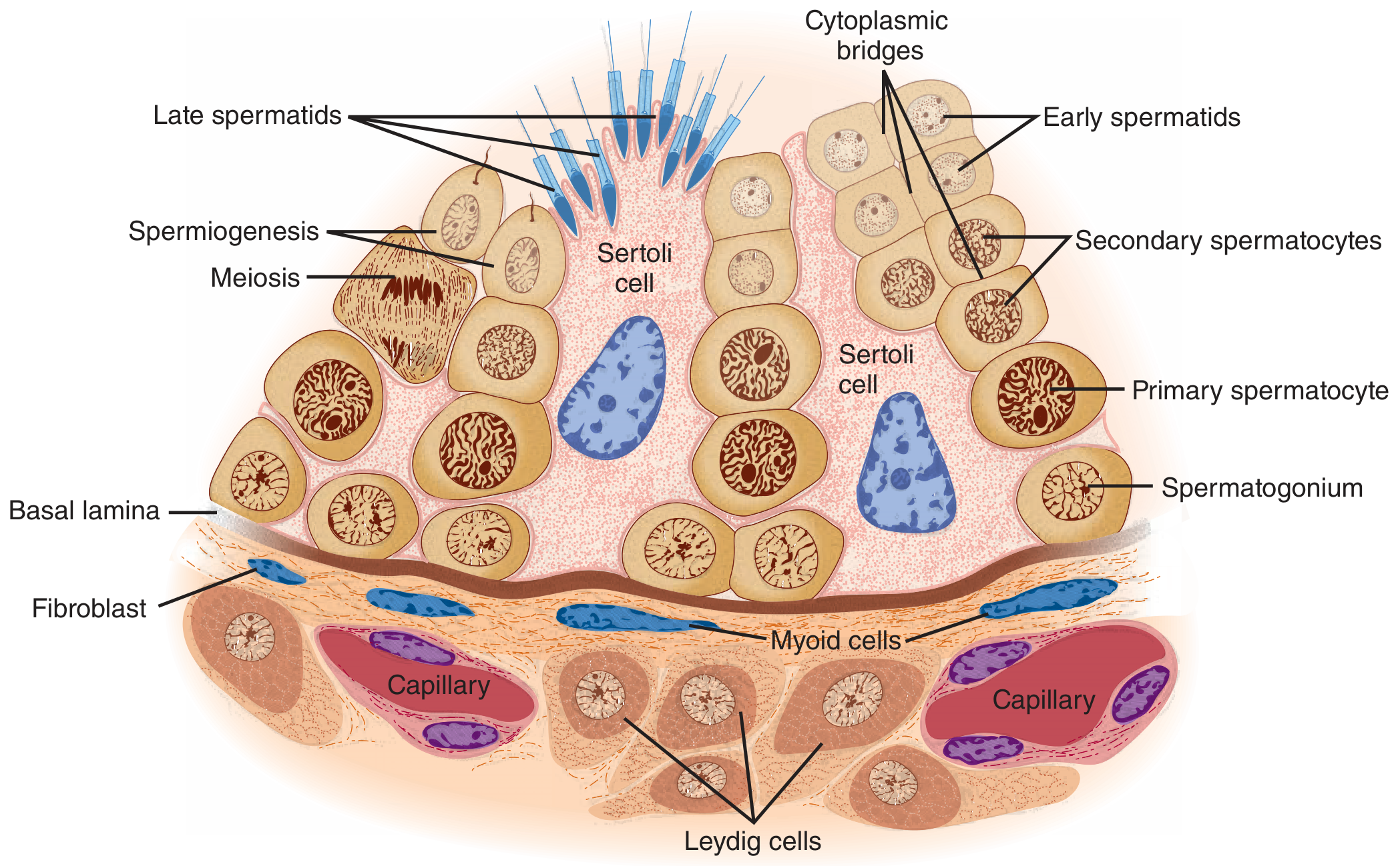
Seminiferous epithelium. Spermatogonia at the basal lamina mature progressively toward the lumen. Leydig cells lie in the interstitium between tubules. - Ganong's Review of Medical Physiology
Scrotal Thermoregulation
Spermatogenesis requires a temperature ~4-7°C below core body temperature (~32°C in humans; ~34°C in bulls). Mechanisms for testicular cooling include:
- Countercurrent heat exchange between the pampiniform plexus (venous) and testicular artery
- Air circulation around the scrotum
- Cremaster muscle reflex (drawing testes closer to/away from body)
In cryptorchid males (undescended testes), the elevated temperature impairs spermatogenesis while Leydig cell function and testosterone secretion are relatively preserved (Sertoli/germ cells are more temperature-sensitive). Bilateral cryptorchidism causes sterility.
Species notes: Boars, stallions, bulls, rams, and bucks - all scrotal; cats and dogs - scrotal; elephants and marine mammals - abdominal testes (adapted to lower core body temperature by other mechanisms).
2. Endocrine Regulation - The HPG Axis
The hypothalamic-pituitary-gonadal (HPG) axis governs testicular function:
| Level | Hormone | Source | Action |
|---|
| Hypothalamus | GnRH (decapeptide) | Pulsatile | Stimulates LH + FSH from anterior pituitary |
| Pituitary | LH (luteinizing hormone) | Anterior pituitary | Stimulates Leydig cells → testosterone synthesis |
| Pituitary | FSH (follicle-stimulating hormone) | Anterior pituitary | Acts on Sertoli cells and spermatocytes → initiates/maintains spermatogenesis |
| Testis | Testosterone | Leydig cells (95%); peripheral conversion (5%) | Spermatogenesis, secondary sex characters, libido |
| Testis | Inhibin B | Sertoli cells | Negative feedback on FSH |
| Testis | Estradiol | Aromatization of androgens (Sertoli + Leydig) | Fluid reabsorption in rete testis; negative feedback on LH |
Key feedback loops:
- Testosterone → negative feedback on hypothalamus (↓GnRH) and pituitary (↓LH)
- Inhibin B → selective negative feedback on FSH
- GnRH pulses must be episodic - continuous GnRH exposure paradoxically suppresses LH/FSH (basis of GnRH agonist-induced desensitization)
Testosterone biosynthesis pathway:
Cholesterol → Pregnenolone (via 20,22-desmolase/StAR) → Progesterone → 17-OH-progesterone → Androstenedione → Testosterone → DHT (via 5α-reductase) or Estradiol (via aromatase/CYP19)
Testosterone is carried in blood bound to sex-hormone binding globulin (SHBG) and albumin; only ~2% is free/biologically active.
3. Spermatogenesis
Spermatogenesis is the process by which spermatogonial stem cells produce haploid spermatozoa. It occurs in three phases and takes approximately 74 days in humans (50-60 days in bulls; ~42 days in stallions; ~49 days in rams).
Phase 1: Spermatogonial Phase (Mitosis)
- Type A spermatogonia (stem cells) at the basal lamina divide mitotically
- Some type A cells self-renew; others differentiate into type B spermatogonia
- Type B spermatogonia divide to yield primary spermatocytes
- Descendants remain connected by cytoplasmic bridges ensuring synchronized differentiation
Phase 2: Spermatocyte Phase (Meiosis)
- Primary spermatocytes (diploid, 2n=46 in humans) undergo Meiosis I → two secondary spermatocytes (haploid, but with paired chromatids)
- Secondary spermatocytes rapidly undergo Meiosis II → four spermatids (haploid, 23 chromosomes, 1 chromatid each)
- One spermatogonium theoretically yields 512 spermatids
Phase 3: Spermatid Phase - Spermiogenesis (Post-meiotic Differentiation)
Spermatids transform into mature spermatozoa without further division. Key events:
- Golgi apparatus forms the acrosome (cap over nucleus, rich in enzymes: acrosin, hyaluronidase, ZP-binding proteins needed for fertilization)
- Nucleus condenses and elongates (chromatin compacted by protamines replacing histones)
- Centrioles form the axoneme (9+2 microtubule arrangement) - the flagellum
- Mitochondria migrate and align around the proximal flagellum forming the midpiece (energy source for motility)
- Residual cytoplasm is shed as residual bodies (phagocytosed by Sertoli cells)
- Spermiation = release of mature spermatids from Sertoli cell cytoplasm into the tubule lumen
Sertoli Cell Functions
Sertoli cells are essential regulators of spermatogenesis:
- Form the blood-testis barrier (tight junctions between adjacent Sertoli cells) - protects haploid germ cells from immune attack
- Phagocytose residual bodies and apoptotic germ cells
- Secrete androgen-binding protein (ABP) - maintains high androgen concentration in tubular fluid
- Secrete inhibin B - FSH feedback suppressor
- Secrete Mullerian inhibiting substance (MIS/AMH) - causes regression of Mullerian ducts in embryo
- Express aromatase (CYP19) - convert androgens to estrogens
- Respond to FSH and testosterone
Hormonal Requirements
| Stage | Primary Regulator |
|---|
| Spermatogonia → spermatocytes | FSH |
| Spermatocytes → spermatids | Both FSH + testosterone |
| Spermatids → spermatozoa | Testosterone (high intra-testicular concentration via ABP) |
| After hypophysectomy | LH (→testosterone) alone can maintain spermatogenesis |
4. Sperm Structure
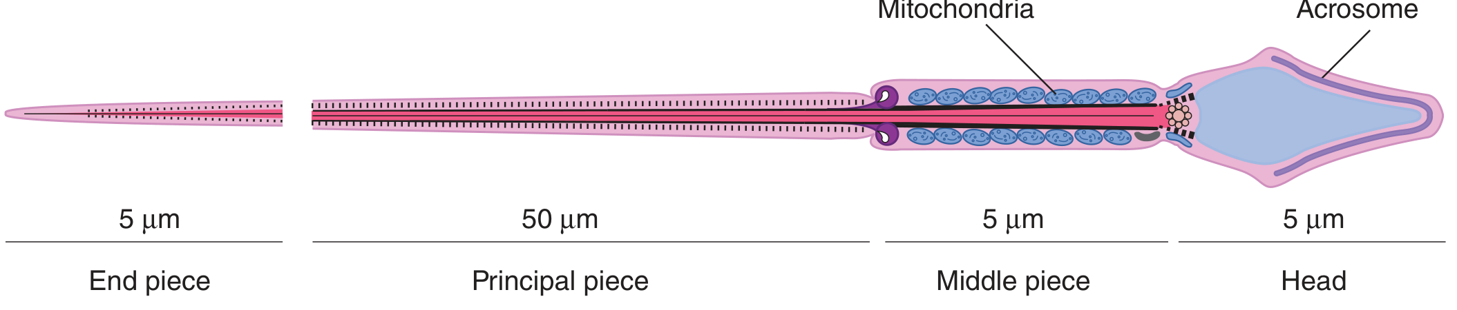
Mature spermatozoon - head (5 μm), middle piece (5 μm), principal piece (50 μm), end piece (5 μm). - Ganong's Review of Medical Physiology
| Region | Structure | Function |
|---|
| Head | Nucleus (condensed DNA) + Acrosome cap | Genetic material; acrosomal enzymes for zona pellucida penetration |
| Neck | Connecting piece | Flexion point |
| Midpiece | Axoneme + mitochondrial sheath | ATP generation via oxidative phosphorylation for motility |
| Principal piece | Axoneme + fibrous sheath | Main propulsive segment |
| End piece | Bare axoneme | Terminal segment |
Species-specific head shapes:
- Cattle/sheep/goats: oval/paddle-shaped head
- Horses: oval, piriform (pear-shaped) common
- Pigs: slightly flattened oval
- Dogs: oval
- Cats: spatula-like with a prominent hook
5. Epididymal Maturation
Spermatozoa leaving the testis are immotile and infertile. The epididymis (a highly coiled duct ~6 meters long in humans, ~50 meters in a ram) is critical for:
| Epididymal Region | Function |
|---|
| Caput (head) | Fluid reabsorption, initiation of maturation |
| Corpus (body) | Membrane remodeling, acquisition of progressive motility |
| Cauda (tail) | Storage of mature sperm (weeks-months in cool conditions) |
Key changes during epididymal transit:
- Surface membrane protein and glycoprotein modifications
- Cytoplasmic droplet migration from neck to midpiece (a sperm maturity marker; excessive proximal droplets indicate immaturity)
- Acquisition of progressive motility via activation of CatSper Ca2+ channels (alkaline-sensitive, located on principal piece)
- Disulfide bond formation in nuclear proteins and structural proteins → increased rigidity and resistance to denaturation
6. Semen Composition and Evaluation
Semen Composition
Ejaculate = spermatozoa + seminal plasma. Seminal plasma contributions by species vary:
| Gland | Contribution | Key Contents |
|---|
| Seminal vesicles | 50-80% volume | Fructose (energy source), prostaglandins, citrate, vesiculase |
| Prostate | 15-30% | Citric acid, zinc, PSA (liquefies coagulum), buffers |
| Bulbourethral (Cowper's) | 5% | Mucoproteins (clears urethral urine), lubricant |
| Epididymis | Minor | Carnitine, glycerophosphocholine |
Semen Analysis Parameters (Standard - WHO 5th Edition as Reference)
| Parameter | Definition | WHO 5th Ed. Reference (Human) |
|---|
| Volume | Total ejaculate volume | ≥1.5 mL |
| Concentration | Sperm per mL | ≥16 million/mL |
| Total sperm count | Vol × concentration | ≥39 million/ejaculate |
| Motility | % moving sperm | ≥42% total motility |
| Progressive motility | % with forward movement | ≥30% |
| Morphology | % normal forms (strict Kruger) | ≥4% |
| Vitality | % live (eosin-nigrosin) | ≥54% |
| pH | | ≥7.2 |
In animals, reference ranges differ substantially by species:
| Species | Volume (mL) | Concentration (×10⁶/mL) | Motility (%) | Morphologically Normal (%) |
|---|
| Bull | 5-10 | 800-2000 | ≥70 | ≥70 |
| Ram | 0.8-1.2 | 2000-5000 | ≥70 | ≥80 |
| Boar | 150-300 | 200-300 | ≥70 | ≥70 |
| Stallion | 50-100 | 100-150 | ≥60 | ≥60 |
| Dog | 5-30 | 200-600 | ≥70 | ≥70 |
| Tom (cat) | 0.05-0.3 | 60-1000 | ≥70 | ≥70 |
Semen Evaluation Techniques
Macroscopic:
- Volume (graduated tube), color (creamy-white; yellow = urine contamination; pink/red = blood), pH, consistency
Microscopic - motility:
- Mass motility (wave motion in concentrated species: bull, ram) - graded 0-5
- Individual progressive motility (%)
- CASA (computer-assisted sperm analysis) - measures VCL (curvilinear velocity), VSL (straight-line velocity), VAP (average path velocity), LIN (linearity), WOB (wobble)
Morphology assessment:
- Wet prep (phase contrast) or stained smear (Diff-Quik, Spermac, eosin-nigrosin)
- Classify: normal vs. primary defects (arising during spermatogenesis - head, neck defects, Dag defect, pyriform head) vs. secondary defects (arising in epididymis - distal droplets, coiled tails) vs. tertiary defects (arising post-ejaculation - minor, bent tails)
- Count minimum 100-200 sperm
Acrosomal integrity:
- Giemsa staining, phase contrast, or FITC-PSA/FITC-PNA lectin staining
- Important for fertility prediction in bulls/stallions
Sperm DNA integrity:
- SCSA (Sperm Chromatin Structure Assay) - DNA fragmentation index (DFI)
- TUNEL assay
- High DFI associated with pregnancy failure, early embryonic death
7. Spermiogram Terminology
| Term | Definition |
|---|
| Normospermia | Normal semen parameters |
| Oligospermia | Low sperm concentration |
| Azoospermia | No sperm in ejaculate (obstructive vs. non-obstructive) |
| Asthenospermia | Reduced motility |
| Teratospermia | High proportion of abnormal morphology |
| Hypospermia | Low volume |
| Necrospermia | High proportion of dead sperm |
| Oligoasthenoteratospermia (OAT) | Combined defects |
8. Breeding Soundness Evaluation (BSE)
BSE is the standard clinical assessment of male reproductive fitness used in livestock:
Components:
- Physical examination - body condition, conformation, locomotion, libido assessment
- Scrotal circumference (SC) - strong positive correlation with sperm output and daily sperm production
- Bulls: minimum 30 cm (yearling), 34 cm (mature)
- SC correlates with testicular parenchyma volume and is the best single predictor of bull fertility
- Internal reproductive tract palpation - rectal palpation (cattle) for ampullae, seminal vesicles
- Semen collection and evaluation - electroejaculation or artificial vagina
- Serving capacity test - libido and mating ability
BSE Classification (Society for Theriogenology):
- Satisfactory: passes all parameters
- Unsatisfactory: fails one or more
- Deferred: temporary problem expected to resolve
9. Semen Collection Methods
| Method | Species | Description |
|---|
| Artificial vagina (AV) | Bull, stallion, ram, boar, dog | Most physiologic; requires trained teaser animal |
| Electroejaculation (EE) | Bull, ram, llama, bison, wildlife | Rectal probe; less selective; suitable for uncooperative animals |
| Digital manipulation | Boar | Manual stimulation |
| Pharmacological | Cats, exotic felids | α2-agonist sedation + urethral catheterization |
| Vaginal aspiration | Small ruminants | Post-coitus |
| Post-mortem cauda epididymis | All | Emergency harvest from recently deceased males |
10. Semen Preservation and Cryopreservation
Chilled/Extended Semen
- Extended in egg yolk-citrate, Tris-citrate, milk-based, or commercial extenders
- Antibiotics added (gentamicin, penicillin/streptomycin) to control bacterial contamination
- Chilled to 4-5°C; shelf life 24-72 hours (species dependent)
- Used in equine industry extensively (stallion transported semen)
Cryopreservation (Liquid Nitrogen, -196°C)
Key steps:
- Semen collection and initial evaluation
- Extension with cryoprotective extender (egg yolk + glycerol most common; DMSO for some species)
- Equilibration period (4°C, 4 hours typically) - allows glycerol penetration
- Packaging (0.25 mL or 0.5 mL plastic straws, or pellets)
- Controlled cooling: room temp → 5°C (slow) → vapor phase LN₂ (-130°C, fast) → plunge into LN₂ (-196°C)
- Thaw protocol: 35-37°C water bath, 30-60 seconds
Cryoinjury mechanisms: ice crystal formation, osmotic shock, reactive oxygen species (ROS), cold shock
Species-specific cryosensitivity:
- Dogs: very sensitive (low post-thaw motility is common; use at 35-37°C)
- Cats: very poor post-thaw quality
- Boar: moderately poor (high lipid content of membrane)
- Bull: best - standard commodity
- Stallion: variable between individuals
Post-thaw acceptance criteria (bulls): ≥30% progressive motility, ≥50% morphologically normal
11. Male Reproductive Abnormalities
Hypogonadism
Hypogonadotropic hypogonadism (central cause - low LH/FSH):
- Kallmann syndrome (GnRH deficiency + anosmia)
- Hyperprolactinemia (prolactinoma; drugs like dopamine antagonists)
- Hypothalamic/pituitary lesions
- Drug-induced (anabolic steroids, GnRH analogs, opiates)
Hypergonadotropic hypogonadism (primary testicular failure - high FSH/LH):
- Klinefelter syndrome (XXY) - most common genetic cause in mammals; occurs in cats, horses, dogs
- Orchitis (viral - canine distemper; bacterial - Brucella, E. coli)
- Testicular torsion
- Irradiation, cytotoxic drugs
- Heat stress
Common Pathologies in Animals
| Condition | Species | Description |
|---|
| Cryptorchidism | All domestic spp; common in horses, dogs | Undescended testis; increased cancer risk (dogs); sterile if bilateral |
| Orchitis/epididymitis | Bulls, rams, dogs | Brucella abortus (cattle), B. ovis (sheep), B. canis (dogs) - major fertility concern |
| Varicocele | Humans, dogs | Dilation of pampiniform plexus; elevated testicular temperature → oligospermia |
| Testicular hypoplasia | Bulls, boars | Reduced SC, low sperm output; heritable in some breeds |
| Retrograde ejaculation | Dogs | Semen enters bladder; post-ejaculation urine centrifugation to recover sperm |
| Persistent penile frenulum | Bulls, dogs, horses | Congenital fibrous band; prevents extension |
| Balanoposthitis | All | Penile/preputial inflammation; infectious or traumatic |
| Dag defect | Bulls | Ruffled/folded flagellar membranes; heritable; leads to poor motility |
| Nuclear vacuoles (knobbed acrosome) | Bulls, boars | Autosomal recessive defect; abnormal acrosome; infertile |
12. Capacitation and Fertilization
After ejaculation, sperm undergo capacitation in the female reproductive tract (or in vitro):
- Removal of cholesterol from sperm plasma membrane (albumin-mediated in vivo)
- Activation of cAMP pathway
- Hyperpolarization then rapid depolarization of membrane potential
- CatSper channel activation → Ca²+ influx → hyperactivated motility (vigorous whipping)
- Prepares sperm for acrosome reaction
Acrosome reaction (triggered by zona pellucida glycoproteins, ZP3 mainly):
- Fusion of outer acrosomal membrane with plasma membrane
- Exocytosis of acrosomal contents (acrosin, hyaluronidase)
- Enables zona penetration and cortical reaction after fertilization
Sperm chemotaxis: Sperm express olfactory-like receptors; oocyte secretes odorant-like molecules guiding sperm toward the oocyte.
13. Key Species Comparisons
| Feature | Bull | Ram | Stallion | Boar | Dog |
|---|
| Ejaculate volume | 5-10 mL | 0.8-1.2 mL | 50-100 mL | 150-300 mL | 5-30 mL |
| Sperm concentration | 800-2000 ×10⁶/mL | 2000-5000 ×10⁶/mL | 100-150 ×10⁶/mL | 200-300 ×10⁶/mL | 200-600 ×10⁶/mL |
| Acrosome shape | Paddle | Paddle | Cap-like | Paddle | Oval |
| Cryopreservation | Excellent | Good | Variable | Poor | Sensitive |
| Seminal vesicles | Yes | Yes | Yes | Yes | Absent |
| Prostate | Yes | Yes | Yes | Yes | Yes (diffuse) |
| Bulbourethral glands | Yes | Yes | Yes | Yes (large) | Yes |
| Coagulum in semen | No | No | No | Yes (gel fraction) | Yes (prostatic fraction) |
14. Assisted Reproductive Technologies (ARTs) in Animal Andrology
- Artificial insemination (AI): most widely used; fresh, chilled, or frozen-thawed semen; improves genetic dissemination
- In vitro fertilization (IVF): used in cattle, horses, primates
- Intracytoplasmic sperm injection (ICSI): for severely oligospermic or post-thaw cauda epididymis sperm in horses, cats, wildlife
- Semen sexing (flow cytometry): separates X- and Y-bearing sperm by DNA content difference (~3.8% in cattle); used commercially in dairy cattle (IntelliGen®/SexedULTRA)
- Sperm banking (genome resource banking): conservation of endangered species
- Testicular sperm extraction (TESE): used in obstructive azoospermia (dogs, cats)
References:
- Ganong's Review of Medical Physiology, 26th Ed.
- Tietz Textbook of Laboratory Medicine, 7th Ed. (Male Reproductive Biology, p. 2355-2360)
- Histology: A Text and Atlas with Correlated Cell and Molecular Biology (Male Reproductive System, p. 2077-2100)
- Campbell-Walsh-Wein Urology (Spermatogenesis and Semen Evaluation)