
Changes occurring in feral circulatin after birth from standard textbooks
Reading File
Fetal circulation changes at birth involve the transition from a placental oxygen source to pulmonary gas exchange, triggering closure of several fetal shunts (foramen ovale, ductus arteriosus, ductus venosus) and a dramatic rise in pulmonary blood flow.
Searching sources...
Finding Sources
Finding Sources
Excellent - I found dedicated sections in multiple authoritative textbooks. Let me read all of them in parallel.
Reading File
Reading File
Reading File
Reading File
I now have excellent content from Guyton, Ganong, Bailey & Love, and Mulholland & Greenfield. Let me read the truncated Guyton section and fetch the key diagrams.
Reading File
Loading Image
Loading Image
I also need to read the end of the Guyton ductus venosus section that was truncated:
Reading File
I now have comprehensive, detailed content from four authoritative textbooks. Here is the complete answer:
Changes in Fetal Circulation After Birth
Fetal Circulation - Background
In fetal life, the right and left ventricles pump in parallel (not in series as in adults), owing to three anatomical shunts: the ductus venosus, foramen ovale, and ductus arteriosus. Only ~12% of blood flows through the unexpanded, non-functional lungs; ~55% passes through the placenta for oxygenation.
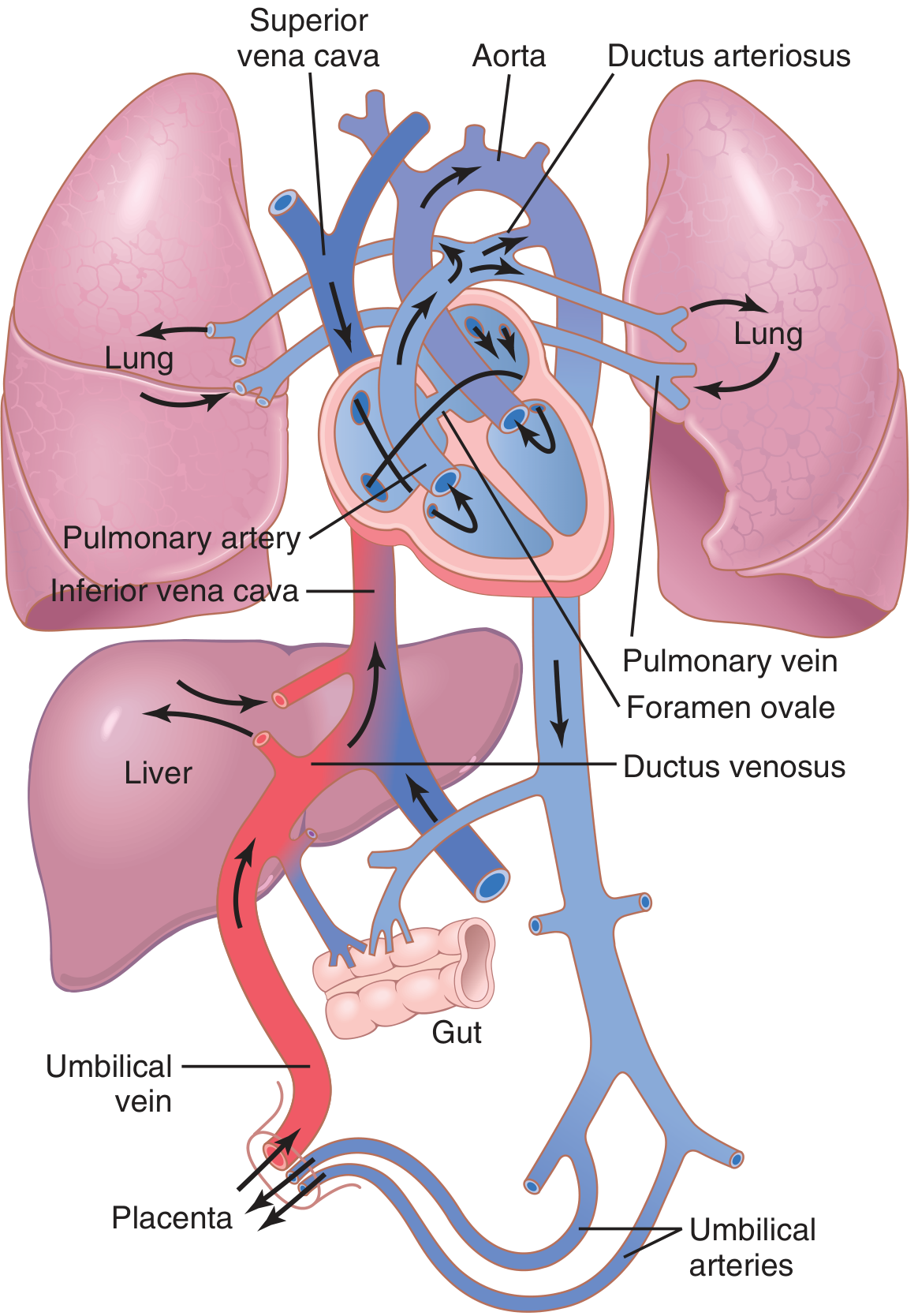
Fig. 84.4 - Organization of the fetal circulation (Guyton & Hall, p.1051)
The Two Primary Vascular Resistance Changes at Birth
At birth, two simultaneous, opposing changes in vascular resistance form the foundation of all subsequent circulatory adjustments:
| Parameter | Change at Birth | Mechanism |
|---|---|---|
| Systemic vascular resistance (SVR) | Doubles | Loss of low-resistance placental circuit after umbilical cord clamping |
| Pulmonary vascular resistance (PVR) | Falls ~5-fold | Lung expansion + relief of hypoxic vasoconstriction + NO-mediated vasodilation |
Why PVR falls so dramatically:
- In utero, compressed, unexpanded lung vessels have high resistance
- Hypoxia of fetal lungs causes tonic vasoconstriction
- At birth: lung expansion physically decompresses pulmonary vessels
- Increased alveolar O2 eliminates hypoxic vasoconstriction
- Oxygen acts as a pulmonary vasodilator via nitric oxide (NO) production
- Bradykinin, released from lungs during initial inflation, also contributes to pulmonary vasodilation
- PVR falls to less than 20% of in-utero values after the first few breaths
"The pulmonary vascular resistance falls to less than 20% of the value in utero after the lungs are expanded by the first few breaths." - Ganong's Review of Medical Physiology, 26th Ed., p.616
Closure of the Three Fetal Shunts
1. Closure of the Foramen Ovale
Mechanism: The foramen ovale closes because of a reversal in the interatrial pressure gradient.
- In fetal life: Right atrial pressure > Left atrial pressure → blood flows R→L through foramen ovale
- At birth:
- SVR rises → left atrial pressure rises
- PVR falls → increased pulmonary venous return → left atrial pressure rises further
- Right atrial pressure falls (less venous return from placenta, lower PVR)
- Result: Left atrial pressure exceeds right atrial pressure by 2-4 mmHg → the valve-like flap (septum primum) on the left side of the interatrial septum is pushed against the opening and functionally closes the foramen ovale
Permanent closure timeline:
- Functional closure: within minutes of birth
- Anatomical (adhesive) closure: within a few months to years in ~2/3 of people
- In ~20% of adults, structural fusion never completes (patent foramen ovale, PFO), but left atrial pressure keeps the valve sealed and it remains functionally closed
(Guyton & Hall, p.1052; Mulholland & Greenfield's Surgery, p.5283)
2. Closure of the Ductus Arteriosus
Mechanism: The ductus arteriosus closes due to smooth muscle vasoconstriction triggered by two key signals:
A. Increased oxygen tension:
- Fetal ductus blood: PO2 only 15-20 mmHg
- After birth: PO2 rises to ~100 mmHg within hours
- High O2 directly contracts ductal smooth muscle
B. Fall in prostaglandins:
- High concentrations of PGE2 (vasodilator) keep the ductus open in fetal life
- After birth, PGE2 synthesis is blocked (cyclooxygenase inhibition at birth), removing this vasodilatory stimulus
- Bradykinin (released from lungs on first inflation) also acts as a vasoconstrictor on the ductus
Timeline:
| Stage | Timing |
|---|---|
| Functional closure (vasoconstriction) | Within 30 minutes - 8 days |
| Anatomical closure (fibrous obliteration) | 1 to 4 months |
Hemodynamic explanation for ductus closure trigger:
- In fetal life: pulmonary artery pressure > aortic pressure → flow goes PA → Aorta (R→L)
- After birth: aortic pressure rises, pulmonary artery pressure falls → flow reverses to Aorta → PA (L→R) briefly before the ductus constricts
Patent ductus arteriosus (PDA): In premature neonates, blood O2 levels within the ductus don't drop enough to trigger remodeling. Treatment with indomethacin (COX inhibitor, blocks PGE2 synthesis) can induce closure.
(Guyton & Hall, p.1052; Ganong's, p.616; Bailey & Love's, p.985)
3. Closure of the Ductus Venosus
Mechanism: Passive then active closure driven by loss of umbilical venous flow.
- In fetal life: oxygenated umbilical venous blood bypasses the liver via the ductus venosus, flowing directly into the IVC
- After birth: umbilical cord clamping stops flow through the umbilical vein → ductus venosus pressure falls
- Within 1-3 hours: smooth muscle in ductus venosus wall contracts strongly → active closure
- Portal venous pressure rises from ~0 to 6-10 mmHg, now sufficient to drive portal blood through the liver sinuses (full hepatic circulation established)
Timeline: Complete closure between 2 to 18 days after birth
(Guyton & Hall, p.1052-1053)
Umbilical Vessels - Closure
- Umbilical arteries: After birth, rising blood O2 (via pulmonary respiration) reaches the umbilical arteries → vasoconstriction. Cold and handling of the cord augment this. Completely constricted within 3-5 minutes.
- Umbilical vein: Flow continues briefly after arterial constriction due to increased placental pressure within the contracting uterus and the baby's negative intrathoracic pressure during breathing efforts (-30 to -50 mmHg). This redistribution is called the "placental transfusion." Flow ceases within ~5 minutes.
(Ganong's, p.616)
Stimulus for First Breath
The stimulus for the newborn to initiate breathing is not primarily hypoxia. The main triggers are:
- Sudden exposure to cold air
- Light, sound, and tactile stimuli
(Ganong's Review of Medical Physiology, 26th Ed.)
Net Circulatory Result: Parallel → Series Transition
| Feature | Fetal Life | Postnatal Life |
|---|---|---|
| Ventricular arrangement | Parallel pumps | Series pumps |
| Gas exchange site | Placenta | Lungs |
| Pulmonary blood flow | ~12% of cardiac output | ~100% of cardiac output |
| Placental blood flow | ~55% | 0% |
| RV output vs LV output | RV slightly > LV | Equal (must be equal in series system) |
| PVR | High | Low |
| SVR | Low (placenta as runoff) | High |
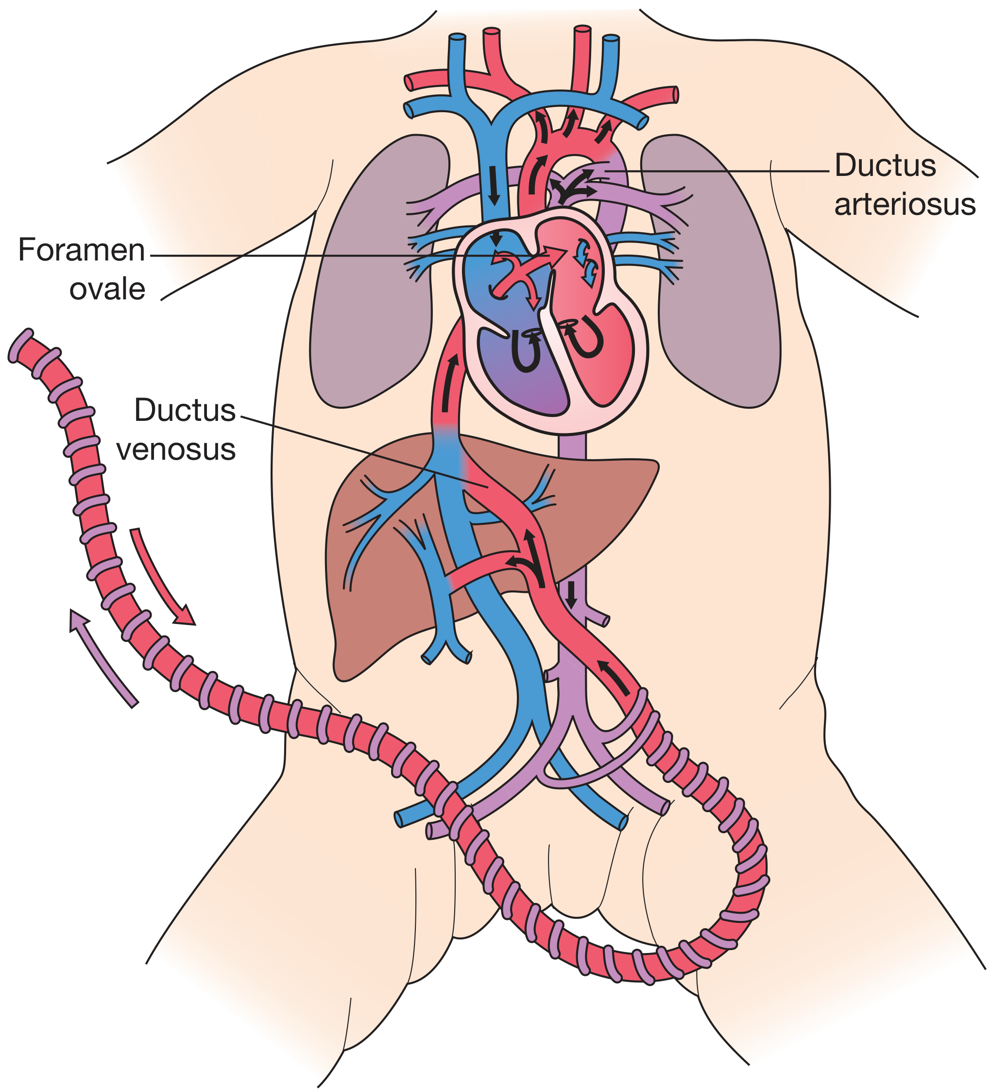
Fetal circulation showing the three shunts: ductus venosus, foramen ovale, and ductus arteriosus (Bailey & Love's, p.985)
Summary of Closure Timeline
| Structure | Functional Closure | Anatomical Closure | Adult Remnant |
|---|---|---|---|
| Umbilical arteries | 3-5 min | Days-weeks | Medial umbilical ligaments |
| Umbilical vein | ~5 min | Days-weeks | Ligamentum teres hepatis |
| Ductus arteriosus | 30 min - 8 days | 1-4 months | Ligamentum arteriosum |
| Foramen ovale | Minutes (valve closure) | Months-years (2/3 of people) | Fossa ovalis |
| Ductus venosus | 1-3 hours | 2-18 days | Ligamentum venosum |
Sources:
- Guyton and Hall Textbook of Medical Physiology, pp. 1051-1053
- Ganong's Review of Medical Physiology, 26th Ed., p. 616
- Bailey and Love's Short Practice of Surgery, 28th Ed., p. 985
- Mulholland and Greenfield's Surgery: Scientific Principles and Practice, 7th Ed., pp. 5283-5284
Other systems
The user is asking about changes in other systems (beyond the cardiovascular/fetal circulation) that occur at birth - respiratory, renal, hepatic, hematological, thermoregulation, GI, neurological, immunological, etc.
Searching sources...
Finding Sources
Finding Sources
Let me search more specifically for each system.
Finding Sources
Finding Sources
Finding Sources
Reading File
Finding Sources
Finding Sources
Excellent content from Guyton. The output was truncated - let me read the rest.
Reading File
Reading File
I have comprehensive Guyton content. Let me also fetch the key image on RBC/bilirubin changes, and read the truncated section on liver function.
Loading Image
Loading Image
Reading File
I now have all the key content from Guyton & Hall covering every major system. Here is the comprehensive answer:
Changes in Other Systems at Birth (Neonatal Physiology)
1. Respiratory System
The transition to breathing is the single most important event at birth.
Initiation of breathing:
- The stimulus is not primarily hypoxia - main triggers are sudden exposure to cold air, light, sound, and tactile stimuli
- The first breath requires a very large negative inspiratory force to overcome fluid surface tension in the airways
- Pulmonary fluid in alveoli is absorbed into the interstitial space and fetal circulation during and after the first breaths
- Delay in this process causes Transient Tachypnea of the Newborn (TTN) - usually resolves within 2-48 hours
Normal neonatal respiratory parameters:
| Parameter | Neonate | Adult (relative to body weight) |
|---|---|---|
| Respiratory rate | ~40 breaths/min | ~12-16 breaths/min |
| Tidal volume | ~16 mL/breath | Proportionally larger |
| Minute ventilation | ~640 mL/min | ~Half of neonatal rate per kg |
| Functional residual capacity | ~Half of adult value (per kg) | Reference |
- The low functional residual capacity means blood gas concentrations fluctuate more widely if breathing slows - the residual air in adult lungs buffers gas changes
- Respiratory rate of ~40 breaths/min and minute respiratory volume ~twice that of an adult per unit body weight
(Guyton & Hall, p.1053)
2. Hematological System
Red Blood Cells
- At birth: RBC count averages ~4 million/mm³
- If umbilical cord blood is stripped into the infant, RBC count rises by 0.5-0.75 million → peak ~4.75 million/mm³ in the first few hours
- In the following weeks: very few new RBCs are formed - the hypoxic stimulus of fetal life (which drove erythropoiesis) is gone
- The old fetal RBCs (which have a shorter lifespan) break down faster than new ones are made
- Result: Physiological Anaemia - RBC count falls to less than 4 million/mm³ by 6-12 weeks of life, then recovers
Haemoglobin Transition
- Fetal haemoglobin (HbF, α₂γ₂) has higher O₂ affinity than adult HbA (α₂β₂) - essential in the low-PO₂ fetal environment
- After birth, O₂ demand increases but HbF's high affinity impairs O₂ delivery to tissues
- During the 1-2 month transition period: rising levels of 2,3-DPG and mild acidosis reduce O₂ affinity and improve tissue delivery
Neonatal Jaundice (Physiological Hyperbilirubinaemia)
- In fetal life, bilirubin crosses the placenta and is excreted by the mother's liver
- After birth, the neonate must rely on its own liver - which is immature and cannot conjugate bilirubin with glucuronic acid adequately for the first week or so
- Result: Plasma bilirubin rises from <1 mg/dL to an average of 5 mg/dL in the first 3 days, causing mild jaundice of skin and sclerae, then falls as liver matures
- Treatment: Phototherapy with blue-green light (peak 478 nm) converts unconjugated bilirubin in skin microcirculation to water-soluble isomers excreted in urine and faeces
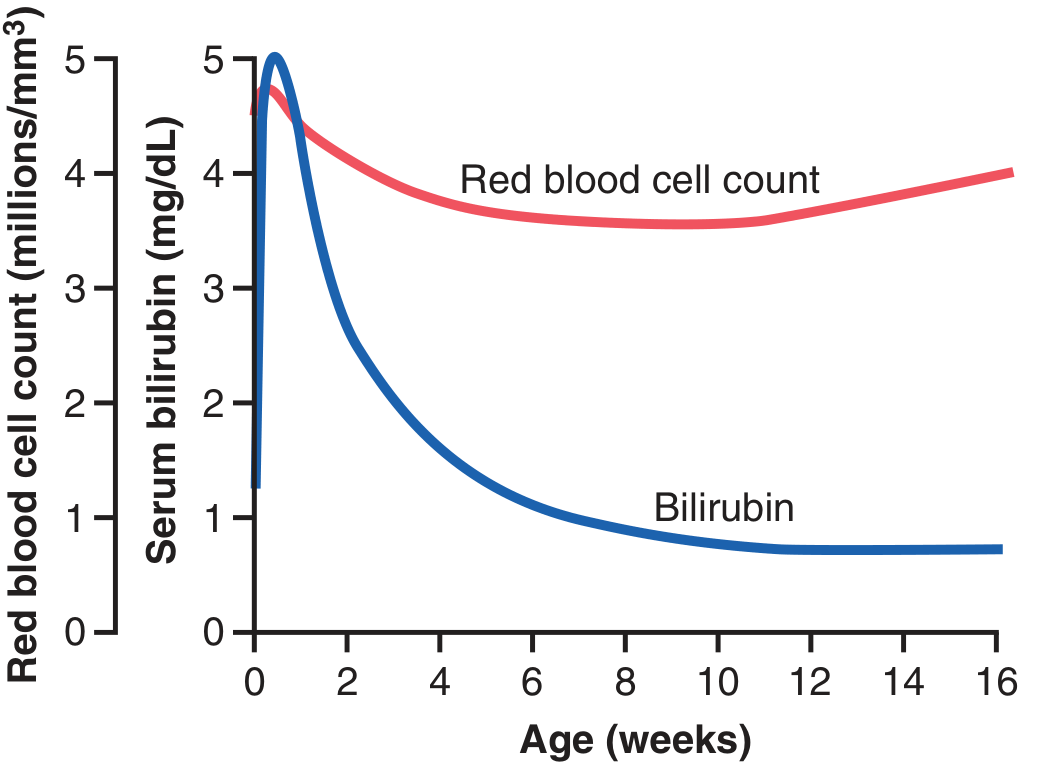
Fig. 84.6 - Physiological anaemia at 6-12 weeks and physiological hyperbilirubinaemia in first 2 weeks (Guyton & Hall, p.1054)
3. Circulatory Parameters (Post-transition)
| Parameter | Neonate | Adult (relative) |
|---|---|---|
| Blood volume | ~300 mL (or 375 mL if cord stripped) | Proportionally less per kg |
| Cardiac output | ~500 mL/min (~twice adult per kg) | Reference |
| Systolic BP (Day 1) | ~70 mmHg | ~115 mmHg |
| Diastolic BP (Day 1) | ~50 mmHg | ~70 mmHg |
- BP gradually rises over months toward ~90/60 mmHg, then slowly to adult values at adolescence
- Cord stripping: the extra 75 mL of blood redistributes - fluid shifts to tissues, raises haematocrit, blood volume returns to ~300 mL. Some risk of mild pulmonary oedema
(Guyton & Hall, p.1053)
4. Liver Function
The neonatal liver is functionally immature in four key ways:
- Poor bilirubin conjugation - cannot conjugate bilirubin with glucuronic acid adequately → physiological jaundice (described above)
- Low plasma protein synthesis - plasma protein concentration falls 15-20% below normal in first weeks → risk of hypoproteinaemic oedema
- Deficient gluconeogenesis - blood glucose falls to 30-40 mg/dL (~40% of normal) in the unfed neonate; the infant depends on stored fat for energy until feeding is established
- Deficient coagulation factor synthesis - liver produces insufficient clotting factors → risk of neonatal bleeding (Vitamin K-dependent factors II, VII, IX, X particularly affected)
(Guyton & Hall, p.1054)
5. Renal Function
- Renal functional development is not complete until the end of the first month of life
- Urine concentrating ability: Neonate can concentrate urine to only 1.5× plasma osmolality vs. adult capacity of 3-4× plasma osmolality
- Fluid turnover: Rate of fluid intake and excretion is 7× greater per unit weight than in adults
- Metabolic rate is 2× greater per kg → 2× more acid produced → tendency toward metabolic acidosis
- Consequences: Neonates are highly vulnerable to:
- Acidosis
- Dehydration
- (Less commonly) Overhydration
(Guyton & Hall, p.1054)
6. Thermoregulation
- Metabolic rate is ~twice that of adults per kg - generates more heat
- BUT body surface area is disproportionately large relative to body mass → heat is rapidly lost
- At birth: Body temperature often falls several degrees in the first few hours
- Recovery: Returns to normal in 7-10 hours
- Temperature regulation remains immature for the early days of life → marked fluctuations continue for days
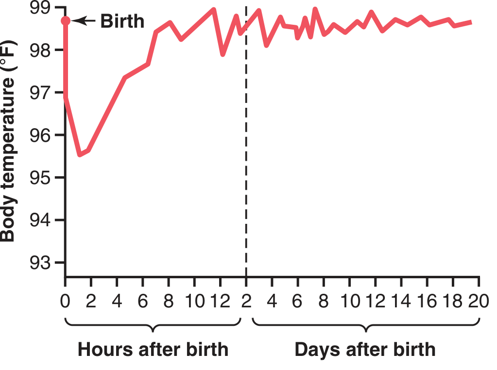
Fig. 84.7 - Temperature falls from ~98.6°F to ~95.5°F in first 2 hours, recovering by 7-10 hours; oscillates for days (Guyton & Hall, p.1055)
- Premature infants are especially vulnerable - less subcutaneous fat, even larger surface-to-mass ratio, poorer thermoregulatory mechanisms
7. Gastrointestinal System
The neonate's GI system is functional but differs in three ways:
- Pancreatic amylase deficiency - starches are poorly digested; not significant if diet is breast milk/formula (low starch)
- Reduced fat absorption - cow's milk (high fat) is less well absorbed; breast milk has lipases that compensate
- Unstable blood glucose - liver dysfunction means glucose cannot be reliably maintained
Meconium: First bowel movement (dark, sterile) typically within 24-48 hours. Absence suggests bowel obstruction (e.g. Hirschsprung disease, meconium ileus).
(Guyton & Hall, p.1054-1055)
8. Nutritional Requirements - Key Deficiency Risks
| Nutrient | Issue | Consequence |
|---|---|---|
| Calcium + Vitamin D | Rapid bone ossification + poor gut Ca absorption without Vit D | Rickets (especially in premature) within weeks |
| Iron | Maternal iron stores in fetal liver last 4-6 months only | Iron-deficiency anaemia after ~3 months if diet inadequate |
| Vitamin C | Not stored significantly in fetal tissues | Scurvy risk if diet lacking (cartilage, bone, capillary fragility) |
9. Hormonal / Neurogenic Control Systems
- All hormonal and neurogenic control systems show instability in the neonate
- Due to: (a) immature organ development AND (b) control systems not yet calibrated to extrauterine life
- Examples: poor blood glucose regulation, poor temperature regulation, immature autonomic reflexes
10. Haemoglobin / Oxygen Delivery - Transition Summary
| Feature | Fetal | Neonatal transition |
|---|---|---|
| Haemoglobin type | HbF (α₂γ₂) - high O₂ affinity | Switches to HbA (α₂β₂) over 1-2 months |
| O₂ delivery mechanism | High affinity needed (low PO₂ environment) | Rising 2,3-DPG + mild acidosis right-shifts curve → better tissue O₂ delivery |
| Erythropoiesis | High (hypoxia-driven) | Falls after birth; physiological anaemia by 6-12 weeks |
Source: Guyton and Hall Textbook of Medical Physiology, pp. 1053-1055 (Chapter 84: Fetal and Neonatal Physiology); Mulholland & Greenfield's Surgery, 7th Ed., p.5283-5284; Ganong's Review of Medical Physiology, 26th Ed., p.616
This is a shared conversation. Sign in to Orris to start your own chat.