
Wound healing
"wound healing"[MeSH Terms]
wound healing phases hemostasis inflammation proliferation remodeling diagram
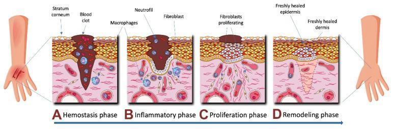
A medical pathophysiology diagram illustrating the four sequential phases of human skin wound healing: Hemostasis (A), Inflammation (B), Proliferation (C), and Remodeling (D). The cross-sectional views depict the stratum corneum, epidermis, and dermis. In the Hemostasis phase (A), a large blood clot fills the deep wound tissue with early macrophage presence. The Inflammatory phase (B) shows a persisting clot with an influx of neutrophils, macrophages, and early fibroblasts. The Proliferation phase (C) illustrates significant fibroblast activity and cellular multiplication beneath a shrinking scab as the epidermal gap begins to close. The final Remodeling phase (D) depicts a freshly healed epidermis and dermis with organized tissue structure and the absence of the initial clot and inflammatory cells. This illustration is an educational resource for understanding dermatological repair mechanisms and cellular recruitment during tissue injury.
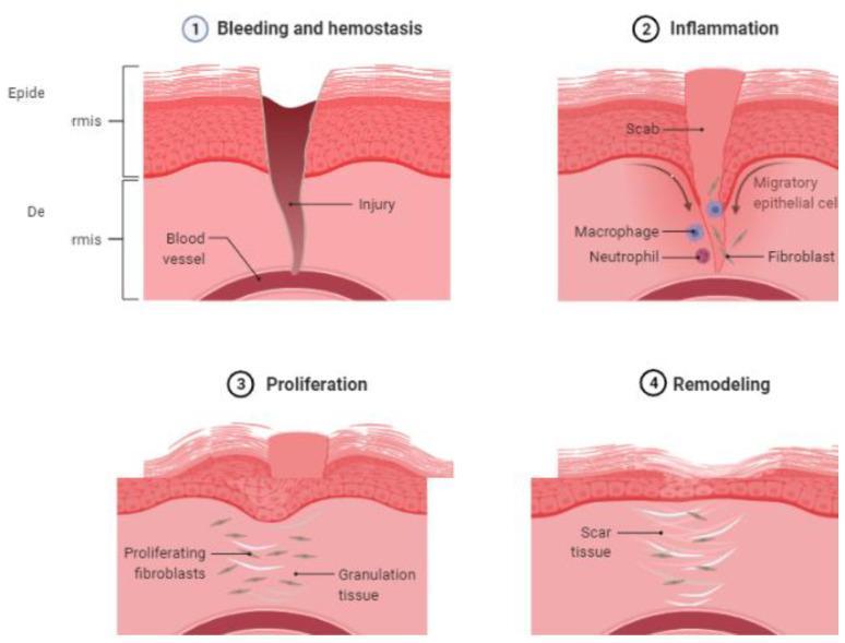
Anatomical diagram illustrating the four sequential phases of cutaneous wound healing: (1) Bleeding and Hemostasis, (2) Inflammation, (3) Proliferation, and (4) Remodeling. The cross-sectional illustrations represent the epidermis and dermis layers. Phase 1 shows a full-thickness injury penetrating through to the underlying blood vessel, depicting acute hemorrhage. Phase 2 (Inflammation) displays the formation of a surface scab, with arrows indicating the movement of migratory epithelial cells, alongside an infiltration of macrophages, neutrophils, and fibroblasts. Phase 3 (Proliferation) demonstrates the development of granulation tissue, characterized by proliferating fibroblasts and the reconstruction of the epidermal layer beneath the residual scab. Phase 4 (Remodeling) illustrates the final stage where the epidermis is fully restored, and the dermis contains organized scar tissue in place of the original injury site. This educational infographic provides a visual timeline of cellular responses and tissue structural changes during the natural course of human skin repair, useful for medical students and clinical practitioners studying pathophysiology.
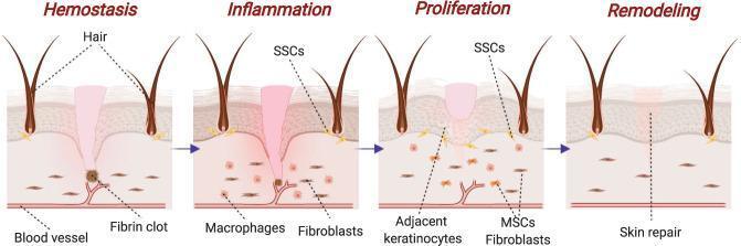
A pathophysiology diagram illustrating the four chronological stages of skin wound healing: Hemostasis, Inflammation, Proliferation, and Remodeling. The visual depicts a cross-section of the integumentary system including the epidermis, dermis, hair follicles, and vasculature. 1) Hemostasis: Shows a fibrin clot formation at the site of vascular injury to stop bleeding. 2) Inflammation: Characterized by the presence of macrophages for phagocytosis and fibroblasts beginning to migrate into the wound site; skin stem cells (SSCs) are indicated near the hair follicles. 3) Proliferation: Displays the migration and activity of adjacent keratinocytes, mesenchymal stem cells (MSCs), and fibroblasts to form a provisional extracellular matrix. 4) Remodeling: Demonstrates the final 'skin repair' phase where tissue integrity is restored, collagen fibers are realigned, and the wound site is closed. This diagram serves as an educational tool for medical students to understand the cellular and structural progression of cutaneous tissue repair.
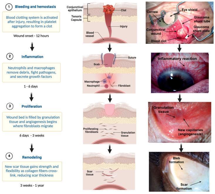
This infographic and clinical photograph series illustrates the four stages of ocular wound healing following glaucoma filtration surgery (e.g., shunt implantation). The diagram is organized into four horizontal rows representing the chronological phases: (1) Bleeding and Hemostasis (0–12 hours), showing platelet aggregation and a visible blood clot at the conjunctival wound site alongside surgical elements like the glaucoma shunt tube and plate. (2) Inflammation (1–6 days), characterized by neutrophil and macrophage recruitment and a clinical appearance of diffuse episcleral/conjunctival injection (redness). (3) Proliferation (6 days–3 weeks), showing fibroblast migration, collagen deposition, and angiogenesis (new capillary formation) clinical visible as fleshy granulation tissue. (4) Remodeling (3 weeks–1 year), depicting scar tissue maturation and the formation of a functional surgical bleb. The figure combines anatomical cross-sections of the conjunctiva and Tenon's capsule with real-world clinical images to demonstrate the physiological transition from acute injury to long-term surgical outcome in ophthalmology.
Wound Healing
Phases of Wound Healing
Phase 1: Hemostasis (Minutes to Hours)
- ADP, serotonin, thromboxane A2 - promote platelet aggregation and vasoconstriction
- PDGF (platelet-derived growth factor) - a powerful chemoattractant for fibroblasts and macrophages
- TGF-β - initiates matrix synthesis
- Fibronectin and fibrinogen - form the provisional matrix (the scaffold for cell migration)
Phase 2: Inflammation (Days 1–5)
- Phagocytosis of bacteria and debris
- Release of proteases (collagenase, elastase) to break down damaged matrix
- Release of reactive oxygen species
- Continue phagocytosis and debridement
- Release growth factors (PDGF, TGF-β, FGF, VEGF) that drive subsequent phases
- Orchestrate the transition from inflammation to proliferation
Phase 3: Proliferation (Days 4–21)
3a. Fibroplasia and Granulation Tissue Formation
- Oxygen (delivered by new blood vessels)
- Cofactors: Vitamin C (for proline/lysine hydroxylation), zinc, iron
3b. Angiogenesis
3c. Epithelialization
- Lose attachment to underlying dermis
- Enlarge and migrate across the provisional matrix
- Fixed basal cells undergo rapid mitotic division
- Cells migrate in a "leapfrog" fashion until the defect is covered
- Once bridged, cells become columnar, increase mitotic activity, and re-establish layering/keratinization
Phase 4: Maturation and Remodeling (Day 21 - Months to Years)
- Type III collagen (weaker, disorganized) is gradually replaced by Type I collagen (stronger, organized along tension lines)
- MMPs (matrix metalloproteinases) degrade old matrix; TIMPs (tissue inhibitors of MMPs) regulate this
- TGF-β increases new collagen transcription AND decreases collagen breakdown by stimulating TIMP synthesis
- Collagen fibers become cross-linked and reoriented along lines of tension
- Cellularity and vascularity decrease - the wound "matures"
- Maximum wound strength (~80% of original) is reached at ~3 months
- Wounds never fully regain 100% of pre-injury tensile strength
Types of Wound Healing
| Type | Description | Example |
|---|---|---|
| Primary intention | Wound edges reapproximated soon after injury | Surgical incision, sutured laceration |
| Secondary intention | Wound left open; heals by granulation tissue, contraction, epithelialization | Abscess cavity, infected wound |
| Tertiary intention (delayed primary) | Wound left open initially, then closed after 4-5 days | Contaminated wound that is later sutured |
Wound Contraction
- Occurs during the proliferative phase
- Mediated by myofibroblasts - modified fibroblasts expressing alpha-smooth muscle actin
- Can reduce wound area by up to 40-80%
- Beneficial in open wounds, but excessive contraction causes contracture (e.g., post-burn)
Growth Factors in Wound Healing
| Growth Factor | Source | Key Role |
|---|---|---|
| PDGF | Platelets, macrophages | Chemotaxis of fibroblasts/macrophages |
| TGF-β | Platelets, macrophages, T-cells | Collagen synthesis, fibroblast recruitment, fibrosis |
| EGF | Platelets, saliva | Epithelialization |
| VEGF | Macrophages, keratinocytes | Angiogenesis |
| bFGF | Macrophages, endothelium | Angiogenesis, fibroblast proliferation |
| IGF-1 | Liver, fibroblasts | Fibroblast proliferation |
Factors Impairing Wound Healing
Local Factors
- Infection - most common cause of delayed healing; biofilm formation in chronic wounds perpetuates non-healing
- Ischemia/hypoxia - oxygen is required for collagen synthesis and bacterial killing
- Foreign body - perpetuates inflammation
- Irradiation - damages vasculature and fibroblasts
- Necrotic tissue - inhibits cell migration
Systemic Factors
- Diabetes mellitus - impairs neutrophil function, microvascular disease, neuropathy
- Malnutrition - Vitamin C deficiency impairs collagen hydroxylation; zinc deficiency impairs fibroblast function
- Corticosteroids - suppress inflammation, inhibit fibroblast proliferation, reduce collagen synthesis
- Chemotherapy/immunosuppression - reduce inflammatory cell numbers
- Age - reduced growth factor production, slower cell turnover
- Anemia/poor perfusion - limits oxygen delivery
Excess Healing (Abnormal Scarring)
| Feature | Hypertrophic Scar | Keloid |
|---|---|---|
| Boundaries | Within wound margins | Extends beyond wound margins |
| Regression | May regress over time | Rarely regresses |
| Skin types | Any | More common in darker skin types |
| Common sites | Any high-tension area | Face, earlobes, deltoid, presternal |
| Treatment | Silicone, steroid injection, compression | Excision + adjuvant radiotherapy; may recur |
Wound Healing in Specific Tissues
- Bone: Heals through callus formation (endochondral ossification) - inflammatory phase, soft callus (cartilage), hard callus (woven bone), remodeling to lamellar bone
- Tendon: Slow and poor due to poor vascularity; intrinsic (tenocyte) and extrinsic (synovial fibroblast) repair mechanisms
- Nerve: Regenerates at ~1 mm/day (axonal regrowth along Schwann cell tubes)
- Cartilage: Very limited repair due to avascularity and lack of progenitor cells
- GI tract: Heals rapidly; submucosal collagen provides strength
Fetal Wound Healing
- High HA (hyaluronic acid) content in fetal ECM
- Low TGF-β signaling
- More robust inflammatory response resolution
- Intrinsic properties of fetal fibroblasts
Treatment Principles
- Moist wound environment - increases epithelialization rate by 50% (Galen first described this; proven scientifically in the 20th century)
- Debridement - removal of necrotic tissue
- Infection control - topical antiseptics, antibiotics when indicated
- Modern dressings - hydrocolloids, foams, alginates, hydrogels (recent systematic review, 2025, PMID 40118421 confirms hydrogel dressings benefit chronic wounds)
- Negative pressure wound therapy (NPWT) - removes exudate, promotes granulation tissue
- Hyperbaric oxygen - improves oxygenation in ischemic/diabetic wounds
- Skin substitutes/cellular products - for chronic wounds and burns
Key Summary Points
- Wound healing is hemostasis → inflammation → proliferation → remodeling (overlapping phases, not sequential)
- Macrophages are the master regulators of wound healing
- Collagen synthesis requires oxygen, Vitamin C, zinc, and iron
- Maximum tensile strength (~80% original) is reached at ~3 months; 100% is never regained
- TGF-β is the most important profibrotic/scarring growth factor
- Moist environment accelerates healing
- Wound strength at 1 week = ~10%; at 3 weeks = ~50%; at 3 months = ~80%
Sources: Schwartz's Principles of Surgery, 11th Ed. | Bailey and Love's Short Practice of Surgery, 28th Ed. | Sabiston Textbook of Surgery
In short phase
Phases of Wound Healing
Phase 1 - Hemostasis (Minutes to Hours)
- Vasoconstriction → platelet aggregation → fibrin clot
- Clot = scaffold for healing + reservoir of growth factors (PDGF, TGF-β)
Phase 2 - Inflammation (Days 1–5)
- Neutrophils first (24–48 hrs) → phagocytosis, debris clearance
- Macrophages next (day 2–3) → master regulators, release growth factors
- Clinical signs: redness, swelling, warmth, pain (normal - not infection)
Phase 3 - Proliferation (Days 4–21)
- Fibroplasia → fibroblasts lay down Type III collagen; granulation tissue forms
- Angiogenesis → new capillaries driven by VEGF
- Epithelialization → keratinocytes migrate and cover the defect
- Contraction → myofibroblasts shrink wound size
Phase 4 - Remodeling (Day 21 → Months/Years)
- Type III collagen replaced by stronger Type I collagen
- MMPs degrade old matrix; TGF-β controls balance
- Maximum tensile strength = ~80% of original (at ~3 months)
- 100% strength is never fully regained
Memory aid: H-I-P-R → Hemostasis, Inflammation, Proliferation, Remodeling