
Posture for 15 marks
posture definition types good bad posture physiotherapy kinesiology 15 marks
https://www.healthline.com/health/bone-health/the-4-main-typ…
posture types kyphosis lordosis scoliosis flatback forward head posture
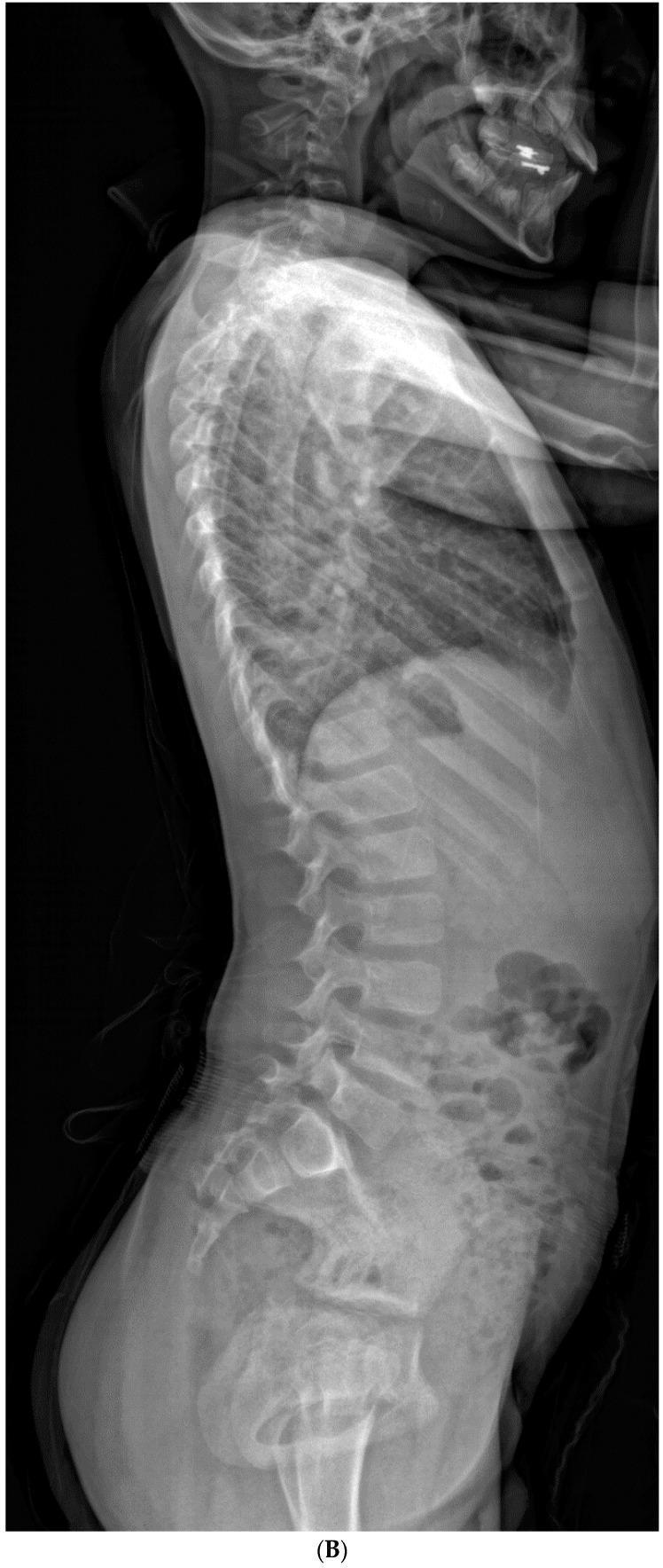
This lateral spinal radiograph of an 8-year-old male demonstrates significant sagittal plane deformity associated with Congenital Cervical Scoliosis (CCS). The image shows a marked cervical kyphosis resulting in a pronounced forward head posture. This cervical deformity is associated with a compensatory increase in lumbar lordosis to maintain sagittal balance. Key anatomical findings include anterior narrowing of intervertebral disc spaces and wedge-shaped vertebral bodies within the cervical region, contributing to the kyphotic curvature. The thoracic spine exhibits a standard kyphotic curve, while the lumbar spine shows increased lordosis. The image illustrates the natural history of spinal compensation where thoracic and lumbar segments adjust their alignment in response to primary cervical vertebral anomalies, such as hemivertebrae or formation failure, in patients with Klippel-Feil syndrome or similar congenital conditions.
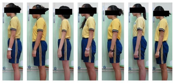
A series of seven lateral clinical photographs showing the habitual standing postures of female adolescents aged 10–13. The image illustrates various postural deviations common in musculoskeletal screening, including forward head posture (anterior translation of the head relative to the shoulders), rounded shoulders, and variations in spinal sagittal alignment such as thoracic kyphosis and flat back. Each subject is viewed from the side to demonstrate the relationship between the cervical, thoracic, and lumbar regions. The visual serves to distinguish between neutral spinal alignment—where the ear is aligned over the shoulder and hip—and postural impairments that may be associated with signs of scoliosis or muscle activity imbalances. Key clinical features shown include the degree of cervical lordosis, thoracic curvature, and shoulder elevation, which are used in orthopedic assessments to screen for spinal conditions and determine the need for further diagnostic intervention like the Adam's forward bending test or angle of trunk rotation (ATR) measurement.
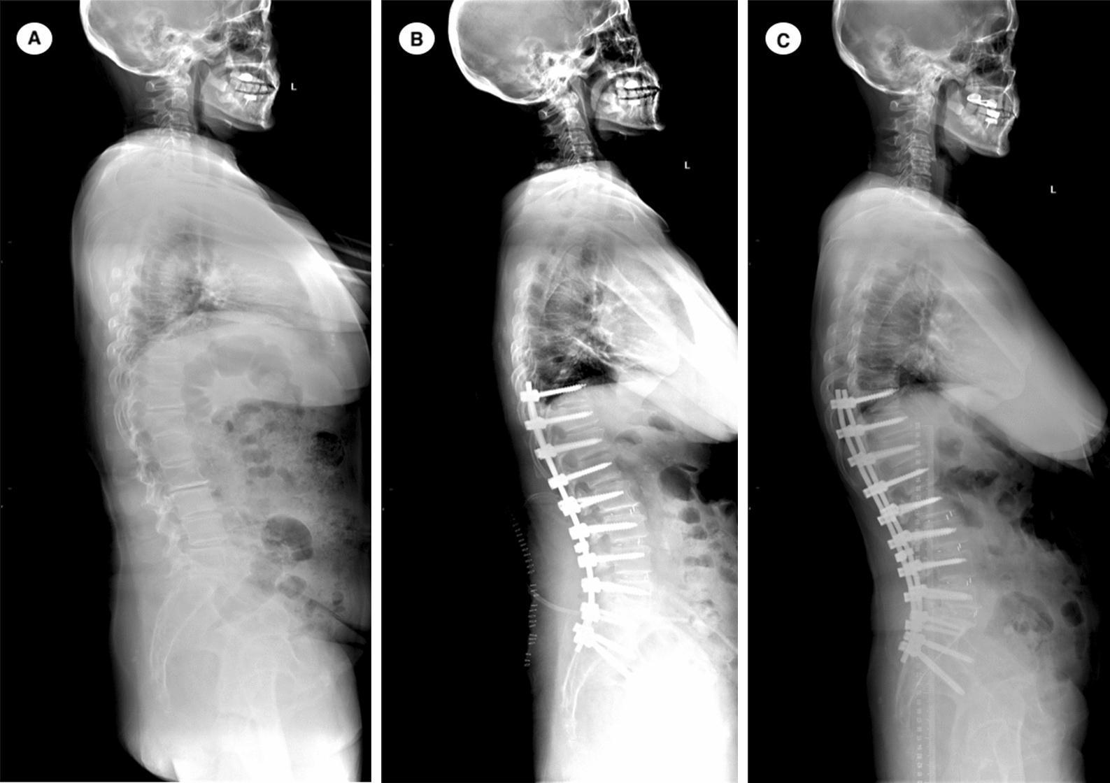
This diagnostic image consists of three side-by-side lateral full-spine radiographs (labeled A, B, and C) illustrating the surgical management of degenerative lumbar scoliosis (DLS). Image A (Preoperative): Shows sagittal imbalance characterized by increased thoracic kyphosis and loss of lumbar lordosis, resulting in a forward-leaning posture. Image B (Postoperative): Displays thoracolumbar fusion and stabilization with spinal instrumentation extending from T10 to S2. Posterior pedicle screws and rods are visible. The sagittal alignment is significantly corrected, restoring lumbar lordosis and achieving a more neutral Sagittal Vertical Axis (SVA), with the head positioned over the pelvis. Image C (18-month Follow-up): Demonstrates a complication known as Proximal Junctional Failure (PJF). While the instrumentation remains intact, there is a pathologic focal kyphosis occurring at the vertebral levels immediately superior to the proximal end of the fusion construct (UIV). This failure results in a progressive forward sagittal tilt compared to the immediate postoperative state. The sequence serves as an educational example of spinopelvic parameter correction and subsequent junctional complications in long-construct adult spinal deformity surgery.
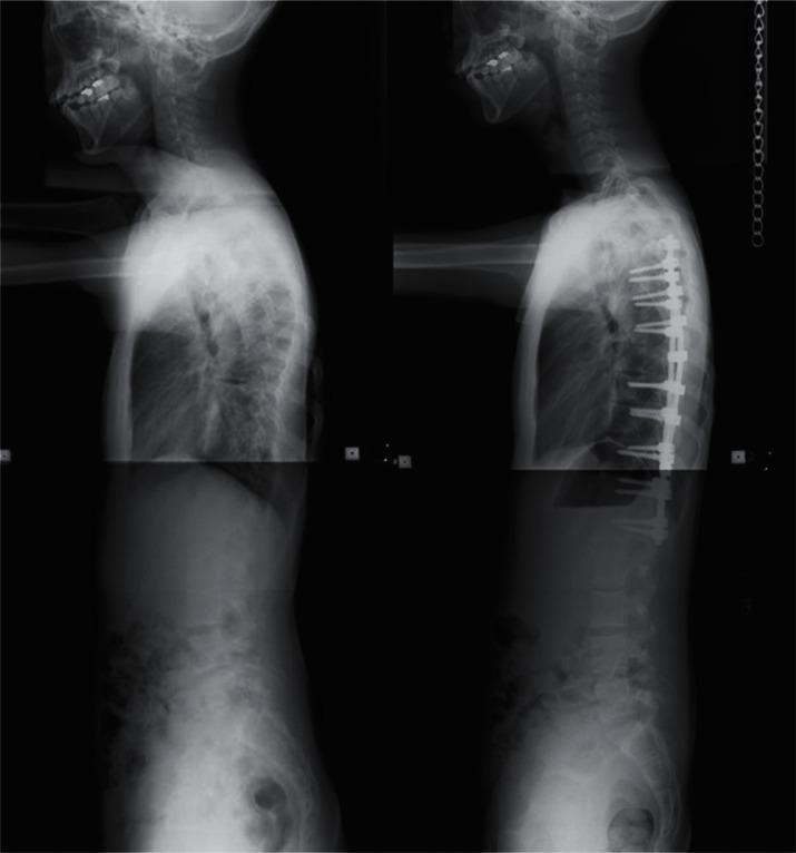
This diagnostic image consists of two side-by-side lateral x-ray radiographs of the human spine, demonstrating a 'before and after' comparison of surgical correction for spinal deformity. The left panel shows a preoperative state characterized by severe thoracic kyphosis and a compensatory forward head posture with loss of normal cervical lordosis. The right panel demonstrates the postoperative outcome following posterior spinal fusion. Visible medical instrumentation includes a segmental pedicle screw construct, featuring multiple pedicle screws anchored into the thoracic and upper lumbar vertebral bodies, connected by longitudinal stabilization rods. This surgical intervention has resulted in significant reduction of the kyphotic curvature and improved sagittal balance, aligning the head more vertically over the pelvis. The images serve as an educational example of orthopedic spinal reconstruction and the use of modern internal fixation systems to treat adolescent or adult idiopathic scoliosis and kyphosis.
POSTURE (15 Marks)
1. Definition
"Posture is the attitude assumed by the body either with support during the course of muscular activity, or as a result of the co-ordinated action performed by muscles working to maintain stability."
2. Types of Posture
A. Static Posture
- Examples: standing at attention, sitting at a desk, lying supine.
B. Dynamic Posture
- Examples: walking, running, lifting, throwing.
3. Ideal / Good Posture
- The body is in optimal alignment with minimal stress on muscles, joints, ligaments, and bones.
- Energy expenditure is minimal.
- The body is in equilibrium - the line of gravity (plumb line) passes through the standard landmarks.
Plumb Line Test (Standard Postural Assessment)
| View | Plumb Line Passes Through |
|---|---|
| Lateral | Ear lobe - Acromion - Greater trochanter - Just posterior to patella - Anterior to lateral malleolus |
| Anterior/Posterior | Midline of the skull, sternum, umbilicus, midline of pelvis, midpoint between feet |
4. Factors Maintaining Posture
- Bony structures - shape of joints, articular surfaces
- Ligaments - passive restraints
- Muscles (postural/antigravity muscles) - active support; key muscles include erector spinae, iliopsoas, quadriceps, soleus, and gluteals
- Fascia - myofascial continuity
- Neurological mechanisms - spinal reflexes, righting reflexes, proprioceptive and vestibular input
5. Bad / Poor Posture and Postural Deformities
A. Kyphosis (Hyperkyphosis)
- Exaggerated convexity of the thoracic spine (hunchback).
- Rounded upper back, shoulders rolled forward.
- Causes: osteoporosis, Scheuermann's disease, muscle weakness, prolonged sitting.
- Effects: back pain, impaired breathing, increased fracture risk (70% higher in older women).
B. Lordosis (Hyperlordosis / Swayback)
- Exaggerated concavity (inward curvature) of the lumbar or cervical spine.
- Hips and pelvis tilt anteriorly.
- Causes: obesity, pregnancy, weak core muscles, tight hip flexors.
- Effects: low back pain, disc degeneration, hip and knee problems.
C. Scoliosis
- Lateral curvature of the spine (sideways C or S curve), often combined with rotation.
- Causes: idiopathic (most common), congenital vertebral anomaly, neuromuscular disease.
- Assessment: Adam's Forward Bend Test, Cobb angle on X-ray.
- Effects: shoulder/pelvic asymmetry, breathing difficulties in severe cases.
D. Flat Back (Decreased Lumbar Lordosis)
- Loss of the normal lumbar curve; spine appears straight.
- Causes: disc degeneration, ankylosing spondylitis, hip flexor weakness.
- Effects: lower back pain, difficulty standing for long periods.
E. Forward Head Posture (FHP)
- Head protrudes anterior to the plumb line.
- For every 1 inch of forward displacement, the load on the cervical spine increases by approximately 10 lbs.
- Causes: prolonged screen/phone use ("text neck"), poor desk ergonomics.
- Effects: neck pain, headaches, reduced cervical mobility, decreased respiratory function.
6. Effects of Poor Posture
- Chronic pain (neck, back, shoulders, knees)
- Muscle imbalances (tight vs. weak muscles)
- Nerve compression and radiculopathy
- Impaired respiratory function
- Reduced balance and increased fall risk
- Joint degeneration and disc damage
- Negative effects on self-confidence and mood
7. Postural Assessment in Physiotherapy
- Observation - lateral, anterior, posterior views against plumb line
- Palpation - identifying bony landmarks, muscle tightness
- Flexibility tests - Thomas test (hip flexor), hamstring length
- Muscle strength testing - core, gluteals, spinal extensors
- Functional movement screening - squat, lunge, overhead reach
8. Management / Correction of Faulty Posture
| Approach | Examples |
|---|---|
| Therapeutic exercises | Core strengthening, stretching tight structures, postural re-education |
| Manual therapy | Soft tissue mobilization, joint manipulation |
| Ergonomic modification | Workplace/desk setup, chair height, screen position |
| Postural aids | Braces, orthotics, taping (e.g., kinesio tape for kyphosis) |
| Patient education | Awareness of posture during ADLs, sleep positions |
9. Summary Table
| Type | Curve Direction | Region Affected | Key Feature |
|---|---|---|---|
| Kyphosis | Increased convexity | Thoracic | Hunchback |
| Lordosis | Increased concavity | Lumbar/Cervical | Swayback |
| Scoliosis | Lateral deviation | Any spinal region | Asymmetry |
| Flat back | Decreased lordosis | Lumbar | Straight back |
| FHP | Anterior translation | Cervical | Head protrusion |
- Posture = Position + Alignment + Balance
- Two types: Static and Dynamic
- Plumb line = key assessment tool
- 5 major deformities: Kyphosis, Lordosis, Scoliosis, Flat back, FHP
- Management = Exercises + Manual therapy + Ergonomics + Education