
List common injury
Musculoskeletal Injuries
- Wrist fractures (Colles' fracture)
- Ankle fractures
- Hip fractures (common in elderly)
- Stress fractures (overuse)
- Ankle sprain (most common joint injury)
- ACL/MCL knee sprain
- Hamstring strain
- Rotator cuff strain (shoulder)
- Shoulder dislocation
- Finger/thumb dislocation
- Patella (kneecap) dislocation
Soft Tissue Injuries
- Contusions (bruises) - direct blunt trauma
- Lacerations - cuts/tears in skin
- Abrasions - skin scraping
- Tendon injuries - Achilles tendon rupture, patellar tendinitis
- Bursitis - inflammation of fluid-filled sacs near joints
Head & Spine Injuries
- Concussion - mild traumatic brain injury
- Whiplash - cervical spine injury from rapid acceleration/deceleration
- Herniated disc - lumbar or cervical spine
- Traumatic brain injury (TBI)
Sports-Specific Common Injuries
| Sport | Common Injury |
|---|---|
| Football/Rugby | Concussion, ACL tear |
| Running | Shin splints, stress fractures |
| Swimming | Rotator cuff injury |
| Basketball | Ankle sprain, finger fractures |
| Cycling | Collarbone fracture, road rash |
Workplace Injuries
- Lower back strain (lifting)
- Carpal tunnel syndrome (repetitive motion)
- Cuts and lacerations
- Falls from height
- Crush injuries
Burns & Thermal Injuries
- First degree - superficial (sunburn)
- Second degree - partial thickness (blistering)
- Third degree - full thickness (deep tissue damage)
By Mechanism
| Mechanism | Typical Injuries |
|---|---|
| Falls | Hip/wrist fractures, head injury |
| Motor vehicle accidents | Whiplash, rib fractures, TBI |
| Overuse | Stress fractures, tendinopathy |
| Direct blow | Contusion, fracture |
| Twisting | Ligament sprain, meniscus tear |
List common injury in sports health
common sports injuries epidemiology
common sports injuries diagram anatomy
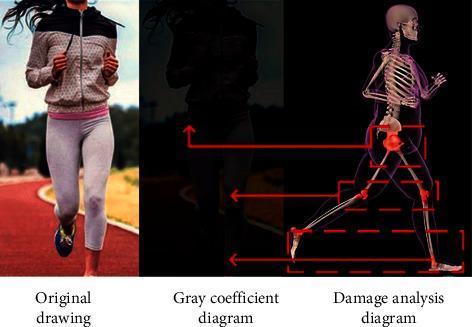
This composite educational graphic illustrates the transition from clinical observation to diagnostic injury analysis in sports medicine. It contains three side-by-side panels: an Original drawing, a Gray coefficient diagram, and a Damage analysis diagram. The 'Original drawing' is a clinical photograph showing a female runner on a track, serving as the baseline for activity monitoring. The 'Gray coefficient diagram' displays a segmented, low-contrast grayscale silhouette used for computer-aided image recognition and motion tracking. The 'Damage analysis diagram' provides a medical illustration of the runner's musculoskeletal system. This panel highlights high-stress anatomical regions susceptible to sports-related injury, specifically the hip joint (acetabulofemoral joint), knee joint (tibiofemoral joint), and ankle joint (talocrural joint). These areas are emphasized with red coloration and bounding boxes, connected via red arrows to the corresponding anatomical locations on the motion-tracking model. The image demonstrates the application of image recognition technology in identifying common student and adolescent sports injuries, focusing on joint biomechanics and physical health assessment.
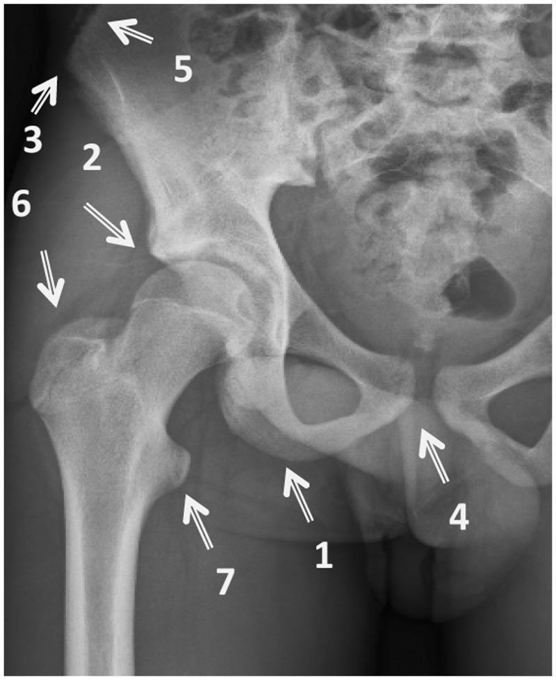
This diagnostic image is an anteroposterior (AP) radiograph of the right hip and pelvis, annotated to demonstrate common sites for avulsion fractures. The radiograph shows the bony anatomy of the pelvis, including the ilium, ischium, and pubis, along with the proximal femur. Seven specific anatomical landmarks where tendons or muscles attach are highlighted with arrows and numbers: (1) Ischial tuberosity (hamstrings), (2) Anterior inferior iliac spine (rectus femoris), (3) Anterior superior iliac spine (sartorius/tensor fasciae latae), (4) Pubic symphysis (adductors/gracilis), (5) Iliac crest (abdominal muscles), (6) Greater trochanter (gluteal muscles), and (7) Lesser trochanter (iliopsoas). The image serves as an educational diagram for sports medicine and orthopedics, illustrating the pathophysiology of traction-related injuries where forceful muscle contraction pulls off a bone fragment at its insertion point. It is intended for medical students and clinicians to aid in the identification of diagnostic hotspots in pelvic trauma cases.
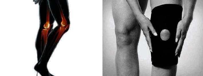
A dual-panel educational graphic illustrating knee anatomy and management of musculoskeletal injuries. The left panel features a stylized anatomical diagram of the lower extremities in profile. The skeletal structure is highlighted in orange against a black silhouette, clearly depicting the femur, tibia, fibula, patella, and the tibiofemoral joint space. The right panel is a clinical photograph in grayscale showing a patient applying or adjusting a neoprene compression knee brace. The brace features an open-patella design (a circular cutout) to stabilize the kneecap and alleviate pressure during physical activity or recovery from sprains, lacerations, or strains. The image demonstrates the relationship between internal joint anatomy and external orthopedic support. This content is relevant to sports medicine, orthopedics, and physical therapy education, focusing on injury prevention and the management of joint instability.
Common Sports Health Injuries
1. Ankle Sprain
- Management: For acute severe sprains, a below-knee cast or Aircast produces faster recovery than compression bandages alone; outcomes are similar at 9 months.
- Return to sport: Guided by functional rehabilitation and proprioceptive training.
Goldman-Cecil Medicine, p. 3848
2. Anterior Cruciate Ligament (ACL) Injury
- Commonly a non-contact injury from sudden deceleration, knee valgus, or rapid change of direction.
- A second blow before full recovery worsens outcomes significantly.
- Recent evidence: balance training programs reduce ACL incidence in soccer players (Su et al., 2025 - Systematic Review & Meta-Analysis, PMID 40051003).
Campbell's Operative Orthopaedics 15th Ed, Block 25
3. Hamstring Strain
- Ranges from mild muscle strain to complete musculotendinous tear.
- Usually occurs at the musculotendinous junction during sudden acceleration/deceleration.
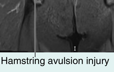
- In adolescents: ischial tuberosity avulsion fracture is more common due to apophyseal weakness.
- Complete recovery can take weeks to months.
- Management: Crutches, protected weight-bearing, sports medicine referral, progressive strengthening.
Gray's Anatomy for Students, p. 3643; Rosen's Emergency Medicine, Table 47.3
4. Quadriceps Strain / Tear
- Ambulation is significantly impaired; patient may be unable to actively extend the knee.
- A palpable depression proximal to the patella suggests a complete tear.
- Ultrasound confirms diagnosis; complete tears require urgent surgical repair.
Rosen's Emergency Medicine, Table 47.3
5. Concussion (Sports-Related TBI)
- Defined as a traumatically induced transient disturbance of brain function.
- More than 90% are NOT associated with loss of consciousness.
- Symptoms: headache, confusion, amnesia, nausea, balance problems, vision changes.
- CT and MRI are normal in concussion.
- Key rule: A player should NEVER return to play while symptomatic.
- Follow stepwise, graded return-to-play protocol when asymptomatic.
Textbook of Family Medicine 9e, p. 1503-1521
6. Rotator Cuff Injury (Shoulder)
- Ranges from impingement to partial/complete rotator cuff tears.
- Recent systematic review found multiple risk factors for shoulder injuries in female overhead athletes (Steele et al., 2025 - PMID 38898813).
Goldman-Cecil Medicine, p. 3520; Rosen's Emergency Medicine, Block 7
7. Muscle Strains - Other Lower Limb
| Muscle Group | Mechanism | Key Feature |
|---|---|---|
| Iliopsoas | Sudden forceful hip flexion | Groin/low back pain; avulsion of lesser trochanter |
| Hip adductors | Forceful abduction (straddle injury) | Medial groin pain; ecchymosis |
| Gluteus | Forced hip extension (jumping) | Less severe pain; tender on extension/abduction |
Rosen's Emergency Medicine, Table 47.3
8. Plantar Fasciitis
- Pain is worst in the first steps after waking, improving then worsening later in the day.
- Tenderness at the anteromedial calcaneal tubercle.
- Treatment: rest, NSAIDs, heel cord stretching, insoles, local corticosteroid injection.
Goldman-Cecil Medicine, p. 3853
9. Knee Dislocation
Summary Table
| Injury | Common Sports | Key Feature |
|---|---|---|
| Ankle sprain | All field sports, basketball | Lateral ligament; most common |
| ACL tear | Soccer, basketball, football | Non-contact; knee valgus |
| Hamstring strain | Sprinting, track | Posterior thigh; MTJ most vulnerable |
| Concussion | Contact sports | 90% without LOC; never RTP while symptomatic |
| Rotator cuff | Swimming, throwing sports | Impingement or tear |
| Plantar fasciitis | Running | Morning heel pain |
| Quadriceps tear | Jumping sports | Palpable defect; surgical if complete |
Describe role of sports health nurse
role of sports health nurse in athlete care responsibilities
sports nursing role athlete health care
https://www.carerev.com/blog/what-is-a-sports-medicine-nurse
Role of the Sports Health Nurse
1. Injury Assessment and Triage
- Conducts initial injury assessment at the sideline, clinic, or emergency setting
- Evaluates and triages sprains, strains, fractures, dislocations, lacerations, and concussions
- Differentiates between injuries requiring immediate medical transfer and those manageable on-site
- Collaborates with sports medicine physicians to develop individualized treatment plans
CareRev Sports Medicine Nursing Guide
2. Concussion Management
- Recognizes signs and symptoms: headache, confusion, amnesia, balance problems, nausea
- Applies standardized sideline assessment tools (symptom checklists, cognitive and balance tests)
- Enforces the cardinal rule: a player must never return to play while symptomatic
- Oversees a graduated return-to-play protocol progressing through:
- No activity
- Light aerobic exercise
- Sport-specific exercise
- Non-contact training drills
- Full-contact practice
- Return to competition (only when fully symptom-free)
- Monitors for second-impact syndrome - a catastrophic, high-mortality condition where a second head injury occurs before the first has resolved
Swanson's Family Medicine Review, p. 7863-7865; Textbook of Family Medicine 9e, p. 1503-1521
3. Preventive Care and Health Promotion
- Conducts or assists with pre-participation physical examinations (PPE) to screen for conditions that may pose risk during competition
- Identifies risk factors for injury (muscle imbalances, prior injury history, biomechanical issues)
- Educates athletes on:
- Proper warm-up and cool-down techniques
- Safe training loads and rest periods
- Correct sport technique and protective equipment use
- Nutrition, hydration, and sleep for performance and recovery
Credenza Health Sports Medicine Nursing Guide
4. Emergency Response
- Responds rapidly to cardiac arrest (AED use, CPR), heat stroke, severe fractures, anaphylaxis, and spinal injuries
- Recognizes and acts on exertional heat illness: any athlete showing mental status changes in a hot environment requires immediate removal from play and active cooling (ice-water immersion)
- Facilitates safe transfer to hospital when needed
Textbook of Family Medicine 9e, p. 1706-1712
5. Rehabilitation Support
- Assists in designing and monitoring progressive exercise rehabilitation programs
- Tracks recovery milestones and communicates progress to the medical team
- Coordinates with physiotherapists, strength coaches, and physicians
- Provides wound care, taping, bracing, and post-operative care
- Supports return-to-sport readiness assessments
6. Chronic Condition Management
- Manages conditions such as asthma, diabetes, hypertension, and overuse injuries (tendinopathy, stress fractures)
- Monitors medications (including ensuring compliance with anti-doping regulations)
- Conducts regular health screening and monitoring
7. Mental Health Support
- Identifies signs of anxiety, depression, eating disorders, and overtraining syndrome
- Provides initial support and facilitates referral to sport psychologists
- Supports athletes during periods of injury-related stress or performance pressure
8. Doping Prevention and Ethics
- Educates athletes about prohibited substances (WADA/national anti-doping codes)
- Ensures medications prescribed are compliant with anti-doping rules
- Maintains confidentiality and ethical standards in athlete health management
9. Documentation and Coordination
- Maintains accurate medical records and injury logs
- Coordinates care between physicians, physiotherapists, coaches, and athletic trainers
- Provides medical clearance documentation and liaison with insurance or governing bodies
Where Sports Health Nurses Work
| Setting | Examples |
|---|---|
| Professional sports teams | NFL, NBA, soccer clubs, cricket boards |
| Collegiate athletics | University sports programs |
| Sports medicine clinics | Outpatient orthopaedic and sports clinics |
| Schools and community sport | Youth leagues, school sports events |
| International events | Olympics, Commonwealth Games, World Cups |
| Military and occupational settings | Defence forces, industrial athletic programs |
Key Skills Required
- Advanced musculoskeletal assessment
- Emergency and trauma response (BLS, ACLS)
- Knowledge of sports physiology and biomechanics
- Communication with athletes, coaches, and multidisciplinary teams
- Cultural sensitivity (working with diverse athlete populations)
- Familiarity with anti-doping regulations