
Renal rickets renal osteosystrophy 15 marks structured answer what examiner expects
I'll start with a quick answer while I research the detailed content from medical textbooks.
Renal osteodystrophy (renal rickets) is the metabolic bone disease seen in chronic kidney disease, resulting from impaired phosphate excretion, reduced 1,25-(OH)₂ vitamin D synthesis, secondary hyperparathyroidism, and metabolic acidosis. A 15-mark structured answer should cover definition, pathophysiology, clinical features, investigations, and management.
Searching sources...
Reading File
Now let me search the medical library in parallel for comprehensive content.
Finding Sources
Finding Sources
Finding Sources
Excellent results. Now let me read the most relevant sections in parallel - the Nephrology textbook, Robbins Pathology, and Campbell's Orthopaedics:
Reading File
Reading File
Reading File
Excellent content from Robbins Pathology and Nephrology textbook. Let me now get the Rheumatology content and more detailed nephrology sections:
Reading File
Reading File
I have rich content. Let me get more on treatment/management and X-ray findings, then fetch the key pathophysiology diagram:
Reading File
Reading File
Loading Image
Excellent! I now have comprehensive content. Let me get the management/treatment and X-ray sections to complete the answer:
Reading File
Reading File
I now have comprehensive content from multiple authoritative textbooks. Let me compile the full structured answer.
Renal Rickets / Renal Osteodystrophy - 15 Mark Structured Answer
Sources: Robbins & Cotran Pathologic Basis of Disease | Comprehensive Clinical Nephrology 7th Ed | Rheumatology 2-Vol Set (Elsevier) | Harrison's Principles 22E
What the Examiner Expects (15 Marks)
A 15-mark answer needs: definition + classification (2 marks), pathophysiology (4 marks - the core differentiator), clinical features (3 marks), investigations (3 marks), management (3 marks). Precision in the pathophysiology cascade (FGF-23, Klotho, 1,25-(OH)₂D₃, PTH) is what separates high scorers.
DEFINITION
Renal osteodystrophy (ROD) is the collective term for all skeletal abnormalities arising from altered mineral and hormonal metabolism in chronic kidney disease (CKD). It represents the bone-specific component of the broader entity called CKD-Mineral and Bone Disorder (CKD-MBD), which also encompasses vascular calcification and cardiovascular disease. In growing children, the rickets-like deformities produced are called renal rickets.
- Robbins Pathologic Basis of Disease, p. 841
CLASSIFICATION (TMV System - KDIGO)
KDIGO proposed the TMV classification based on iliac crest bone histomorphometry, assessing three parameters:
| Parameter | Type | Features |
|---|---|---|
| Turnover (T) | High-turnover (osteitis fibrosa cystica) | Increased osteoclasts/blasts, peritrabecular fibrosis due to excess PTH |
| Low-turnover / Adynamic bone disease | Little osteoclast/osteoblast activity; seen with over-suppression of PTH | |
| Mineralization (M) | Osteomalacia | Excess osteoid accumulation, impaired mineralization |
| Volume (V) | Osteopenia/Osteoporosis | Diminished bone quantity |
A mixed pattern (osteitis fibrosa + osteomalacia together) = mixed uremic osteodystrophy.
PATHOPHYSIOLOGY
This is the highest-yield section. The diagram below illustrates the cascade:
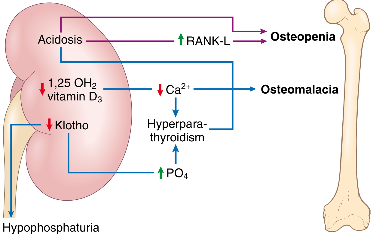
Four Interrelated Mechanisms:
1. Phosphate Retention and FGF-23 Axis
- As GFR declines, renal phosphate excretion falls → hyperphosphatemia
- Osteocytes respond by secreting FGF-23 (fibroblast growth factor-23) - the earliest biochemical change, rising from CKD stage 2
- FGF-23 normally acts via the co-receptor Klotho on renal tubules to promote phosphaturia (downregulating Na/Pi cotransporters IIa and IIc)
- In CKD, decreased renal Klotho expression blunts FGF-23 effectiveness → persistent phosphate retention
- Hyperphosphatemia itself stimulates PTH secretion, completing a vicious cycle
2. Impaired Vitamin D Activation
- The kidney is the primary site of conversion of 25-(OH)D → 1,25-(OH)₂D₃ (calcitriol) via 1α-hydroxylase
- In CKD: (a) GFR fall reduces delivery of substrate to proximal tubule 1α-hydroxylase; (b) FGF-23 directly inhibits 1α-hydroxylase and activates 24-hydroxylase (catabolizes active vitamin D); (c) phosphate retention further suppresses 1α-hydroxylase
- Calcitriol deficiency → reduced intestinal Ca²⁺ absorption → hypocalcemia → PTH stimulation
- Low calcitriol also releases the PTH gene from VDR-mediated suppression and reduces parathyroid VDR and CaSR expression, facilitating parathyroid cell proliferation
3. Secondary Hyperparathyroidism
- Driven by: hypocalcemia + low calcitriol + hyperphosphatemia + skeletal PTH resistance
- Parathyroid gland hyperplasia begins early (diffuse → nodular/monoclonal)
- PTH resistance in bone: down-regulation of PTH receptors, reduced calcitriol, phosphate retention
- Compensatory supranormal PTH is initially adaptive (promotes phosphaturia) but sustained high PTH → osteitis fibrosa cystica with peritrabecular fibrosis and increased fracture risk
- iPTH >600 pg/mL on dialysis is characteristic of osteitis fibrosa
4. Metabolic Acidosis
-
Renal tubular dysfunction → systemic acidosis
-
Acidosis upregulates RANKL expression on osteoblasts → increased osteoclast activity → bone matrix resorption and osteopenia
-
This mechanism is independent of PTH
-
Comprehensive Clinical Nephrology 7th Ed, pp. 1180-1184; Rheumatology 2-Vol Set, pp. 1862-1863
CLINICAL FEATURES
In Children (Renal Rickets):
- Growth retardation (common; partial improvement with calcitriol)
- Rachitic features: widened epiphyses, genu varum/valgum, metaphyseal fraying, frontal bossing
- Slipped capital femoral epiphysis
- Bone pain and tenderness
- Muscle weakness
In Adults:
-
Bone pain - nonspecific; lower back, hips, and legs; aggravated by weight bearing; can be debilitating
-
Pathological fractures - long bones, axial skeleton; kyphoscoliosis or chest wall deformity from vertebral fractures
-
Proximal myopathy - muscle weakness
-
Periarthritis - acute, localized pain from periarticular calcium phosphate crystal deposition (especially with marked hyperphosphatemia); mimics gout or pseudogout
-
Pruritus - due to hyperparathyroidism and calcium-phosphate deposition in skin
-
Calciphylaxis (calcific uremic arteriolopathy) - rare but life-threatening; ischemic skin necrosis from vascular calcification
-
Extraskeletal calcifications: vascular (arterial), pulmonary, myocardial, periarticular
-
Comprehensive Clinical Nephrology 7th Ed, p. 1181
INVESTIGATIONS
Biochemistry:
| Test | Finding |
|---|---|
| Serum phosphate | Elevated (normal until CKD stage 3-4) |
| Serum calcium | Low (or normal early); may be elevated with over-treatment |
| Intact PTH (iPTH) | Elevated; >600 pg/mL suggests osteitis fibrosa in dialysis patients |
| 25-(OH)D₃ | Reduced |
| 1,25-(OH)₂D₃ (calcitriol) | Reduced (not routinely used to differentiate lesion type) |
| Serum FGF-23 | Elevated early (CKD stage 2 onwards) |
| Alkaline phosphatase / Bone-specific ALP | Elevated (bone formation marker; useful when PTH result inconclusive) |
| Serum Al³⁺ | If aluminum-related bone disease suspected |
| ABG / serum bicarbonate | Metabolic acidosis |
Radiology:
- Subperiosteal erosions - most characteristic; radial side of middle phalanx of middle finger
- Rugger-jersey spine - alternating sclerosis and lucency of vertebral bodies
- Brown tumors - lytic lesions; can mimic metastases (see Fig. 88.10 from nephrology textbook)
- Looser's zones (pseudofractures) - in osteomalacia component; bilateral, symmetrical
- Rachitic changes in children: metaphyseal fraying, widening/cupping
- Vascular/soft tissue calcification on plain X-rays
- DXA scan - used for fracture risk assessment (DEXA Z-score <-2.5 seen in >50% of young adults with prior pediatric ESRD)
Bone Biopsy (Gold Standard):
-
Iliac crest biopsy with double tetracycline labeling (tetracycline given in two separated courses, biopsy 4 days after second course)
-
Tetracycline fluoresces at mineralization fronts - distance between bands = bone formation rate
-
Histology distinguishes: osteitis fibrosa, osteomalacia, adynamic bone disease, mixed pattern
-
Aluminum staining >15% of trabecular surface + bone formation rate <220 mm²/day = aluminum-related bone disease
-
Not done routinely; indicated when biochemistry is inconclusive and treatment decisions depend on histology
-
Comprehensive Clinical Nephrology 7th Ed, pp. 1182-1185
MANAGEMENT
Prevention is the primary goal - therapy should begin at CKD stage 3.
1. Phosphate Control
- Dietary phosphate restriction: limit dairy, meat, dark sodas, whole grains, processed foods
- Phosphate binders (taken with meals):
- Calcium-based: calcium carbonate, calcium acetate - affordable but risk of hypercalcemia and vascular calcification
- Non-calcium-based (preferred when hypercalcemia risk): sevelamer carbonate/hydrochloride
- Iron-based: sucroferric oxyhydroxide, ferric citrate - additional benefit of improving anaemia and lowering FGF-23
2. Correction of Hypocalcemia
- Calcium supplementation (elemental calcium 1-1.5 g/day) - but avoid in patients with vascular calcification risk
- Normalize calcium before starting vitamin D analogues
3. Vitamin D Supplementation
- Nutritional vitamin D (cholecalciferol/ergocalciferol): replenish 25-OH-D₃ stores
- Active vitamin D analogues: calcitriol (1,25-OH₂D₃), alfacalcidol (1α-OH-D₃), or paricalcitol
- Suppress PTH transcription via VDR, increase CaSR expression on parathyroid
- Paricalcitol has relatively less hypercalcemic effect than calcitriol
- Target: suppress secondary hyperparathyroidism, prevent parathyroid hyperplasia
4. Calcimimetics
- Cinacalcet - allosteric activator of calcium-sensing receptor (CaSR) on parathyroid cells → PTH suppression at lower calcium doses → less hypercalcemia risk
- Useful in patients with hypercalcemia or where vitamin D analogues are insufficient
5. Correction of Metabolic Acidosis
- Oral sodium bicarbonate supplementation
- Targets serum bicarbonate ≥22 mmol/L
6. Management of Adynamic Bone Disease
- Reduce/stop calcium-based binders and active vitamin D analogues
- Allow PTH to rise to physiological levels to restore bone turnover
7. Surgical: Parathyroidectomy
- Indications: severe, refractory secondary hyperparathyroidism; marked hypercalcemia despite medical therapy; calciphylaxis; very high PTH with nodular hyperplasia unresponsive to medical treatment
- Types: subtotal or total parathyroidectomy ± autotransplantation
8. In Children (Renal Rickets)
- Address underlying renal disease (transplantation is the definitive treatment)
- Calcitriol for growth improvement (partial)
- Orthopedic management for deformities: guided growth, corrective osteotomy, Ilizarov technique for severe angular deformity
HIGH-YIELD SUMMARY FOR EXAMINER
| Feature | Key Point |
|---|---|
| Earliest biochemical change | Elevated FGF-23 (CKD stage 2) |
| Gold standard diagnosis | Iliac crest bone biopsy with double tetracycline labeling |
| Most characteristic X-ray sign | Subperiosteal erosions (radial side of middle phalanx) |
| High-turnover vs. low-turnover PTH | >600 pg/mL = osteitis fibrosa; Low PTH = adynamic bone disease |
| Preferred phosphate binder (hypercalcemia) | Sevelamer (non-calcium-based) |
| Drug that works via CaSR | Cinacalcet (calcimimetic) |
| Definitive treatment in children | Renal transplantation |
| New terminology | CKD-MBD (broader); ROD (bone-specific component) |
Key sources consulted:
- Robbins & Cotran Pathologic Basis of Disease, p. 841-865
- Comprehensive Clinical Nephrology 7th Ed, pp. 1180-1190
- Rheumatology 2-Volume Set (Elsevier 2022), pp. 1862-1866
- Harrison's Principles of Internal Medicine 22E (2025)
This is a shared conversation. Sign in to Orris to start your own chat.