
from Guyton and Hall textbook of medical physiology, 4th South Asia edition, write a 5-mark short note on Circulatory Changes at Birth
Reading File
Finding Sources
Finding Sources
Finding Sources
Finding Sources
Finding Sources
Reading File
Reading File
Loading Image
I now have all the content needed from Guyton and Hall. Here is the complete short note:
Short Note: Circulatory Changes at Birth
(Guyton and Hall Textbook of Medical Physiology, 4th South Asia Edition - Chapter 84)
Fetal Circulation - A Brief Background
In fetal life, the lungs are non-functional and the placenta serves as the organ of gas exchange. Three special shunts characterize fetal circulation: the foramen ovale (between atria), the ductus arteriosus (between pulmonary artery and aorta), and the ductus venosus (bypassing the liver). Only about 12% of cardiac output goes to the lungs, while 55% flows to the placenta.
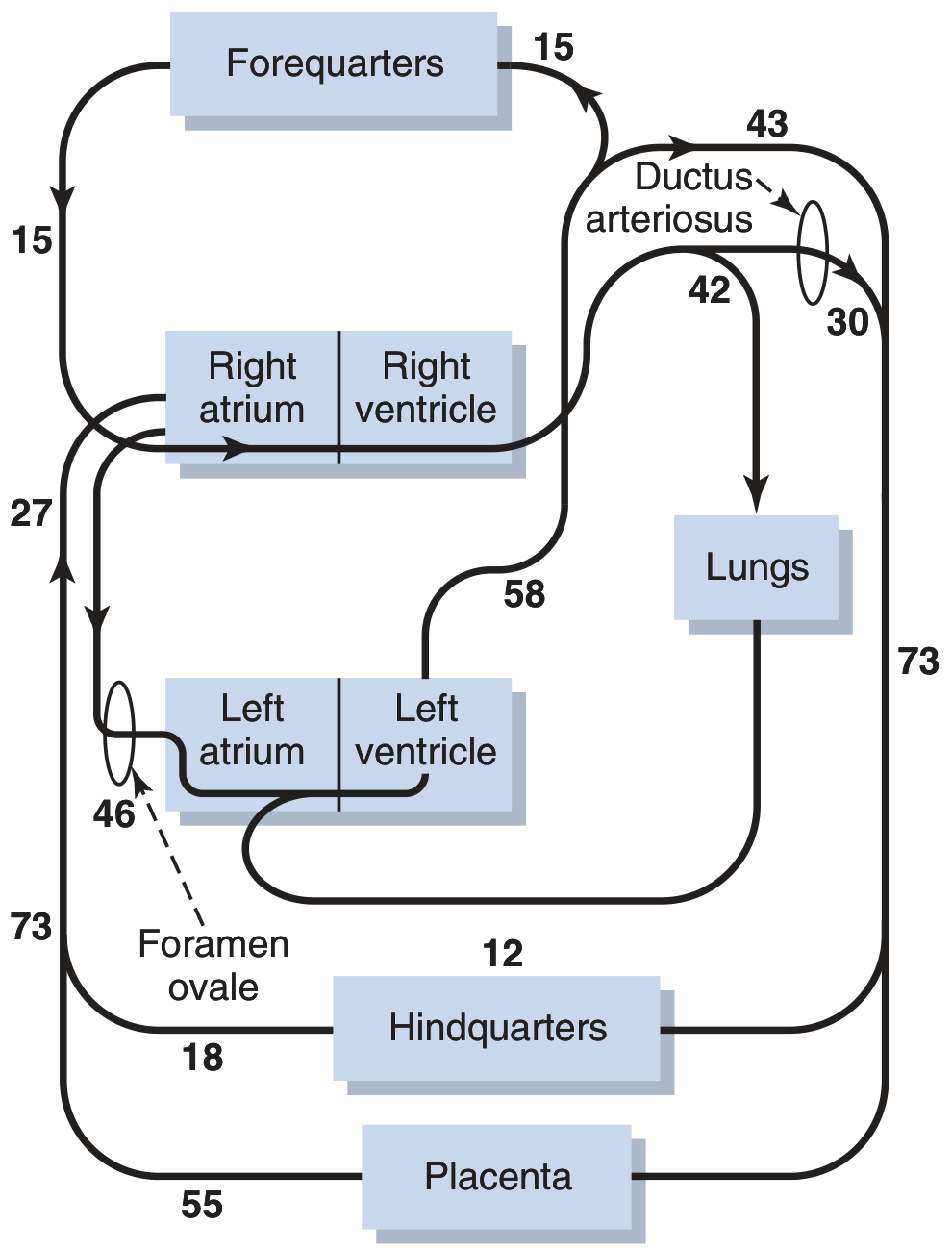
Figure 84.5 - Diagram of the fetal circulatory system. Numerals represent the percentage of total output from both sides of the heart flowing through each vascular area.
1. Changes in Pulmonary and Systemic Vascular Resistance
At birth, two major vascular resistance changes occur simultaneously:
- Systemic vascular resistance doubles - Loss of the large-volume, low-resistance placental circulation approximately doubles systemic vascular resistance. This raises aortic pressure and left atrial/ventricular pressure.
- Pulmonary vascular resistance decreases 5-fold - Expansion of the lungs decompresses previously compressed pulmonary vessels. Additionally, fetal hypoxia had caused tonic pulmonary vasoconstriction; aeration eliminates the hypoxia, causing vasodilation. Together, these reduce pulmonary arterial pressure, right ventricular pressure, and right atrial pressure.
2. Closure of the Foramen Ovale
In fetal life, blood flows from the right atrium to the left atrium through the foramen ovale (right atrial pressure > left atrial pressure). At birth, the reversal in pressure gradients - high left atrial pressure and low right atrial pressure - causes blood to attempt backward flow from left to right. This pushes a small valve (located on the left side of the atrial septum) over the foramen ovale, sealing it.
- In two-thirds of individuals, the valve becomes permanently adherent within a few months to years.
- In the remainder, the valve remains un-fused but stays closed as long as left atrial pressure remains 2-4 mm Hg above right atrial pressure (a condition termed patent foramen ovale, which is usually functionally insignificant).
3. Closure of the Ductus Arteriosus
In fetal life, blood flows from the pulmonary artery to the aorta via the ductus arteriosus (pulmonary artery pressure > aortic pressure). At birth, this gradient reverses - aortic pressure rises and pulmonary artery pressure falls - so blood now flows backward (aorta to pulmonary artery). This reversal triggers closure by two mechanisms:
- Oxygen effect: Fetal PO2 in the ductus is only 15-20 mm Hg. After birth, it rises to ~100 mm Hg. The elevated PO2 causes marked contraction of the ductal smooth muscle.
- Loss of PGE2: Prostaglandin E2 (PGE2) maintains ductal dilation in fetal life. After birth, PGE2 levels fall, removing this vasodilatory influence.
Functional closure occurs within 1-8 days. Anatomical (permanent) closure occurs within 1-4 months as fibrous tissue replaces the ductal wall. Failure to close results in a patent ductus arteriosus (PDA), treatable with indomethacin (a prostaglandin synthesis inhibitor).
4. Closure of the Ductus Venosus
In fetal life, the ductus venosus allows umbilical venous blood to bypass the liver and drain directly into the inferior vena cava. Immediately after birth, cessation of umbilical blood flow reduces ductal pressure and lumen diameter. Within 1-3 hours, the ductal smooth muscle contracts strongly, redirecting portal blood through the liver sinuses (portal venous pressure rises from ~0 to 6-10 mm Hg). Complete closure occurs within 2-18 days after birth.
Summary Table
| Structure | Fetal Function | Stimulus for Closure at Birth | Timeline |
|---|---|---|---|
| Foramen ovale | R→L atrial shunt | Reversal of atrial pressure gradient | Functional: immediate; Permanent: months-years |
| Ductus arteriosus | PA→Aorta shunt | ↑PO2 + ↓PGE2 | Functional: 1-8 days; Anatomical: 1-4 months |
| Ductus venosus | Bypasses liver | Loss of umbilical flow | 2-18 days |
- Guyton and Hall Textbook of Medical Physiology, pp. 1051-1052 (Chapter 84: Fetal and Neonatal Physiology)
This is a shared conversation. Sign in to Orris to start your own chat.